
Diagram of female reproductive system from netter's anatomy
Reading File
query_images_db
Finding Sources
Searching Images
labeled diagram of female reproductive system uterus ovaries fallopian tubes Netter anatomy
Searching PubMed
female reproductive anatomy
Searching the Web
Netter Atlas human anatomy female reproductive system diagram
A Netter illustration of the female reproductive system is available here:
For a more detailed pelvic view, see the Netter Anatomy Atlas entries for “Adult Uterus, Ovaries, and Uterine Tubes” and “Female Pelvic Relationships” listed in the Netter Anatomy Atlas 7e.
Compare Netter’s view with a simplified diagram
Reading File
Finding Sources
Searching the Web
Netter female reproductive system adult uterus ovaries uterine tubes illustration
Searching PubMed
female reproductive anatomy
Generating Image
A simple clean educational labeled diagram of the internal female reproductive system in front view, showing left and right ovaries, fimbriae, uterine tubes, uterus with fundus and uterine cavity, cervix, and vagina. Anatomically accurate but simplified, flat textbook-style illustration.
Netter anatomical view vs simplified diagram
| Feature | Netter view | Simplified diagram |
|---|---|---|
| Purpose | Detailed anatomical study | Rapid orientation and basic learning |
| Structures included | Uterus, uterine tubes, ovaries, fimbriae, ligaments, blood vessels, pelvic relationships, and surface detail | Main pathway only: ovaries, tubes, uterus, cervix, vagina |
| Anatomical realism | Three-dimensional, textured, and spatially realistic | Flat, schematic, and intentionally not to scale |
| Best for | Understanding location, attachments, relations, and dissection anatomy | Memorizing the sequence and identifying key organs |
| Potential difficulty | Extra structures can obscure the main reproductive pathway | May falsely suggest that the organs lie in one flat, perfectly symmetric plane |
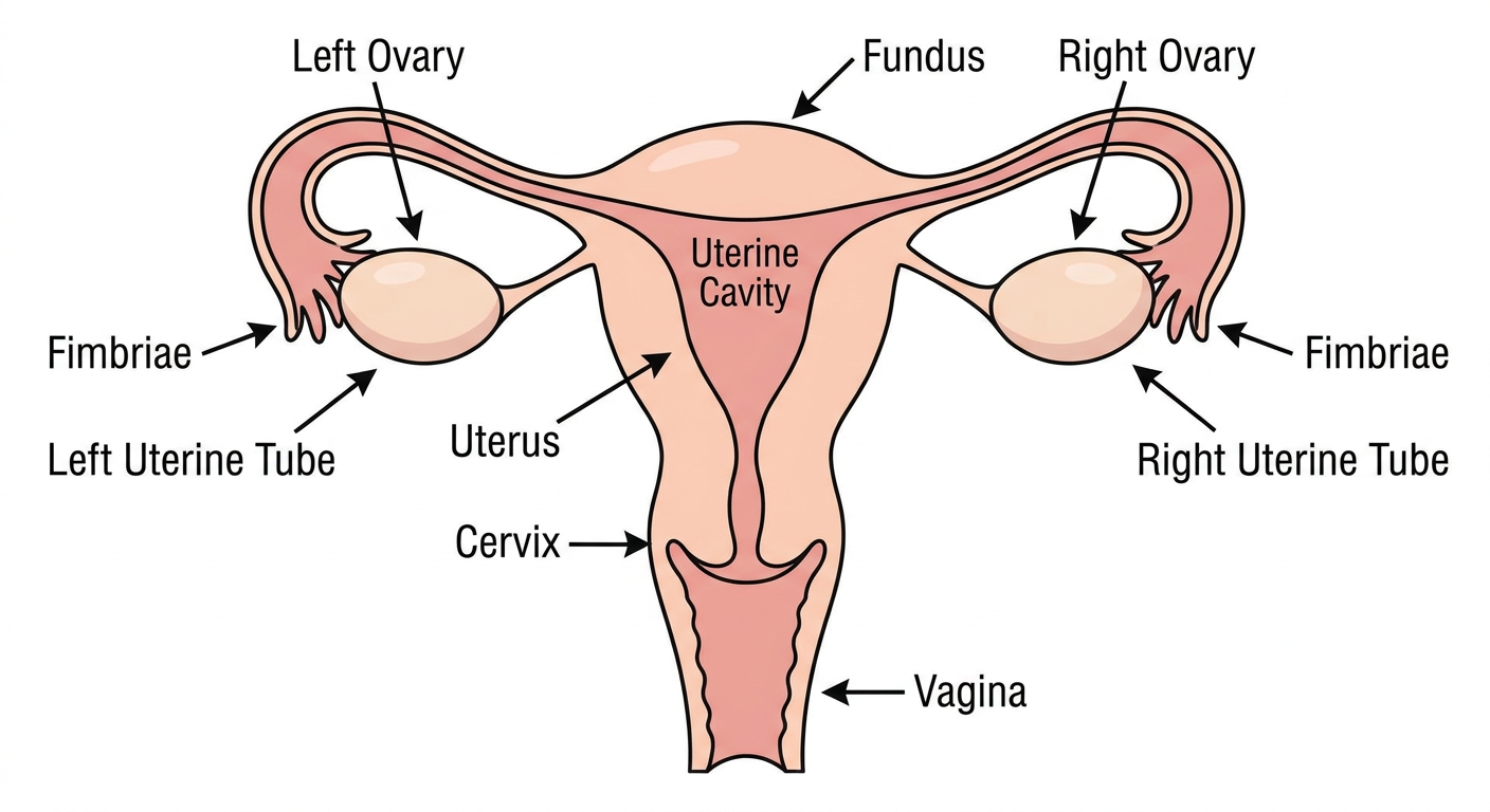
How to read them together: Use the simplified diagram first to follow the route: ovary -> fimbriae/uterine tube -> uterus -> cervix -> vagina. Then use Netter’s illustration to add the realistic spatial relationships, especially the lateral position of the ovaries and the detailed uterine-tube segments. The core organs in both views are the ovaries, uterine tubes, uterus, and vagina, as described in Guyton and Hall Textbook of Medical Physiology (female reproductive anatomy section).
The relevant Netter plate is “Adult Uterus, Ovaries, and Uterine Tubes,” available through Netter Images.
This is a shared conversation. Sign in to Orris to start your own chat.