
Stab injury for pedagogy for FMT viva
stab wound skin forensic autopsy
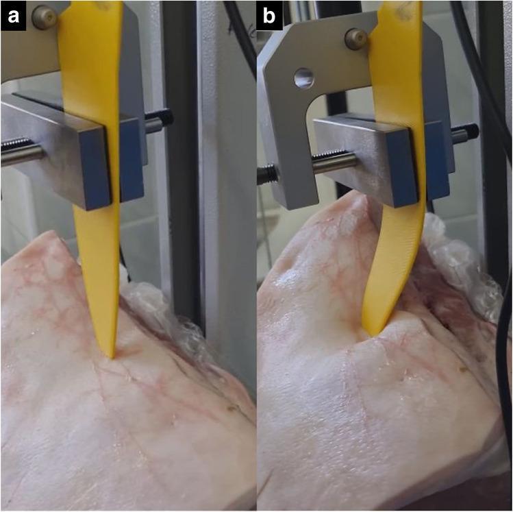
Two clinical photographs (a and b) demonstrate a dynamic stabbing force experiment using a 3D-printed yellow polylactic acid (PLA) replica knife and biological tissue (pork loin) to simulate forensic pathology scenarios. The setup utilizes a motorized test stand with a metallic clamping mechanism to control the downward displacement of the replica blade into a pre-existing stab wound. In image 'a', the replica knife is shown during initial entry, maintaining a straight, rigid vertical orientation. In image 'b', as the oversized replica reaches the boundaries of the original entrance wound, it exhibits significant lateral buckling and deformation due to tissue resistance. The pork loin displays visible skin tension and surface distortion around the wound site in response to the increased mechanical load. This experimental model is used to investigate the relationship between stabbing force, tool morphology, and wound characteristics in forensic medicine, specifically testing the fit and potential for wound alteration during autopsy reconstructions.
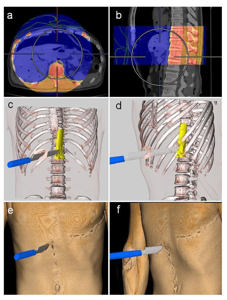
This composite image illustrates the use of post-mortem computed tomography (PMCT) and 3D volume rendering for forensic reconstruction of a fatal stab wound. Panels A and B show 2D axial and sagittal CT views, respectively, using a pseudocolor overlay to align pre-autopsy and intra-autopsy data at the level of the T11-T12 vertebrae. Panels C and D provide 3D volume-rendered reconstructions with transparent skin, highlighting the skeletal structures in white and the abdominal aorta in yellow. A digital model of the weapon (a knife) is superimposed, demonstrating its trajectory through the right hypochondriac region and its direct impact on the ruptured aorta anterior to the first lumbar vertebra. Panels E and F show the external 3D surface rendering with brown skin tones, depicting the entry site of the knife in the right hypochondrium, along with surgical markers of resuscitation, including a midline laparotomy scar and a left-sided thoracotomy scar. This visualization aids in defining the wound path and explaining the mechanism of exsanguination.
"stab wound" forensic pathology characteristics
Stab Injury — FMT Viva Notes
1. Definition
2. Key Terminology (always asked)
| Term | Meaning |
|---|---|
| Penetrating wound | Weapon enters a body cavity (thorax/abdomen) but does not exit |
| Perforating (transfixing) wound | Weapon passes through and through, creating both entry and exit |
| Incised-stab wound | Starts as incised, ends as stab (or vice versa) - occurs when the blade is thrust in and then withdrawn at an angle, or a nick/tail is seen at one end |
| Punctured wound | Umbrella term used interchangeably with stab wound in Indian texts (Dikshit) |
3. Description of a Stab Wound (systematic viva answer structure)
- Usually larger than exit (tapering weapon)
- Shape reflects blade cross-section:
- Single-edged knife → wedge-shaped, one angle acute + one blunt/notched
- Double-edged knife → elliptical/spindle-shaped, both angles acute
- Round pointed weapon → circular
- Square/cruciform weapon (e.g., screwdriver) → cruciate wound
- Two-pronged weapon (fork, scissors) → paired wounds (number of wounds ≠ number of thrusts)
- Margins clean-cut with sharp weapon; irregular/ragged with blunt-pointed weapon (e.g., screwdriver)
- Hilt/guard mark: a bruise or abrasion at the wound margin indicates the entire blade length entered ("full-thrust" mark) - important for estimating minimum blade length and force
- Wound gapes if it crosses Langer's lines transversely/obliquely; appears slit-like if parallel to them
- Probe gently (never a rigid metal probe before organ exam - risk of dislodging clots/false track); use a soft catheter or radio-opaque dye + X-ray to trace track
- Track length may exceed external wound depth if abdominal wall is compressed at the time of stabbing, or may be shorter than blade length if only partial penetration occurred
- Note organs transfixed, direction (upward/downward/medial/lateral), relation to body position (supine at autopsy vs upright at time of injury - respiration/posture changes internal organ relationships)
4. Estimating the Weapon from the Wound
- Length of wound ≈ breadth of blade (may be slightly less if blade entered obliquely, or more if blade was rocked/withdrawn at an angle)
- Both angles acute → double-edged weapon; one acute + one blunt/squared → single-edged
- Depth of track ≈ length of blade (if hilt mark present, at least full blade length penetrated)
- Skin elasticity means wound width can under- or over-estimate true blade width
5. Medicolegal Classification - Homicidal, Suicidal, Accidental (core viva topic)
| Feature | Homicidal | Suicidal | Accidental |
|---|---|---|---|
| Number/site | Multiple, scattered, on any accessible part incl. back | Single or few, over "sites of election" - precordium, left chest (heart), wrist, neck, abdomen | Usually single, situation inconsistent, no site of election |
| Clothing | Cut/damaged over the wound | Clothing often lifted/opened before self-stabbing, wound may not match cut in clothes | Clothing damage corresponds to accident circumstances |
| Defense wounds | Often present | Absent | Absent |
| Direction | Variable, often from front/side/back depending on assailant position | Fairly regular, controlled, matches hand used | Variable but weapon usually found anchored/embedded (fell onto object) |
| Site accessibility | Anywhere | Accessible to the person's own hand | Accessible per circumstance (fall, gore injury) |
| Weapon | Usually absent from scene or hidden | Often found in hand ("cadaveric spasm") or nearby | Found impaling/embedded at scene |
| Hesitation/tentative cuts | Absent | May be present nearby (trial cuts) | Absent |
6. Defense Wounds (frequently asked)
- Active defense wounds: victim grasps at the blade/weapon → cuts on palms, flexor surface of fingers
- Passive defense wounds: victim raises arms/legs to shield the body → cuts/stabs on extensor forearms, backs of hands, outer arms, lower legs
7. Postmortem vs Antemortem Stab Wounds
- Antemortem: bruised/hemorrhagic margins, everted/gaping edges, clotted blood
- Postmortem: pale, yellowish/tan, "parchmented," sharply defined margins, no true bruising (no vital reaction), minimal/no bleeding
8. Special/Named Wounds and Pitfalls
- Therapeutic/surgical artifact: a genuine homicidal stab wound may be incorporated into a surgical incision (e.g., thoracotomy) or a chest drain-site may mimic a stab wound - always correlate with hospital records before autopsy.
- Tailing: a small linear extension at one end of a stab wound from blade withdrawal at an angle - helps determine direction of withdrawal.
- Fatal stab wounds require surprisingly little force in areas overlying vital structures with thin overlying tissue (e.g., inter-costal spaces) - "force of stabbing" cannot be reliably quantitated from wound characteristics alone (DiMaio).
9. Sample Viva Questions
- Define a stab wound; differentiate it from an incised wound.
- Differentiate penetrating from perforating wounds with examples.
- How would you differentiate homicidal, suicidal, and accidental stab wounds?
- What is a defense wound? Classify it.
- What does the direction of a stab wound indicate? (Answer: relative position of assailant and victim)
- What is a hilt mark/guard mark and its significance?
- How do you estimate the dimensions of the weapon from a stab wound?
- What precautions are taken while removing a knife embedded in a body? (Grasp handle by thumb and index finger near the blade to preserve assailant's fingerprints)
- Describe "tailing" and incised-stab wounds.
- Short note on Hara-kiri.