
Classification and clinical features of cl and cp
I'll look up the classification and clinical features of cleft lip (CL) and cleft palate (CP) from the medical library.
Searching sources...
Reading File
Finding Sources
Finding Sources
Good matches found. Let me read the most comprehensive sources - Bailey & Love, Cummings, Scott-Brown, and KJ Lee in parallel.
Reading File
Reading File
Reading File
Good data from Bailey & Love and Cummings. Let me read more of Bailey & Love for clinical features, and fetch the key classification images.
Reading File
Loading Image
Loading Image
Excellent content gathered. Let me read more of the Bailey & Love text on clinical features of cleft palate and get the submucous cleft image from Cummings.
Reading File
Finding Sources
I now have comprehensive content from multiple authoritative sources. Let me compile the full answer.
Cleft Lip (CL) and Cleft Palate (CP) - Classification and Clinical Features
Cleft lip and/or palate (CL/P) is the most common congenital orofacial abnormality, occurring in approximately 1:600 live births. They may occur in isolation or as part of over 300 recognized syndromes.
Incidence Distribution
| Type | Frequency |
|---|---|
| Cleft lip alone | ~15% |
| Cleft lip + palate | ~45% |
| Isolated cleft palate | ~40% |
- Cleft lip ± palate is more common in males
- Isolated cleft palate is more common in females
Classification
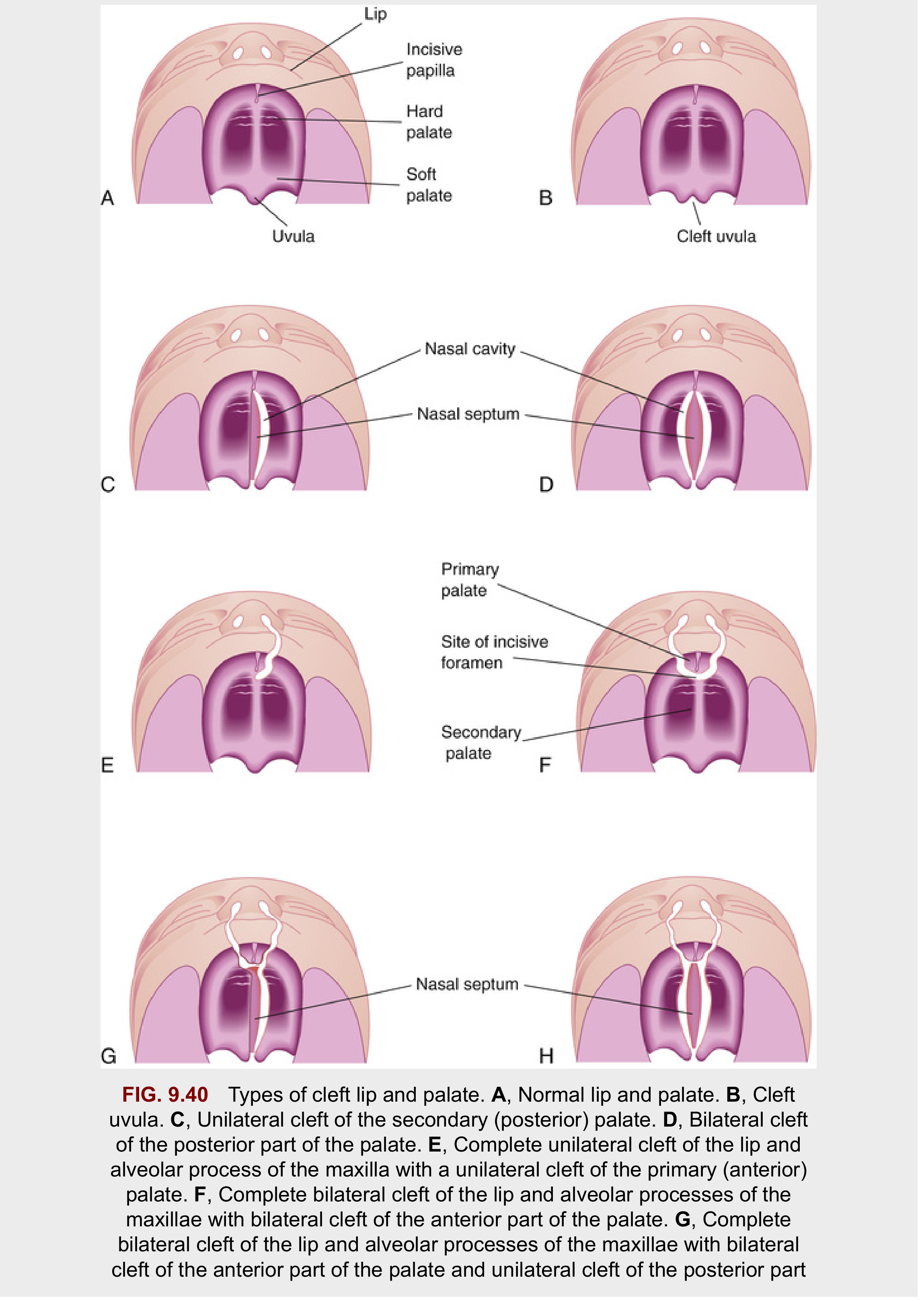
Anatomical Landmark - The Incisive Foramen
The incisive foramen is the key embryologic reference point dividing the palate into:
- Primary palate (anterior to incisive foramen): premaxilla, lip, nasal tip, and columella
- Secondary palate (posterior to incisive foramen): hard palate + soft palate, extending to the uvula
Two Major Phenotypic Groups
Group 1 - Cleft Lip with or without Cleft Palate (CL±P)
Involves structures anterior to the incisive foramen (primary palate), with or without extension posteriorly.
Group 2 - Isolated Cleft Palate (CP)
Involves the secondary palate only; embryologically and epidemiologically distinct from Group 1.
Classification of Cleft Lip
| Type | Description |
|---|---|
| Unilateral | One side only (specify left or right) |
| Bilateral | Both sides |
| Complete | Involves entire vertical height of upper lip, often with alveolar cleft |
| Incomplete | Involves only a portion of the lip height; a variable band of continuity remains across the cleft |
| Forme fruste / Microform | Minimal cleft - muscular diastasis with intact overlying skin only |
| Simonart band | A bridge of lip tissue (usually skin, sometimes with muscle) crossing an otherwise complete cleft gap |
(Source: Cummings Otolaryngology Head & Neck Surgery)
Classification of Cleft Palate
| Type | Description |
|---|---|
| Cleft uvula | Mildest form - bifid uvula only |
| Incomplete cleft palate | Soft palate ± part of hard palate; hard palate remains attached to nasal septum/vomer |
| Complete cleft palate | Full separation; nasal septum/vomer completely separated from palatine processes |
| Unilateral | Maxillary process on one side fused with nasal septum |
| Bilateral | No fusion of maxilla to nasal septum on either side |
| Submucous cleft palate (SMCP) | Least severe; intact mucosa but deficient/misdirected underlying musculature |
| Primary palate cleft | Anterior to incisive foramen (CL ± alveolus) |
| Secondary palate cleft | Posterior to incisive foramen (soft and/or hard palate) |
| Combined | Involves both primary and secondary palate |
(Source: Cummings Otolaryngology; Bailey & Love's Surgery)
Classification Diagram (The Developing Human - Moore)
Clinical Features
Cleft Lip
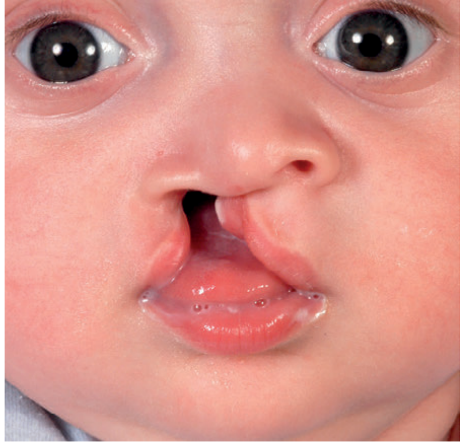
Unilateral Cleft Lip:
- Disruption of the nasolabial and bilabial muscle chains (orbicularis oris)
- The orbicularis muscle fibres, instead of running transversely, are misdirected and insert into the base of the columella on the cleft side and into the alar base
- Nasal deformity: the alar cartilage on the cleft side is displaced downward and outward; the columella is deviated away from the cleft
- The nasal tip is asymmetric, with a widened alar base on the cleft side
Bilateral Cleft Lip:
- Disruption is greater but often symmetrical
- Bilateral flaring of the nose (absence of nasolabial muscle continuity)
- Protrusive premaxilla - often projecting significantly forward
- Prolabium - a strip of skin in front of the premaxilla devoid of muscle
- Cartilaginous and skeletal deformities influence the overlying mucocutaneous tissues
Cleft Palate
Hard palate features:
- Failure of fusion of the two palatine shelves
- In complete cleft: the median part of the palatal vault is absent; palatal fibromucosa is reduced in size
- Communication between oral and nasal cavities
Soft palate features:
- In a normal palate, levator veli palatini fibres run transversely with no significant attachment to hard palate
- In a cleft palate, muscle fibres are oriented anteroposteriorly, inserting abnormally into the posterior edge of the hard palate
- This misdirected musculature critically impairs velopharyngeal closure
Submucous Cleft Palate (SMCP) - Classic Triad:
- Bifid uvula
- Zona pellucida - a bluish midline streak from deficient muscle under intact mucosa
- Notch in the posterior hard palate (palpable rather than visible)
(Note: Diagnosis of SMCP does not require all three features)
Functional Consequences
| Problem | Mechanism |
|---|---|
| Feeding difficulty | Inability to create negative intraoral pressure for suckling |
| Speech disorders | Velopharyngeal insufficiency; hypernasal speech |
| Recurrent otitis media / hearing loss | Eustachian tube dysfunction due to levator veli palatini abnormality |
| Dental abnormalities | Missing, supernumerary, or malformed teeth; malocclusion |
| Nasal deformity | Alar cartilage displacement, septal deviation |
| Psychosocial impact | Altered facial appearance and speech |
Associated Syndromes
Isolated cleft palate is more commonly syndromic than CL±P. Frequently associated syndromes include:
- Stickler syndrome (ophthalmic + musculoskeletal features)
- DiGeorge syndrome (cardiac + thymic anomalies; 22q11 deletion)
- Down syndrome (Trisomy 21)
- Apert syndrome (craniosynostosis + syndactyly)
- Treacher Collins syndrome (mandibulofacial dysostosis)
- Pierre Robin sequence (micrognathia + glossoptosis + CP)
Over 150 named syndromes are associated with cleft lip and palate. All children born with a cleft should be screened for other congenital abnormalities and offered genetic counselling where syndromic cause is suspected.
Sources: Bailey & Love's Short Practice of Surgery 28e, p. 762-765 | Cummings Otolaryngology Head & Neck Surgery, Ch. 188 | The Developing Human: Clinically Oriented Embryology, p. 537-541
This is a shared conversation. Sign in to Orris to start your own chat.