
Cholelithiasis - etiology pathogenesis morphology clinical features according to robbins textbook of pathology
Reading File
Finding Sources
Finding Sources
Searching PubMed
cholelithiasis
Searching the Web
Robbins Cotran Pathologic Basis of Disease cholelithiasis etiology pathogenesis morphology clinical features
Reading File
Reading File
Reading File
Reading File
Cholelithiasis (Gallstones)
Cholelithiasis is the presence of stones in the gallbladder. Robbins classifies them into:
- Cholesterol stones: contain >50% crystalline cholesterol monohydrate
- Pigment stones: composed mainly of calcium salts of bilirubin
More than 95% of biliary tract disease is attributable to gallstones. Robbins, Cotran & Kumar Pathologic Basis of Disease, pp. 810-812.
1. Etiology and risk factors
A. Cholesterol stones
They are most prevalent in the United States and Western Europe and make up about 90% of stones in these populations.
Risk factors
- Increasing age, especially middle to older age
- Female sex
- Estrogen exposure:
- Pregnancy
- Oral contraceptive use
- Obesity and metabolic syndrome
- Rapid weight loss
- Gallbladder stasis, whether neurogenic or hormonal
- Hyperlipidemic states
- Inborn disorders of bile acid metabolism
- Genetic predisposition, including variants in biliary lipid transport genes, especially ABCG8
- Higher prevalence in northern Europeans, North and South Americans, and Native Americans
Estrogen increases hepatic cholesterol uptake and biosynthesis, leading to excess cholesterol secretion into bile. Obesity and rapid weight loss similarly increase biliary cholesterol secretion.
B. Pigment stones
Pigment stones occur predominantly with increased unconjugated bilirubin in bile.
Risk factors
- Chronic hemolytic anemias
- Biliary tract infection
- Parasitic infestation of biliary tract
- Ileal disease, such as Crohn disease
- Ileal resection or bypass
- Cystic fibrosis with pancreatic insufficiency
Biliary infection with organisms such as Escherichia coli, Ascaris lumbricoides, and Clonorchis sinensis favors pigment-stone formation. Robbins, Cotran & Kumar Pathologic Basis of Disease, pp. 810-811.
2. Pathogenesis
A. Cholesterol stones
Normally, cholesterol remains soluble in bile by forming micelles with bile salts and lecithin. Gallstones form when bile becomes supersaturated with cholesterol, so cholesterol precipitates as cholesterol monohydrate crystals.
Four main factors are involved:
- Supersaturation of bile with cholesterol
- Gallbladder hypomotility or stasis
- Accelerated nucleation of cholesterol crystals
- Gallbladder mucus hypersecretion
Mucus traps the nucleated cholesterol crystals, allowing progressive deposition of cholesterol and formation of macroscopic stones.
Sequence:
Excess biliary cholesterol
→ cholesterol supersaturation
→ crystal nucleation
→ mucus trapping plus gallbladder stasis
→ accretion of cholesterol
→ gallstone formation
→ cholesterol supersaturation
→ crystal nucleation
→ mucus trapping plus gallbladder stasis
→ accretion of cholesterol
→ gallstone formation
B. Pigment stones
Pigment stones are mixtures of insoluble calcium salts of unconjugated bilirubin with inorganic calcium salts.
Mechanisms include:
- Hemolysis: increased bilirubin delivery and secretion into bile. A small proportion of bilirubin glucuronides is deconjugated in the biliary tree; during chronic bilirubin overproduction, enough unconjugated bilirubin becomes available to precipitate.
- Biliary infection: microbial beta-glucuronidase hydrolyzes bilirubin glucuronides to insoluble unconjugated bilirubin.
- Ileal dysfunction or bypass: increases risk through altered bile salt metabolism and bilirubin handling.
Robbins, Cotran & Kumar Pathologic Basis of Disease, p. 811.
3. Morphology
A. Cholesterol stones
- Arise exclusively in the gallbladder
- Range from nearly pure cholesterol to stones containing about 50% cholesterol
- Pure stones are:
- Pale yellow
- Round to ovoid
- Hard with a finely granular surface
- On cut section, show a glistening radiating crystalline palisade
- With calcium carbonate, calcium phosphate, and bilirubin incorporation:
- Become gray-white to black
- May be laminated
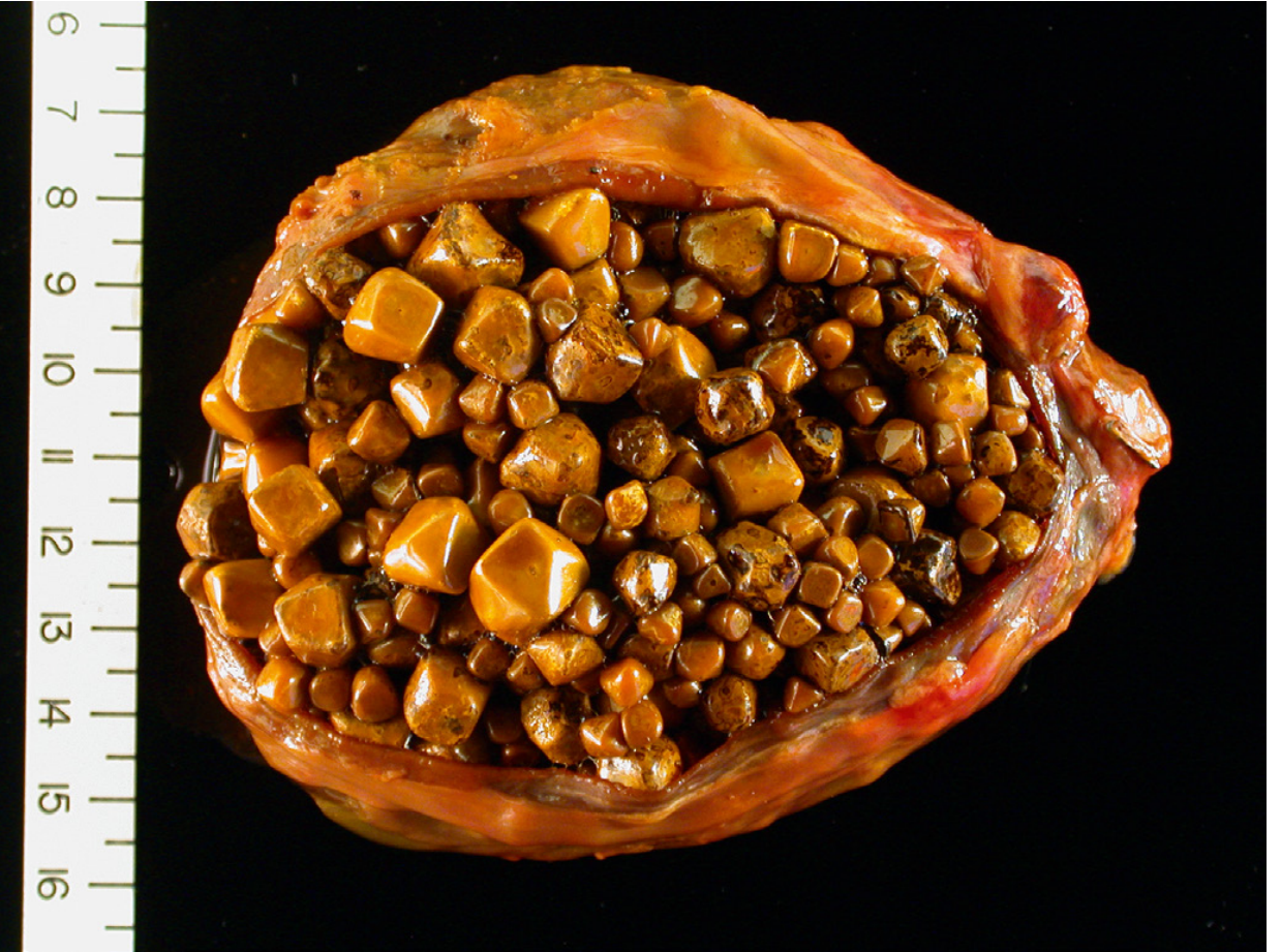
- Usually multiple and may reach several centimeters in diameter
- May have rounded or faceted surfaces due to close apposition with other stones
- Usually radiolucent
- About 10%-20% have enough calcium carbonate to become radiopaque
- Cholesterol-rich bile may also produce cholesterolosis, with foamy macrophages in the lamina propria of gallbladder mucosa.
B. Pigment stones
Pigment stones are brown to black.
| Feature | Black pigment stones | Brown pigment stones |
|---|---|---|
| Typical location | Sterile gallbladder bile | Infected large bile ducts |
| Composition | Oxidized polymers of calcium salts of unconjugated bilirubin, calcium carbonate/phosphate, mucin, minor cholesterol | Similar constituents plus cholesterol and calcium salts of palmitate and stearate |
| Gross appearance | Small, numerous, friable, spiculated/molded | Soft, laminated, greasy or soap-like |
| Size | Usually <1.5 cm | Variable |
| Radiology | 50%-75% radiopaque | Usually radiolucent |
Mucin glycoproteins form the structural scaffold and interparticle cement in all gallstones. Robbins, Cotran & Kumar Pathologic Basis of Disease, pp. 811-812.
4. Clinical features
- Gallstones can remain present for decades without symptoms.
- 70%-80% of affected individuals remain asymptomatic for life.
- Asymptomatic patients become symptomatic at an average rate of up to 4% per year, with decreasing risk over time.
Biliary colic
The usual symptomatic presentation is biliary “colic,” though the pain is typically constant rather than truly colicky.
Typical features
- Often precipitated by a fatty meal
- Gallbladder contraction forces a stone against the gallbladder outlet
- Causes increased intraluminal pressure and severe pain
- Pain occurs in the:
- Right upper quadrant, or
- Epigastrium
- May radiate to the:
- Right shoulder
- Back
Complications
- Acute or chronic cholecystitis
- Empyema of gallbladder
- Perforation
- Cholangitis
- Obstructive cholestasis
- Acute pancreatitis
- Cholecystoenteric fistula
- Gallstone ileus, including Bouveret syndrome
- Increased risk of gallbladder carcinoma
Small stones are more dangerous for ductal obstruction because they can enter the cystic duct or common bile duct. A large stone can erode into adjacent bowel and cause intestinal obstruction.
Robbins, Cotran & Kumar Pathologic Basis of Disease, p. 812.
A recent review topic that may be relevant only in selected settings is ceftriaxone-associated cholelithiasis in children, reported in a 2025 systematic review; it does not alter the core Robbins pathology framework above.
This is a shared conversation. Sign in to Orris to start your own chat.