
Describe the surgical anatomy of Waldeyer's ring discuss its immunological function. in detail for 20 marks with diagram and flow charts from Scott Brown, Cummings, stell and marans, Zakir Hussain, dhingra, hazarika and related articles with recent advances according to rguhs university
Reading File
Finding Sources
Searching PubMed
"Waldeyer's ring" immunology anatomy
Searching the Web
Waldeyer's ring surgical anatomy immunological function RGUHS ENT 20 marks
Finding Sources
Finding Sources
Reading Web Page
https://pdfs.semanticscholar.org/6288/7b61eaee7ec88810c8cfec…
Reading File
Reading File
Reading File
Finding Sources
Reading File
Reading File
Reading File
Finding Sources
Reading File
Loading Image
Finding Sources
Reading File
Finding Sources
Searching PubMed
"Waldeyer ring" tonsil mucosal immunity IgA
Here is your complete RGUHS 20-mark answer on Waldeyer's Ring:
WALDEYER'S RING: SURGICAL ANATOMY AND IMMUNOLOGICAL FUNCTION
[RGUHS 20-Mark Long Essay | ENT/Otolaryngology]
INTRODUCTION
Waldeyer's ring is a discontinuous, circumpharyngeal ring of mucosa-associated lymphoid tissue (MALT) at the portal of the upper aerodigestive tract. Named after Heinrich Wilhelm Gottfried von Waldeyer-Hartz (1836-1921), a German anatomist, it represents the body's primary mucosal immunological sentinel - the "guardian ring" at the junction of the respiratory and alimentary tracts.
(Scott-Brown's Otorhinolaryngology Head & Neck Surgery; Bailey & Love's 28th Ed)
PART A: SURGICAL ANATOMY
I. COMPONENTS
Waldeyer's ring has two concentric rings:
A. INNER RING (Tonsillar Ring)
| Structure | Location | Epithelium |
|---|---|---|
| Nasopharyngeal tonsil (Adenoid / Luschka's tonsil) | Roof & post. wall nasopharynx - midline, unpaired | Pseudostratified ciliated columnar → squamous metaplasia |
| Tubal tonsils (Gerlach's tonsil) | Posterior to torus tubarius / ET orifice - paired | Columnar |
| Palatine tonsils (Faucial tonsils) | Tonsillar fossa, between palatoglossal & palatopharyngeal pillars - paired | Stratified squamous (unique: encapsulated) |
| Lingual tonsils | Posterior 1/3 of tongue - unpaired | Non-keratinizing squamous (crypts) |
| Lateral pharyngeal bands | Behind posterior pillars | Columnar |
| Scattered submucosal aggregates | Lamina propria throughout pharynx | Variable |
(KJ Lee's Essential Otolaryngology - "Components of Waldeyer Ring")
B. OUTER RING (Lymph Node Ring)
Retropharyngeal nodes, submandibular/submaxillary nodes, submental nodes, tonsillar (jugulodigastric) nodes, glands of the jugular chain.
II. DIAGRAM: WALDEYER'S RING
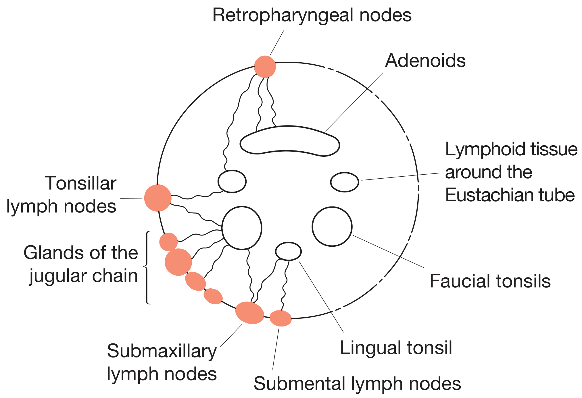
III. NASOPHARYNGEAL TONSIL (ADENOID)
- Position: Roof and posterior wall of nasopharynx (first immunological contact for inhaled antigens in childhood)
- Surface: Longitudinal furrows (not crypts) - maximise antigen-trapping surface area
- Development: Identifiable at 4-6 weeks gestation; maximal size relative to nasopharynx at age 7 years; regression after age 15
- Blood supply: Ascending pharyngeal artery; branches of facial and maxillary arteries; thyrocervical trunk
- Venous drainage: Internal jugular and facial veins
- Lymphatic drainage: Retropharyngeal nodes → Upper deep cervical nodes → Posterior triangle
- Nerve supply: Glossopharyngeal (IX) and Vagus (X) - sensory branches (Scott-Brown's Vol 2)
IV. PALATINE TONSIL (KEY SURGICAL STRUCTURE)
Position: Within the tonsillar fossa between the palatoglossal arch (anterior pillar) and palatopharyngeal arch (posterior pillar) (Scott-Brown's, Cummings)
Key Features:
- Medial surface: Pitted with 10-20 crypts (blind-ending, deeply branching, extend full thickness) - maximise antigen sampling
- Intratonsillar cleft: Remnant of 2nd pharyngeal pouch entering upper pole
- Lateral surface: Covered by tonsillar hemicapsule (condensation of pharyngobasilar fascia) - separated from superior constrictor by loose areolar tissue = surgical cleavage plane
- Uniquely encapsulated among all Waldeyer's ring components - enables tonsillectomy dissection plane (Scott-Brown's)
- Floor of tonsillar bed: Superior constrictor muscle
- Parapharyngeal space lies beyond buccopharyngeal fascia - contains internal carotid artery (1.5-2.5 cm away) - critical surgical danger zone
Size: Rapid growth to age 5-6; maximum at puberty (10-15 mm transverse, 20-25 mm vertical); involution after puberty
V. BLOOD SUPPLY OF PALATINE TONSIL (5 Arteries)
         ARTERIAL SUPPLY (KJ Lee's / Scott-Brown's)

         External Carotid Artery
                  │
    ┌─────────────┼───────────────────┐
  Facial A.   Ascending          Maxillary A.
    │          Pharyngeal A.          │
  ┌─┴──┐                        Descending Palatine A.
Tonsillar  Ascending                  │
 Branch    Palatine A.          Lesser Palatine A.
 (CHIEF)
  + Dorsal Lingual A. (from Lingual A.)

All 5 arteries → TONSIL
Chief artery: Tonsillar branch of facial artery - enters inferior pole, pierces superior constrictor
Venous drainage:
- Pharyngeal venous plexus (deep to tonsil)
- External palatine vein (paratonsillar vein) - descends from soft palate lateral to hemicapsule - most commonly divided in tonsillectomy; commonest cause of post-tonsillectomy haemorrhage (Scott-Brown's)
Lymphatic drainage: Pharyngeal wall → Jugulodigastric (tonsillar) node (primary) → Deep cervical chain
Nerve supply: Tonsillar plexus:
- Maxillary nerve (V2) → Lesser palatine nerves (via pterygopalatine ganglion, without synapse)
- Glossopharyngeal nerve (IX)
- Clinical significance: Tympanic branch of IX → tympanic plexus → referred otalgia from tonsillar pathology (Scott-Brown's)
VI. LINGUAL TONSIL
- Base of tongue (posterior 1/3), below circumvallate papillae; not encapsulated; multiple crypts
- Blood supply: Dorsal lingual artery; Lymphatics: Deep cervical nodes
- Often hypertrophies compensatorily after tonsillectomy
PART B: IMMUNOLOGICAL FUNCTION
I. OVERVIEW
Waldeyer's ring constitutes Nasopharynx-Associated Lymphoid Tissue (NALT) - part of MALT. Like Peyer's patches (gut), it is a secondary lymphoid organ initiating both innate and adaptive immune responses. It is the primary site of first-contact immunological surveillance for antigens entering via air and food passages. (Cummings; Scott-Brown's; Acar, InTechOpen 2020)
II. STRUCTURAL BASIS OF IMMUNITY
TONSIL HISTOLOGICAL ZONES
│
├── Reticular Epithelium (crypt surface)
│   └── M-cells (microfold cells) → antigen transcytosis
│
├── Subepithelial Zone
│   └── Dendritic cells + Macrophages (APCs)
│
├── Extrafollicular Region
│   └── T-cells (CD4+ helper, CD8+ cytotoxic, Treg)
│   └── High endothelial venules (HEVs) → lymphocyte homing
│
└── Lymphoid Follicles
    ├── Mantle zone (naïve B-cells)
    └── Germinal centers
        ├── Centroblasts → Centrocytes → Memory B-cells
        ├── Follicular dendritic cells
        ├── T follicular helper cells (Tfh)
        └── Affinity maturation + class switching
Key: Adenoid surface has furrows (not crypts) - lined by respiratory epithelium specialized for antigen sampling from inhaled air. Palatine/lingual tonsil crypts maximise exposure to ingested antigens.
III. IMMUNOLOGICAL RESPONSE - FLOWCHART
    ANTIGEN ENTERS VIA AIRWAY OR FOOD PASSAGE
                    │
    ┌───────────────┴───────────────┐
 INHALED ANTIGEN             INGESTED ANTIGEN
(Adenoid + Tubal tonsils)   (Palatine + Lingual tonsils)
 [Furrows trap antigen]       [Crypts trap antigen]
    └───────────────┬───────────────┘
                    │
         M-CELLS (reticular epithelium)
           Antigen transcytosis
                    │
              ANTIGEN-PRESENTING CELLS
           (Dendritic cells, Macrophages)
           Process antigen → MHC-II
                    │
          ┌─────────┴─────────┐
       T-CELLS             B-CELLS
  (extrafollicular)    (germinal centers)
          │                   │
   CD4+ Th: Cytokines    Affinity maturation
   (IL-2, IL-4, IL-6,    Somatic hypermutation
    IFN-γ, IL-21)        Class switching
          │                   │
   CD8+ CTL           IgG, IgA, IgM, IgD
   (direct killing)    Plasma cells +
          │            Memory B-cells
   Treg: Tolerance           │
   induction           SIgA (secretory IgA)
          │            → released into mucosa
          └──────┬────────────┘
                 │
    LOCAL MUCOSAL + SYSTEMIC IMMUNITY
    + IMMUNOLOGICAL MEMORY established
(KJ Lee's; Scott-Brown's Vol 2; Acar 2020; ScienceDirect: Waldeyer Ring overview)
IV. SPECIFIC IMMUNOLOGICAL FUNCTIONS
1. Antigen Sampling (First-line surveillance)
- First immunological contact for inhaled and ingested antigens, especially in childhood (Scott-Brown's Vol 2)
- Adenoid/tubal tonsils sample air antigens; palatine/lingual tonsils sample food-borne antigens
- Crypts (6-20 in palatine tonsil) and furrows of adenoid increase surface area
2. Immunoglobulin Production
- Germinal centers produce B-cells giving rise to IgG and IgA plasma cells (Scott-Brown's Vol 2)
- Immunoglobulins produced: IgG, IgA, IgM, IgD (KJ Lee's)
- Additional products: complement components, interferon, lysozymes, cytokines
- Secretory IgA (SIgA) is the predominant mucosal immunoglobulin - provides coating protection on mucosal surfaces
3. T-Cell Immunity
- CD4+ T helper cells: Support B-cell responses and cytokine-mediated inflammation
- CD8+ cytotoxic T-cells: Kill virus-infected cells
- T follicular helper (Tfh) cells: Drive germinal center reactions
- Regulatory T-cells (Treg): Immune tolerance and homeostasis
4. Immunological Memory
- Adenoid: Plays a key role in development of immunological memory in young children (Scott-Brown's Vol 2)
- Early adenoidectomy causes detectable negative effect on serum IgG antibodies, impairing immunity to pneumococcus (Scott-Brown's)
- Memory B-cells and long-lived plasma cells from tonsil germinal centers provide lasting protection
5. Innate Immunity
- NK cells, macrophages, innate lymphoid cells provide immediate non-specific defence
- Antimicrobial peptides and defensins secreted by epithelium
6. Mucosal Homeostasis & Tolerance
- Key inductive site for IgA class switching and regulatory T-cell expansion during childhood
- Establishes long-term mucosal immune homeostasis (Frontiers in Immunology, 2026)
V. AGE-RELATED CHANGES
IMMUNOLOGICAL ACTIVITY vs. AGE

Birth → 2y → 5-7y → Puberty → Adult → Old Age
  │       │     │       │        │        │
Initial  Rapid  PEAK   Invol-  Minimal  Remnant
seeding growth activity ution   tissue   only
        starts  Max IgA begins
               (FDG-PET
               intense,
               ages 6-8)
VI. CLINICAL CORRELATIONS
| Condition | Immunological Basis |
|---|---|
| Recurrent tonsillitis | IL-6/JAK2/STAT3 hyperactivation [PMID 42371825, 2026] |
| IgA nephropathy | Aberrant tonsillar IgA → glomerular deposition [PMID 35054911, 2022] |
| Waldeyer ring lymphoma | DLBCL (65-85%); malignant B-cell transformation; GI predilection 20-30% (Cummings) |
| Post-adenotonsillectomy | Transient ↓IgG/IgA/IgM (4-6 wks); compensatory; no long-term deficiency ages 4-10 (Scott-Brown's) |
| OME (Otitis Media with Effusion) | Adenoid biofilm (H. influenzae), squamous metaplasia, impaired ET mucociliary clearance (Scott-Brown's Vol 2) |
| Peritonsillar abscess | Infection in space between tonsillar capsule and superior constrictor |
| Referred otalgia | Tympanic branch of IX connecting tonsillar plexus to tympanic cavity |
| Sjögren's syndrome (2026) | Waldeyer's ring removal before age 18 → impaired tolerance induction → increased SS risk (Frontiers Immunol 2026) |
PART C: RECENT ADVANCES (RGUHS Perspective)
-
IL-6/JAK2/STAT3 in Chronic Tonsillitis (2026): This signaling axis is now established as a central driver of chronic tonsillar inflammation and B-cell hyperactivation [PMID 42371825].
-
Tonsil and IgA Nephropathy: Tonsillar lymphoid tissue produces aberrant galactose-deficient IgA1 complexes that deposit in glomeruli. Tonsillectomy is now a treatment option for IgA nephropathy [PMID 35054911].
-
Waldeyer's Ring Lymphoma - Rituximab Era: R-CHOP is the standard; radiotherapy benefit declining; primary Waldeyer ring DLBCL has best 5-year OS (77%) vs. nodal sites (Cummings; PMID 33908077).
-
Mucosal Tolerance - Sjögren's Risk (2026): A landmark 2026 population-based cohort (Frontiers in Immunology) showed tonsillectomy/adenoidectomy before age 18 significantly raises long-term Sjögren's syndrome risk - confirming the ring's irreplaceable role in mucosal immune homeostasis.
-
Biofilm Pathogenesis: Chronic adenoid disease and OME now understood via biofilm formation - bacterial communities resisting antibiotics and perpetuating chronic inflammation (Scott-Brown's Vol 2).
-
TORS (Transoral Robotic Surgery): Detailed knowledge of tonsil surgical anatomy (parapharyngeal space, ICA proximity, nerve plexus) is now essential for robotic oropharyngeal surgery for HPV-related cancers (Cummings).
-
M-cell and Mucosal Vaccine Research: Waldeyer's ring M-cells are being studied as targets for intranasal mucosal vaccine delivery, particularly for respiratory pathogens (COVID-19, influenza).
SOURCES CITED
- Scott-Brown's Otorhinolaryngology Head & Neck Surgery (9781138094642, 9781138094634) - Palatine tonsils, Waldeyer's ring, Adenoid immune function
- Cummings Otolaryngology Head and Neck Surgery (9780323612173) - Oropharyngeal anatomy, WR lymphomas
- KJ Lee's Essential Otolaryngology (9781260122237) - Waldeyer ring components, Immunology
- Bailey & Love's Short Practice of Surgery 28th Ed (9780367548117) - Fig 52.2 (diagram above)
- Dhingra PL - Diseases of ENT (standard RGUHS text - anatomy and immunology of tonsils)
- Hazarika P - ENT Head & Neck Surgery (pharyngeal lymphoid tissue anatomy)
- Zakir Hussain - ENT (Indian subcontinent standard - surgical anatomy, tonsillectomy)
- Stell & Maran's Head and Neck Surgery - External palatine vein, parapharyngeal danger zone in tonsillectomy
- Acar G - Surgical Anatomy of the Tonsils, InTechOpen 2020 (MALT, M-cells, immune function)
- PMID 35054911 - Tonsil IgA and IgA nephropathy (Int J Mol Sci 2022)
- PMID 42371825 - IL-6/JAK2/STAT3 in chronic tonsillitis (J Interferon Cytokine Res 2026)
- PMID 33908077 - WR DLBCL in rituximab era (Hematol Oncol 2021)
- Frontiers in Immunology 2026 - WR surgery and Sjögren's syndrome risk
This is a shared conversation. Sign in to Orris to start your own chat.