
IMRT VS 3D conformal Radiotherapy in detail in simplified manner also give detailed simplified answer on IMRT AND EBRT separately frpm standard ENT textbook
I'll start by reading the medical library skill, then search for detailed content on IMRT, 3D-CRT, and EBRT from ENT textbooks.
Reading File
Now let me run parallel searches across the library for IMRT, 3D-CRT, and EBRT in ENT/head and neck contexts.
Finding Sources
Finding Sources
Finding Sources
Excellent! Found key sections in Scott-Brown's. Now let me read the most relevant blocks in parallel.
Reading File
Reading File
Finding Sources
Now let me read the core sections - the physics/planning chapter (block4) for detailed technical content on IMRT vs 3D-CRT, and the EBRT section.
Reading File
Reading File
Reading File
Now let me read the key physics section on IMRT vs 3D-CRT from block4, and also read more of block3 for IMRT planning details.
Reading File
Reading File
Excellent content found. Now let me also read the block7 section on radiotherapy (which mentions PARSPORT and parotid-sparing) and look for a dedicated EBRT section.
Reading File
Finding Sources
Good - found the Grainger & Allison's radiology textbook that has a table listing types of radiotherapy including 3D-CRT and IMRT. Also found the Campbell Walsh Urology section. Let me read the Grainger table and the urology physics section for good 3D-CRT vs IMRT technical content.
Reading File
Reading File
I now have comprehensive content. Let me also quickly fetch the IMRT plan image from Scott-Brown's and the Grainger IMRT image to include in the answer.
Loading Image
Loading Image
I now have all the information needed for a comprehensive, well-illustrated answer. Let me compose the full detailed response.
IMRT, 3D Conformal Radiotherapy, and EBRT - A Detailed Simplified Guide
Sources: Scott-Brown's Otorhinolaryngology Head & Neck Surgery, 8th Ed; Grainger & Allison's Diagnostic Radiology; Campbell-Walsh Urology
Part 1: External Beam Radiotherapy (EBRT) - The Big Picture
What is EBRT?
EBRT means radiation is generated outside the body by a machine (a linear accelerator, or LINAC) and directed as a beam into the patient. This is the opposite of brachytherapy (where radioactive seeds/sources are placed inside the body).
A LINAC works by accelerating electrons at high speed into a tungsten target, which then emits high-energy X-ray photons (typically 6 megavolts for head and neck). These photons penetrate deep into body tissue and deposit energy, causing DNA double-strand breaks in tumour cells, which kills them.
Key concept: Therapeutic ratio
The goal of all EBRT is to maximize dose to the tumour while minimizing dose to surrounding normal tissue. The ratio of benefit (tumour kill) to harm (normal tissue damage) is the "therapeutic ratio." Every advance in radiotherapy technology has been aimed at improving this ratio.
- Scott-Brown's Otorhinolaryngology Head & Neck Surgery, chap. 19
EBRT in Head and Neck Cancer (ENT Context)
In ENT/head and neck surgery, EBRT is used for:
- Primary treatment of cancers (larynx, oropharynx, nasopharynx, hypopharynx, oral cavity)
- Post-operative (adjuvant) treatment after surgery
- Palliative treatment for advanced disease
- Treatment of carcinoma of unknown primary (CUP) - where EBRT fields cover bilateral neck nodes and mucosal sites
Standard dose: Radical (curative) dose is typically around 65-70 Gy delivered in 30-35 daily fractions of about 2 Gy each (conventionally fractionated).
Typical pre-treatment workup before EBRT includes:
- Dental assessment and OPT X-ray (extraction must happen at least 2 weeks before RT to prevent osteoradionecrosis)
- Nutritional assessment (>10% weight loss = early feeding tube)
- Speech and swallowing assessment with proactive swallowing exercises
- Custom thermoplastic immobilization shell (head, neck, and both shoulders - 5-point system)
- Contrast-enhanced planning CT scan co-registered with staging MRI and/or PET-CT
Side effects of EBRT in head and neck:
- Acute (<90 days): Mucositis (may need strong opioids), xerostomia, taste loss, hair loss, dysphagia, skin reactions
- Late (>90 days): Osteoradionecrosis, permanent xerostomia, stricture formation, fibrosis, long-term tube feeding dependence, aspiration pneumonia
- A US SEER-Medicare study showed over 25% of oropharyngeal cancer patients had at least one episode of aspiration pneumonia within 10 years of treatment
- Scott-Brown's Otorhinolaryngology Head & Neck Surgery, chap. 13
Part 2: 3D Conformal Radiotherapy (3D-CRT)
What does "conformal" mean?
"Conformal" simply means the radiation field is shaped to conform to (match) the 3D shape of the tumour. Before 3D-CRT, radiotherapy was delivered using simple rectangular or standard-shaped beams. The problem: these beams covered a lot of normal tissue around the tumour.
How 3D-CRT works
- CT-based planning: A planning CT scan is obtained. The oncologist draws ("delineates") the tumour and nearby critical organs on every CT slice.
- 3D reconstruction: Computer software reconstructs the tumour and normal structures in 3 dimensions.
- Beam shaping: Multiple beams are aimed at the tumour from different angles. Each beam is shaped using custom-cut lead blocks or, more modernly, a multi-leaf collimator (MLC) - a set of movable tungsten "leaves" inside the treatment machine that can be arranged to match the exact 2D projection of the tumour from each beam angle.
- Dose calculation: The 3D dose distribution is calculated and reviewed before treatment starts.
Key features of 3D-CRT
| Feature | Detail |
|---|---|
| Beam intensity | Uniform - each beam delivers the same dose throughout its cross-section |
| Beam shaping | Shaped to the tumour's edge from each angle, but dose within each beam is flat/uniform |
| Planning approach | Forward planning (oncologist sets beam angles and computer calculates dose) |
| Dose conformality | Good conformality to simple/convex-shaped tumours |
| Normal tissue sparing | Better than conventional 2D RT, but limited when tumour wraps around critical structures |
Limitation of 3D-CRT
The key weakness of 3D-CRT is that while the edges of the beam can be shaped, the intensity (dose rate) across the beam is uniform. This is a problem when a tumour is concave or wraps around a critical structure (like the spinal cord, brainstem, or parotid gland) because you cannot selectively reduce dose to just that part of the beam - the whole beam is uniform.
Part 3: IMRT - Intensity Modulated Radiotherapy
The key difference - "Intensity Modulated"
IMRT takes 3D-CRT one giant step further. Instead of just shaping the edge of each beam, IMRT varies the intensity (dose rate) within each beam across its cross-section. Think of it as going from a uniform shower to a precision sprinkler system where each nozzle can be individually controlled.
How IMRT works - Step by step
- The same planning CT is used as in 3D-CRT
- Target volumes are defined (see below)
- Inverse planning (computer-driven): Instead of the oncologist choosing beam angles and intensities, the oncologist sets goals - "deliver 65 Gy to the tumour, keep parotid gland below 26 Gy, keep spinal cord below 45 Gy" - and the computer's optimization algorithm works backwards to determine how to achieve this. This is called inverse planning.
- Multi-leaf collimators (MLCs) move continuously or in steps during beam delivery to modulate the intensity across the beam cross-section
- The result: each beam is divided into hundreds of small "beamlets" each with a different intensity
IMRT delivery techniques
As described in Scott-Brown's:
- Fixed-beam step-and-shoot IMRT: Gantry is stationary at 5-7 set beam angles; MLCs move in steps between delivery (beam is off while leaves reposition)
- Dynamic IMRT: MLCs move continuously while the beam is on
- VMAT (Volumetric Modulated Arc Therapy): The gantry rotates continuously 360° around the patient while simultaneously the beam intensity and MLC shape change continuously - more efficient delivery
- Tomotherapy: Radiation is delivered in a helical (spiral) pattern as the gantry rotates and the couch moves - similar to a CT scanner in reverse
Target Volume Definitions in IMRT (ENT)
IMRT requires precise volume definitions:
- GTV (Gross Tumour Volume): The visible primary tumour (GTVp) and involved lymph nodes (GTVn) on imaging
- CTV1 (High-dose CTV): GTV + ~1 cm margin for microscopic spread + the whole involved nodal level - receives radical dose (~65-70 Gy)
- CTV2 (Intermediate dose CTV): Adjacent nodal levels at risk for subclinical spread (~56-60 Gy)
- CTV3 (Low dose CTV, prophylactic): Further elective nodal regions (~50-54 Gy)
- PTV: Each CTV + a small setup margin (usually 3-5 mm) to account for day-to-day positioning variation
- OARs (Organs At Risk): Parotid glands, spinal cord, brainstem, mandible, cochlea, pharyngeal constrictors - each given dose constraints
This ability to deliver different doses to different volumes simultaneously (a technique called Simultaneous Integrated Boost or SIB) is a major advantage of IMRT over 3D-CRT.
IMRT Plan Images
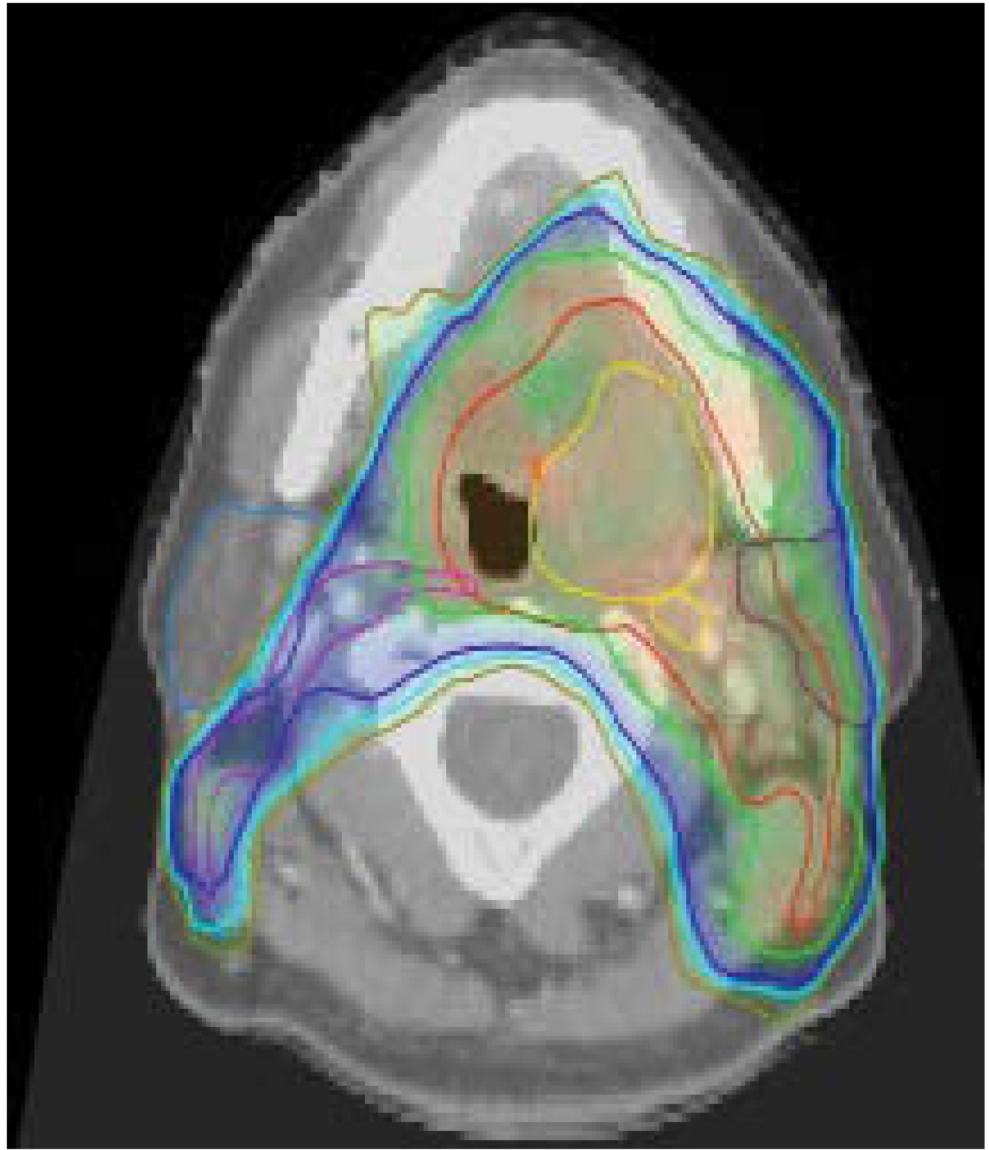
Here is what an actual IMRT plan looks like for a left tonsillar carcinoma (Scott-Brown's):
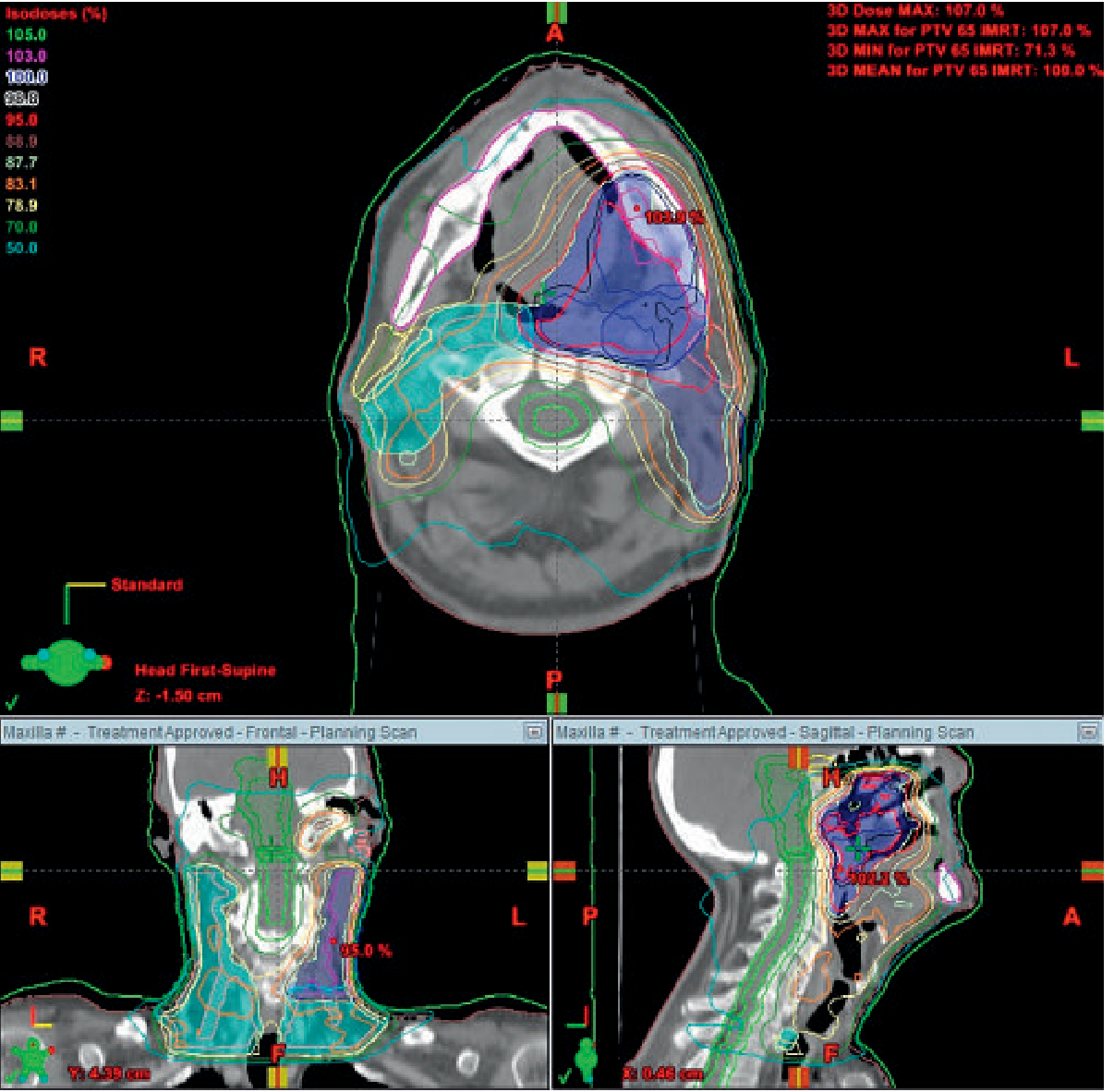
And an IMRT plan for left maxillary tumour (Grainger & Allison's):
Part 4: IMRT vs 3D-CRT - Head-to-Head Comparison
| Feature | 3D Conformal RT (3D-CRT) | IMRT |
|---|---|---|
| Planning approach | Forward planning (oncologist sets beams, computer calculates) | Inverse planning (oncologist sets dose goals, computer optimizes beams) |
| Beam intensity | Uniform across each beam (flat dose profile) | Non-uniform, varies continuously across beam (modulated) |
| Dose conformality | Good for convex tumours | Excellent even for concave/complex tumours |
| OAR sparing | Limited - hard to spare structures adjacent to or behind the tumour | Superior - can sculpt dose around critical structures |
| Simultaneous dose levels | Usually one dose level at a time | Multiple dose levels delivered simultaneously (SIB technique) |
| Treatment time per session | Shorter (fewer, simpler beams) | Longer (more complex beam sequences) |
| Planning complexity | Moderate | High - requires advanced software and physicist involvement |
| Quality assurance (QA) | Moderate | Intensive - each IMRT plan must be independently verified |
| Xerostomia risk | High - parotids usually receive full dose | Significantly reduced - parotids can be spared |
| Current status in ENT | Largely replaced by IMRT for head & neck | Current standard of care for head and neck cancers |
Part 5: The Clinical Evidence - Why IMRT Replaced 3D-CRT in ENT
The PARSPORT Trial (landmark evidence)
The UK PARSPORT (Parotid-Sparing Radiation Therapy vs. Conventional RT) trial was a prospective, phase III randomized controlled trial that directly compared IMRT with conventional/conformal RT in head and neck cancer:
- IMRT reduced grade 2+ xerostomia rates from 83% down to 29% at 24 months after treatment (p = 0.0001) - an extraordinary reduction
- PARSPORT confirmed that parotid-sparing IMRT is now the standard of care; wherever IMRT planning is used, parotid-sparing is mandatory
- Scott-Brown's Otorhinolaryngology Head & Neck Surgery, chap. 13
Dysphagia-Optimized IMRT (Do-IMRT)
Even within IMRT, further refinements are possible:
- Late dysphagia correlates with mean radiation dose to pharyngeal constrictor muscles, glottic/supraglottic larynx, oral cavity, and cervical oesophagus
- Mean doses of 50-60 Gy to pharyngeal musculature are in the critical range for late dysphagia
- Dysphagia-optimized IMRT (Do-IMRT) attempts to reduce dose to swallowing structures further - being evaluated in the UK DARS RCT (ISRCTN:25458988)
CUP (Carcinoma of Unknown Primary) - IMRT advantage
For bilateral neck EBRT in CUP patients:
- Bilateral EBRT gives better locoregional control than unilateral
- But bilateral conventional RT causes severe xerostomia and dysphagia
- IMRT with bilateral neck radiation allows parotid gland sparing while maintaining excellent mucosal coverage - the best of both worlds
- Scott-Brown's Otorhinolaryngology Head & Neck Surgery, chap. 29
Part 6: Summary - The Simple Version
Think of it this way:
Conventional RT = Shining a torch directly at the tumour. Simple, but lights up everything around it.
3D-CRT = Shining a shaped spotlight at the tumour. Better - you can cut the light to fit the tumour shape, but the brightness across the spot is uniform.
IMRT = A precision LED array where each tiny LED can be dimmed or brightened individually. You can now make the light bright in the tumour and dim exactly over the nerve, artery, or salivary gland right next to it - even if they are partly overlapping from every angle.
EBRT = The category that includes all of the above - conventional, 3D-CRT, and IMRT are all forms of EBRT (as opposed to brachytherapy).
Part 7: Beyond IMRT - Future Directions
- IMPT (Intensity Modulated Proton Therapy): Protons have a unique physical property called the Bragg peak - they deposit almost all their energy at a specific depth and essentially stop, with minimal exit dose beyond the tumour. This reduces the "low dose bath" of radiation that IMRT still delivers to surrounding normal tissue. Prospective studies comparing IMPT vs IMRT for head and neck cancer are underway.
- Scott-Brown's Otorhinolaryngology Head & Neck Surgery, chap. 19
Key Takeaways:
- EBRT is the broad term for all externally delivered radiation (linear accelerator-based)
- 3D-CRT uses CT-based 3D planning to shape beams to the tumour outline, but beam intensity is uniform
- IMRT modulates the intensity within each beam using moving multi-leaf collimators and inverse planning, allowing dose sculpting around critical structures
- IMRT is the current standard of care for head and neck cancer radiotherapy, having replaced 3D-CRT
- The PARSPORT trial showed IMRT reduced severe xerostomia from 83% to 29% compared to conventional/conformal RT
- Dysphagia-optimized IMRT is the next refinement, aiming to reduce long-term swallowing problems
This is a shared conversation. Sign in to Orris to start your own chat.