Describe Ganglia And Compare Differences Between Its Types 5 marks
Reading File
Finding Sources
Finding Sources
Reading File
Reading File
Reading File
Loading Image
Loading Image
Here is a comprehensive 5-mark answer on Ganglia:
Ganglia — Description and Types
Definition
Ganglia (singular: ganglion) are ovoid structures in the peripheral nervous system (PNS) containing clusters of neuronal cell bodies and their surrounding glial satellite cells, supported by delicate connective tissue and enclosed by a denser capsule. They function as relay stations for nerve impulses — at least one nerve enters and another exits from each ganglion. The direction of impulse transmission determines whether a ganglion is sensory or autonomic.
— Junqueira's Basic Histology, Text and Atlas, 17e
Types of Ganglia
Ganglia are broadly divided into two main categories based on the direction and nature of the nerve impulses they transmit:
1. Sensory (Afferent) Ganglia
Sensory ganglia receive afferent impulses directed toward the CNS. They are associated with:
- Cranial nerves → cranial ganglia
- Dorsal roots of spinal nerves → dorsal root (spinal) ganglia
Key features:
- Large neuronal cell bodies with prominent nuclei
- Pseudounipolar neurons — relay information from peripheral nerve endings to the gray matter of the spinal cord
- Each cell body is completely enveloped by satellite cells (thin, sheetlike glial cells)
- Well-developed, distinct connective tissue capsule continuous with the epineurium of the nerve
- No blood-nerve barrier equivalent to the blood-brain barrier
- Fascicles of nerve fibers enter and leave the ganglion
Histology:
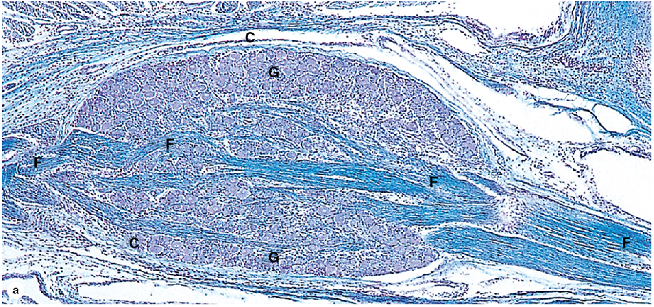
Fig (a): Sensory ganglion (G) showing distinct connective tissue capsule (C) and nerve fiber fascicles (F) — Kluver-Barrera stain ×56
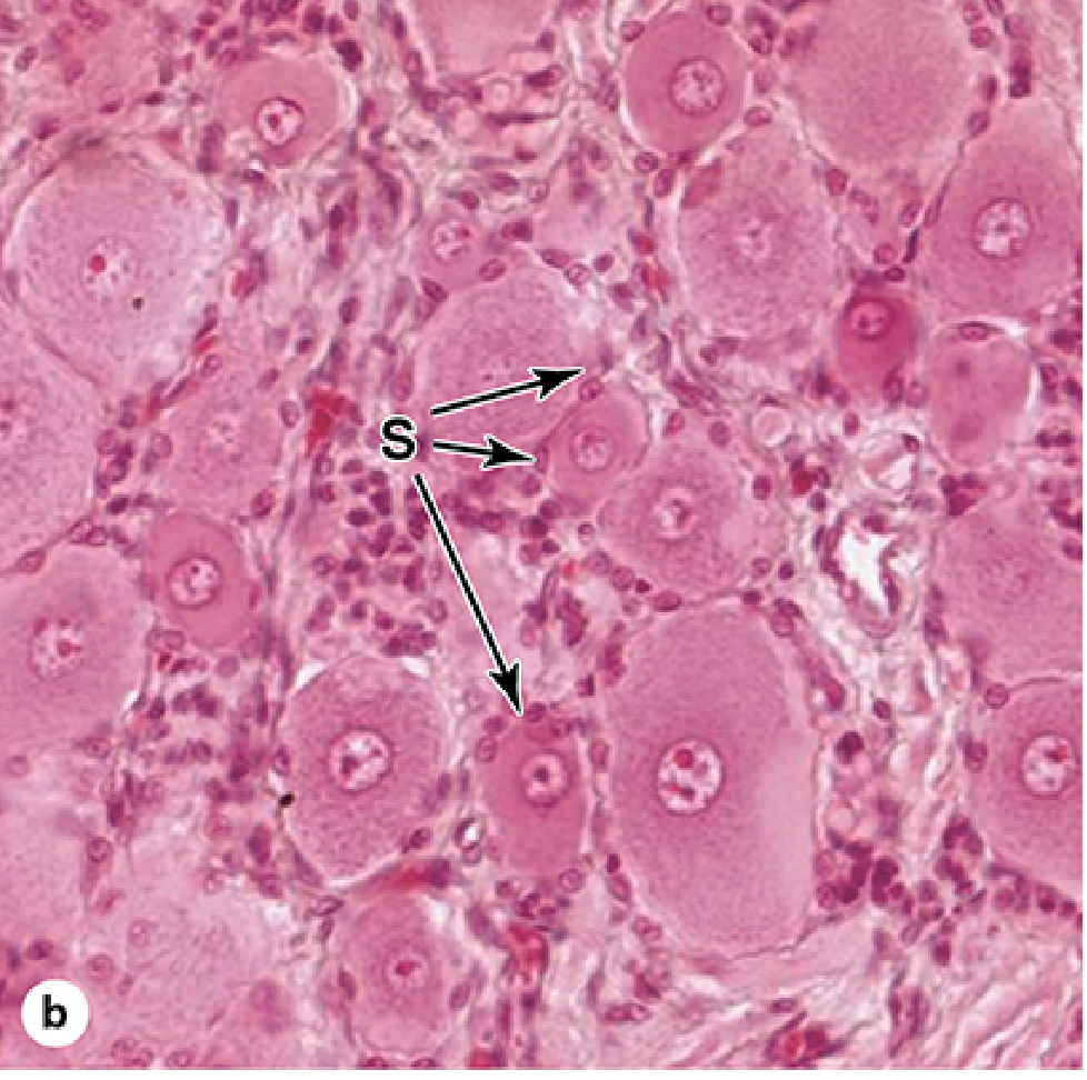
Fig (b): Satellite cells (S) forming sheetlike envelopes around large sensory neuronal cell bodies — H&E ×400
2. Autonomic (Efferent) Ganglia
Autonomic ganglia are efferent — they regulate smooth muscle, cardiac muscle, glands, and visceral organs, maintaining homeostasis.
They use a two-neuron chain:
- Preganglionic neuron — cell body in the CNS; axon uses acetylcholine as the neurotransmitter at the synapse
- Postganglionic (second) neuron — multipolar, located in the peripheral ganglion; axon travels to the effector organ
Autonomic ganglia are subdivided into:
a) Sympathetic Ganglia
- Cell bodies of preganglionic neurons arise from thoracic and lumbar spinal cord segments (T1–L2)
- Second-order multipolar neurons located in small ganglia along the vertebral column (paravertebral chain ganglia) or prevertebral ganglia
- Functionally: "fight or flight" responses
- Cervical sympathetic ganglia include the Superior, Middle, and Inferior (Stellate) cervical ganglia, which supply the heart, head, and neck — Gray's Anatomy for Students
- Satellite cells are present but may differ slightly from sensory ganglia
b) Parasympathetic Ganglia
- Cell bodies of preganglionic neurons in medulla, midbrain, and sacral spinal cord (S2–S4)
- Second-order neurons found in very small ganglia located near or within the effector organs (e.g., walls of the stomach and intestines) — these are called intramural ganglia
- Capsules may be poorly defined or absent; perikarya and satellite cells form a loosely organized plexus within surrounding connective tissue
- Functionally: "rest and digest"
Comparison Table: Sensory vs. Autonomic Ganglia
| Feature | Sensory Ganglia | Autonomic Ganglia |
|---|---|---|
| Impulse direction | Afferent (to CNS) | Efferent (from CNS to effector) |
| Neuron type | Pseudounipolar | Multipolar |
| Location | Dorsal root / cranial nerve roots | Paravertebral, prevertebral, or intramural |
| Capsule | Well-developed, distinct | Less developed; may be absent in parasympathetic |
| Satellite cells | Prominent, sheet-like, envelop each cell body | Present but may be inconspicuous (intramural) |
| Neurotransmitter | — (sensory relay) | Acetylcholine (preganglionic); NE or ACh (postganglionic) |
| Examples | Dorsal root ganglion, trigeminal ganglion | Cervical sympathetic ganglia, ciliary ganglion, enteric ganglia |
Sources:
- Junqueira's Basic Histology, Text and Atlas, 17e — Ganglia, Sensory Ganglia, Autonomic Ganglia
- Gray's Anatomy for Students — Cervical Ganglia
This is a shared conversation. Sign in to Orris to start your own chat.