
Write a short note on malignant fibrous histocytoma for Ms orthopedics theory examination
malignant fibrous histiocytoma histology storiform pattern
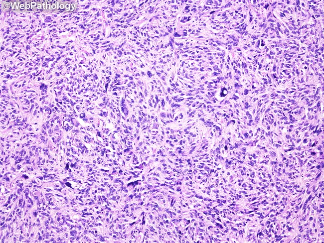
Imaging modality: Light microscopy of Hematoxylin and Eosin–stained renal tumor tissue section, bright-field microscopic view of a storiform-pleomorphic spindle cell neoplasm arising in the kidney. Precise anatomic localization: renal parenchyma (kidney cortex/medulla) with involvement of perivascular stroma. Morphologic features: sheets and short fascicles of plump spindle cells arranged in a storiform (cartwheel) pattern around small-to-medium blood vessels; prominent cellular pleomorphism with irregular, hyperchromatic nuclei; abundant mitotic figures including atypical mitoses; admixed inflammatory cells, notably lipid-laden macrophages (xanthoma cells); background myxoid change may be present; necrosis may be focal in higher grade areas. Immunophenotype is variable; tumor demonstrates undifferentiated pleomorphic sarcoma pattern rather than lineage-specific differentiation; differential diagnosis includes sarcomatoid renal cell carcinoma, fibrosarcoma, malignant fibrous histiocytoma (older term), leiomyosarcoma, and rhabdomyosarcoma. Clinical significance: UPS of the kidney is aggressive with potential for local invasion and metastasis; diagnosis requires correlation with immunohistochemistry and exclusion of specific lineages. Utility: this histology image is essential for educational demonstration of storiform pattern, spindle cell pleomorphism, xanthoma cells, and mitotic activity in renal UPS; useful for pathology teaching, differential diagnosis discussions, and research on soft tissue sarcomas of renal origin. This image also exemplifies the fibrous stroma and inflammatory milieu that may complicate grading.
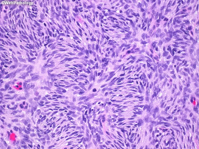
Imaging modality and technique: Light microscopy of a formalin-fixed skin biopsy stained with Hematoxylin and Eosin, imaged at high power (HPF) to highlight cellular detail. Anatomical location: dermis with superficial to mid-dermal spindle cell proliferation infiltrating the subcutaneous fat in a common dermatofibrosarcoma protuberans (DFSP) pattern. Morphology: uniform slender spindle cells arranged in storiform (cartwheel) pattern with interlacing rows and a lightly basophilic, fibrillary stroma; nuclei elongated, with mild to moderate hyperchromasia and inconspicuous mitotic figures. Inset image notes a mitotic figure near the center, illustrating active proliferation in some fields. Immunohistochemical context: classic DFSP demonstrates strong CD34 positivity in tumor cells (CD34+), aiding distinction from benign fibrous histiocytoma, which is typically CD34 negative or variably positive. Lack of significant pleomorphism and absence of prominent necrosis argue against higher-grade sarcoma. Diagnostic significance: this morphological constellation supports DFSP, a locally aggressive dermal/subcutaneous spindle cell neoplasm with infiltrative margins that requires wide excision with clear margins to reduce recurrence risk. Clinical correlation: in suspected DFSP, correlate with CD34 immunostaining, patient age, and tumor location, and consider differential diagnoses including dermatofibroma, malignant fibrous histiocytoma, and cutaneous spindle cell sarcomas. Educational use: representative high-power features useful for histology training and differential diagnosis in dermatopathology.
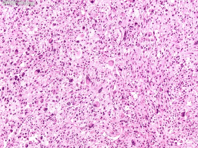
This histopathology image depicts a prostatic malignant mesenchymal neoplasm consistent with undifferentiated pleomorphic sarcoma (formerly malignant fibrous histiocytoma). Acquired from prostate tissue and stained with Hematoxylin and Eosin, the section shows high cellularity with marked nuclear pleomorphism and bizarre multinucleated tumor giant cells scattered haphazardly within a variably fibrous stroma. Tumor cells range from spindle-shaped to polygonal, with hyperchromatic, irregular nuclei and conspicuous nucleoli; cytoplasm is sometimes abundant and vacuolated. Notably, there is an absence of a classic storiform-pleomorphic growth pattern; cells are arranged in diffuse sheets without a defined fascicular architecture. Immunophenotype, based on the described panel, shows positivity only for vimentin, with lack of epithelial (cytokeratin, EMA), muscle (desmin, smooth muscle actin), melanocytic, or neural markers, supporting a diagnosis of a high-grade, undifferentiated sarcoma. Clinically, prostatic UPS is rare and aggressive, requiring immunohistochemical confirmation and exclusion of sarcomatoid carcinoma. Diagnostic significance lies in separating mesenchymal prostatic neoplasms from epithelial malignancies; differential diagnoses include sarcomatoid carcinoma, rhabdomyosarcoma, leiomyosarcoma, and other pleomorphic sarcomas. This image is educational for pathology grading, differential diagnosis, and correlating histology with prognosis and therapeutic planning.
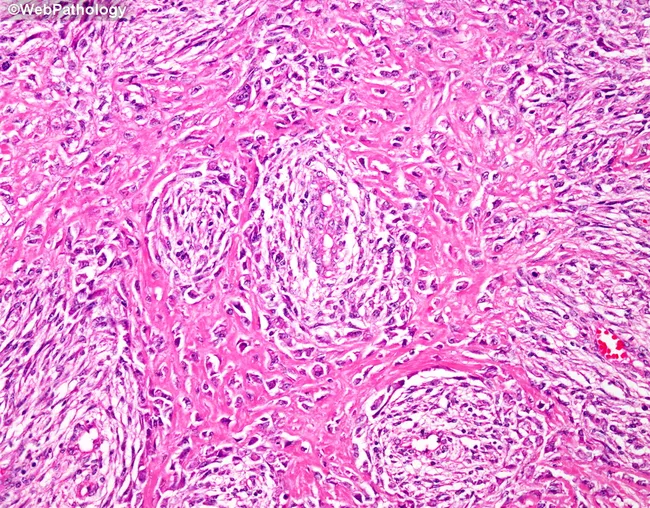
Imaging modality: histopathology section of bone tissue; Technique: hematoxylin and eosin staining; Magnification: not specified. The image depicts a malignant spindle cell neoplasm arising in osseous tissue, consistent with a fibroblastic osteosarcoma, fibroblastic subtype. Tumor cells are predominantly elongated, spindle-shaped, with oval to elongated nuclei and minimal cytoplasm. The cellular arrangement shows interlacing fascicles with a characteristic herringbone or storiform pattern, embedded in a densely eosinophilic collagenous stroma. Osteoid production is focal and variably present, with calcified, lace-like deposits evidencing osteogenic activity; in some regions osteoid is conspicuous, while in others it is scant, reflecting the reported variability of this subtype. The lesion is highly vascular, with numerous capillary and hemangiopericytoma-like vessels interspersed among the spindle cells, contributing to the conspicuous, vascularized stroma. Mitotic activity may be present but is not uniformly high across the field. These features distinguish fibroblastic osteosarcoma from fibrous histiocytoma and benign fibrous lesions; osteoid presence, even if focal, supports osteogenic differentiation. Clinically, this histology carries malignant potential with risk for local recurrence and metastatic spread, especially to the lungs; surgical excision with adjuvant therapy is typically indicated. This image exemplifies the diagnostic pathology that guides staging, prognosis, and multidisciplinary management of osteosarcoma for ongoing study.
malignant fibrous histiocytoma undifferentiated pleomorphic sarcoma orthopedics
Malignant Fibrous Histiocytoma (MFH) / Undifferentiated Pleomorphic Sarcoma (UPS)
Definition
Epidemiology
- Most common soft-tissue sarcoma in adults, accounting for 5-10% of all soft tissue sarcomas
- Peak incidence in older adults (50-70 years), with a slight male predominance
- As a bone tumor, it is often called "nonosteogenic osteosarcoma" and tends to occur in an older age group than conventional osteosarcoma
Site
- Soft tissue MFH/UPS: deep soft tissues of the extremities, especially the thigh, followed by trunk and retroperitoneum
- Bone MFH: commonly affects the distal femur, proximal tibia, proximal femur, ilium, and proximal humerus - metaphyseal location, similar to osteosarcoma
Clinical Features
- Painless or slowly enlarging soft tissue mass (soft tissue form)
- Pain and swelling with a destructive bone lesion (osseous form)
- May present as a pathological fracture in advanced bone lesions
- Can arise secondarily in areas of prior irradiation, bone infarct, Paget's disease, or fibrous dysplasia
Imaging
- Bone lesion: purely lytic, destructive lesion or a mixed lytic-sclerotic pattern, with cortical destruction and soft tissue extension; no significant tumor bone/osteoid formation (distinguishing it from osteosarcoma)
- Soft tissue lesion: large, heterogeneous mass on MRI, often with areas of necrosis and hemorrhage
Histopathology
- Pleomorphic spindle cells and histiocyte-like cells arranged in a storiform ("cartwheel") pattern
- Marked nuclear pleomorphism, bizarre multinucleated tumor giant cells, high mitotic activity (including atypical mitoses)
- May contain lipid-laden macrophages (xanthoma cells) and inflammatory infiltrate
- Diagnosis of exclusion: immunohistochemistry is essential to rule out other pleomorphic sarcomas (dedifferentiated liposarcoma, leiomyosarcoma, malignant peripheral nerve sheath tumor, sarcomatoid carcinoma); tumor cells typically stain only for vimentin and lack epithelial, muscle, or melanocytic markers
Differential Diagnosis
Management
- Core needle biopsy for histologic confirmation before definitive treatment (must not compromise future surgical margins)
- Wide surgical excision with negative margins (limb-salvage surgery where feasible) is the mainstay of treatment
- Adjuvant/neoadjuvant radiotherapy for large, high-grade, or deep lesions to reduce local recurrence
- Chemotherapy (e.g., doxorubicin-based regimens) considered for high-grade, large tumors or metastatic disease
- Amputation reserved for cases not amenable to limb-sparing resection
Prognosis
Write a short note on advances in tumor endoprosthesis and Limb salvage surgery
"limb salvage" AND endoprosthesis
advances tumor endoprosthesis limb salvage surgery 3D printed custom implants growing prosthesis
PMID: 38461457
PMID: 38606257
Advances in Tumor Endoprosthesis and Limb Salvage Surgery
Background
Key Advances
1. 3D-printed, patient-specific implants
- Custom-made titanium (Ti6Al4V) prostheses manufactured by selective laser melting/3D printing now allow anatomically precise reconstruction of complex defects, especially in the pelvis and periacetabular region where standard modular implants fit poorly
- Porous, textured, lattice surfaces improve osseointegration and soft-tissue attachment while reducing implant weight
- Design and manufacture take about 3-4 weeks, enabling truly individualized reconstruction with cutting guides for more accurate resection margins
- Complication rates remain higher in technically difficult sites (pelvis) despite these gains
2. Modular and "off-the-shelf" endoprosthetic systems
- Modular segmental systems (interchangeable stems, bushings, and body segments) allow intraoperative flexibility to match the defect length, reducing the need for custom manufacturing in long bone reconstructions
- Rotating-hinge knee designs have reduced dislocation and improved durability for distal femur/proximal tibia replacements
3. Pelvic reconstruction advances
- The LUMiC endoprosthesis (modular hydroxyapatite-coated stem with acetabular cup) has become a leading option for periacetabular reconstruction after internal hemipelvectomy, with favorable implant survival, although dislocation, infection, and soft-tissue failure remain the main complications (per Henderson classification)
- Navigation-assisted and patient-specific instrumentation improve accuracy of pelvic osteotomies and cup positioning
4. Growing/expandable prostheses for pediatric patients
- Extendible (non-invasive, magnetically or electromagnetically driven) prostheses such as the Juvenile Tumor System allow outpatient, non-invasive lengthening of the implant as a child grows, avoiding repeated open lengthening surgeries and helping preserve limb-length equality into adulthood
5. Infection-resistant and "smart" implant technology
- Antibacterial coatings (e.g., silver-based or nano-textured surfaces) are being incorporated into endoprosthetic stems to reduce the historically high risk of periprosthetic infection, the most common cause of endoprosthesis failure
- Compress/compliant pre-stress fixation (osseointegration via mechanical compression rather than cement or screws) is an alternative fixation method with reduced stress-shielding
6. Biological and adjunct reconstructive techniques
- Distraction osteogenesis and vascularized fibular autografts are increasingly combined with endoprosthetic or allograft reconstruction (e.g., allograft-prosthesis composites) to restore biological bone stock and improve long-term durability, particularly in younger patients
- Orthoplastic (combined orthopedic-plastic surgery) approaches with free flap coverage have expanded the boundaries of what is salvageable, particularly for compromised soft-tissue envelopes
7. Precision oncology integration
- Genomic/molecular tumor profiling, targeted intraoperative imaging, and image-guided/navigated resections are improving the accuracy of surgical margins, which directly affects local recurrence rates and the feasibility of limb-sparing resection
- AI-assisted preoperative planning and robotics are emerging areas expected to further refine resection accuracy and implant fit
Outcomes and Ongoing Challenges
- Functional outcomes after modern endoprosthetic LSS are generally superior to amputation for oncologic indications, though complication rates (aseptic loosening, infection, structural failure) accumulate with longer follow-up, especially in pediatric and pelvic reconstructions
- Cost, accessibility of 3D-printing technology, and long-term durability data remain limitations
- Decision-making between amputation and salvage is more nuanced in trauma (mangled extremity) than in oncologic cases; oncologic literature strongly favors salvage when negative margins are achievable, whereas trauma literature shows more mixed functional and cost outcomes between amputation and complex reconstruction
Summary for exam purposes
Write a short note on 3-D printing and patient specific implants
"3D printing" AND "patient-specific implant" AND orthopedic
3D printing patient specific implants orthopedic surgery advances titanium lattice
3D Printing and Patient-Specific Implants in Orthopedics
Definition
Workflow
- Imaging: High-resolution CT/MRI of the affected region
- Virtual planning: 3D reconstruction of the anatomy; surgeon plans resection margins, osteotomy cuts, or implant placement virtually
- Design: Cutting/drilling guides and/or implant are digitally designed to match the patient's unique anatomy
- Printing: Guides are usually printed in biocompatible polymer (for single use); implants are printed in metal (titanium alloy Ti6Al4V, or occasionally tantalum/cobalt-chromium) using Selective Laser Melting (SLM) or Electron Beam Melting (EBM)
- Sterilization and intraoperative use: The guide envelops the bone/implant and directs drill/screw trajectories or osteotomy cuts exactly as planned
Techniques of Manufacture
- SLM/EBM (powder-bed fusion): most common for metal implants; allows fabrication of complex porous/lattice internal architecture
- Fused deposition modeling (FDM): used for polymer anatomical models and disposable surgical guides
- Lattice/trabecular internal structures mimic cancellous bone porosity, reducing implant stiffness (limiting stress-shielding) while promoting bone ingrowth
Clinical Applications
1. Patient-specific surgical guides
- Fracture reduction: drilling guides for complex fractures (e.g., tibial plateau) allow pre-planned screw trajectories, improving accuracy of plate positioning and reduction
- Corrective osteotomies: for post-traumatic 3D deformities/malunions, cutting/reduction guides improve predictability where 2D imaging planning is unreliable
- Oncologic resection guides: used to achieve pre-planned tumor-free margins during bone tumor resection (e.g., osteosarcoma around the knee)
2. Patient-specific implants
- Musculoskeletal oncology: custom endoprostheses for pelvic/sacral and periacetabular defects after tumor resection, where standard modular implants fit poorly
- Revision arthroplasty: custom acetabular cages/cups for massive bone loss
- Complex deformity/arthrodesis: custom titanium truss implants (e.g., tibiotalocalcaneal arthrodesis after failed total ankle replacement)
- Spine surgery: patient-specific cages and pre-bent titanium plates for complex deformity and oncologic reconstruction
- Pediatric orthopedics: modeling for complex congenital deformity correction and, combined with expandable prosthesis technology, growth-accommodating implants
Advantages
- High anatomical accuracy and fit, especially in geometrically complex regions (pelvis, spine, craniofacial-adjacent regions)
- Reduced operative time and blood loss in several series (per RCT scoping review data)
- Improved osseointegration via porous lattice structures that mimic cancellous bone
- Reduced soft-tissue disruption; guides simplify sterilization compared with reusable jigs
- Enables surgery in cases previously considered unreconstructable
Limitations / Challenges
- Manufacturing lead time (typically 3-4 weeks), unsuitable for emergent cases
- High cost and limited accessibility outside specialized/tertiary centers
- Complication rates remain relatively high in technically demanding sites (e.g., pelvic reconstruction) - infection, aseptic loosening, wound dehiscence
- Long-term durability data are still limited; heterogeneous reporting across studies
- Regulatory and material characterization standards are still evolving (per 2025 review, custom implants "require additional characterization" before wide adoption can be considered equivalent to conventional implants)
Future Directions
- Integration with AI-assisted planning and robotics/navigation for real-time intraoperative accuracy
- Smart implants with embedded sensors for monitoring loading/osseointegration
- Novel biomaterials (magnesium, zinc-based bioresorbable metals) for temporary fixation
- Point-of-care 3D printing labs (e.g., hospital-based printing units) to reduce lead time and cost
- Expanded use in pediatric growth-accommodating and combined oncologic-reconstructive (orthoplastic) surgery
Summary for exam purposes

Write a gold medal level answer for second question
"regenerative medicine" AND cartilage AND "bone repair"
regenerative orthopaedics cartilage bone repair stem cell PRP tissue engineering scaffolds clinical outcomes ethical considerations 2025
Q2. Role of Regenerative Orthopaedics in Cartilage and Bone Repair (25 Marks)
Introduction
1. Stem Cell Therapy, PRP, and Growth Factors
A. Stem Cell Therapy
- Mesenchymal stem cells (MSCs) are the workhorse of orthopaedic regenerative medicine due to their capacity to differentiate into chondrocytes, osteoblasts, and tenocytes, along with strong immunomodulatory and anti-inflammatory properties
- Sources:
- Bone marrow-derived MSCs (BM-MSCs) - most studied; harvested via Bone Marrow Aspirate Concentrate (BMAC) from the iliac crest
- Adipose-derived stem cells (ADSCs) - abundant, easy harvest, good chondrogenic potential
- Synovium-derived and umbilical cord-derived MSCs - emerging sources
- Induced pluripotent stem cells (iPSCs) - experimental, high plasticity but tumorigenicity concerns
- Clinical uses: intra-articular injection for early osteoarthritis, augmentation of cartilage repair procedures (microfracture + BMAC), avascular necrosis of femoral head (core decompression + stem cell injection), nonunion fracture repair, rotator cuff tear augmentation
- Limitation: autologous stem cell number and regenerative capacity decline sharply with age (functionally negligible beyond 70-80 years), creating a ceiling effect in elderly patients
B. Platelet-Rich Plasma (PRP)
- Autologous blood product centrifuged to concentrate platelets (3-5x baseline), rich in growth factors: PDGF, TGF-beta, VEGF, EGF, IGF-1
- Mechanism: stimulates angiogenesis, chemotaxis, and proliferation of local progenitor cells rather than directly providing stem cells
- Leucocyte-rich vs leucocyte-poor PRP: leucocyte-poor preferred for intra-articular use (less pro-inflammatory)
- Clinical applications: knee osteoarthritis (pain relief, especially early-stage), lateral epicondylitis, patellar/Achilles tendinopathy, rotator cuff repair augmentation, and as an adjunct during arthroscopic debridement
- Evidence is strongest for short-term pain relief in tendinopathy and early OA; results in chronic/advanced disease are inconsistent, partly due to lack of standardized preparation protocols
C. Growth Factors and Biologics
- BMP-2 and BMP-7 (OP-1): FDA-approved for spinal fusion and long-bone nonunion; potent osteoinductive agents
- FGF-18: shows promise in preclinical/early clinical studies for cartilage healing augmentation after surgical repair
- PDGF, TGF-beta superfamily: used in combination products and scaffold-bound delivery systems
- Delivery is increasingly scaffold-mediated to achieve sustained local release and avoid systemic effects/heterotopic ossification (a known BMP-2 complication)
2. Tissue Engineering and Scaffolds
A. Scaffold Types
| Category | Examples | Features |
|---|---|---|
| Natural/biologic | Collagen, hyaluronic acid, chitosan, decellularized/acellular extracellular matrix | Biocompatible, bioactive, but weaker mechanical strength |
| Synthetic polymers | PLA, PGA, PLGA, PCL | Tunable degradation and mechanical properties |
| Ceramic/inorganic | Hydroxyapatite, tricalcium phosphate | Osteoconductive, used for bone defects |
| Composite/hybrid | Collagen-hydroxyapatite, 3D-printed lattice titanium-polymer | Combine mechanical support with biologic activity |
| Hydrogels | Alginate, fibrin, PEG-based | Injectable, good for irregular cartilage defects, allow cell encapsulation |
B. Key Techniques
- Autologous Chondrocyte Implantation (ACI) / Matrix-induced ACI (MACI): chondrocytes harvested, expanded in vitro, and re-implanted on a collagen scaffold/membrane for full-thickness chondral defects
- Osteochondral scaffolds (bilayer): mimic native osteochondral unit - a bone-facing calcium phosphate layer fused to a cartilage-facing collagen/hyaluronic layer
- 3D bioprinting: patient-specific, personalized scaffolds with lattice architecture that mimic native trabecular bone porosity, promoting osseointegration and enabling precise anatomical fit; increasingly used for cartilage regeneration research and complex bone defects
- Acellular matrices: decellularized cartilage/dermal matrices used as off-the-shelf scaffolds, avoiding donor-site morbidity and immune reaction concerns of cellular grafts
- Vascularized bone tissue engineering: addressing the critical limitation of poor vascular ingrowth in large scaffolds by incorporating vascularized grafts or angiogenic growth factors (VEGF)
3. Clinical Outcomes and Ethical Considerations
A. Clinical Outcomes
- MSC therapy: consistent evidence of symptomatic improvement and possible structural benefit in early knee OA; results are dose- and source-dependent; long-term (>5 year) structural regeneration data remain limited
- PRP: systematic reviews show significant short-term pain/functional improvement in tendinopathy and early OA, but effect sizes diminish over time and vary with preparation protocol
- ACI/MACI: good long-term (10-year) outcomes for large, focal, full-thickness chondral defects in young, active patients; superior durability compared to microfracture for defects >2-4 cm²
- Scaffold-augmented cartilage repair (BMAC + scaffold): meta-analyses suggest modest but statistically significant improvement in short-term patient-reported outcomes compared to scaffold alone
- BMP-2/7: proven efficacy in accelerating union in long-bone nonunion and open tibial fractures, though use is now more selective due to complication profile
- Overall: regenerative techniques currently function best as adjuncts to established surgical procedures rather than complete replacements, with the strongest evidence in early-stage disease and focal defects rather than advanced/diffuse degeneration
B. Ethical and Practical Considerations
- Regulatory ambiguity: many stem cell/PRP products are minimally regulated "autologous" preparations, allowing clinics to market unproven therapies directly to patients without robust trial evidence - a major source of ethical concern
- Embryonic vs adult stem cells: use of embryonic/fetal-derived stem cells raises bioethical objections; most orthopaedic clinical practice has shifted to autologous adult MSCs (BMAC, adipose-derived) specifically to avoid this controversy
- Informed consent: patients must be counselled that many regenerative treatments remain "off-label" or experimental, with variable and sometimes overstated efficacy claims in direct-to-consumer marketing
- Cost and equity: high cost of cell processing, expansion, and scaffold fabrication limits access, raising concerns about equitable availability
- Standardization: lack of uniform preparation protocols (PRP classification systems, MSC dosing, culture-expansion conditions) hampers reproducibility of trial results and complicates informed clinical decision-making
- Long-term safety: concerns regarding tumorigenic potential of expanded/iPSC-derived cells, and heterotopic bone formation with BMP use, mandate long-term surveillance
- Donor-site morbidity: even autologous harvest (iliac crest BMAC, adipose harvest) carries procedural risk
Conclusion
Induced membrane techniques in ortho short note