
Respiratory Distress Syndrome
"respiratory distress syndrome, newborn" AND surfactant
Respiratory Distress Syndrome neonatal overview pathophysiology treatment 2025
neonatal respiratory distress syndrome chest x-ray ground glass air bronchograms
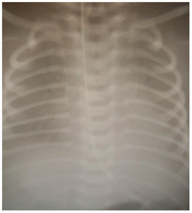
This diagnostic image is an anteroposterior chest X-ray of a neonatal patient. The most striking visual feature is a significant reduction in lung volume, characterized by diminished lung fields and low lung expansion. There is a diffuse, ground-glass opacity throughout the bilateral lung fields, consistent with Respiratory Distress Syndrome (RDS) or surfactant deficiency. Visible anatomical structures include the thoracic ribs and the vertebral column. A centrally located radio-opaque vertical line is observed, indicating the presence of an indwelling medical device, likely an endotracheal tube or a nasogastric feeding tube. The diaphragmatic domes appear elevated due to the decreased lung volume. Educational focus includes the recognition of severe neonatal respiratory failure, the presence of air bronchograms (though limited by image resolution), and the visual manifestations of pathology requiring intensive respiratory support and surfactant administration in a pediatric intensive care setting.
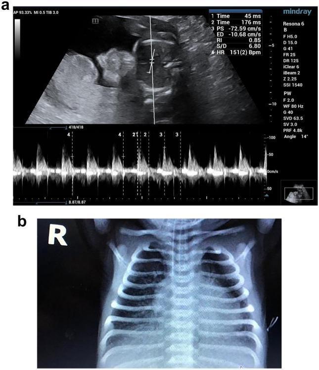
This multi-panel figure demonstrates fetal pulmonary hemodynamics and corresponding neonatal clinical findings. Panel A is a fetal Doppler ultrasound image of the main pulmonary artery (MPA) at 35 weeks and 2 days of gestation. The spectral Doppler waveform exhibits a characteristic 'spike and dome' morphology, featuring a sharp systolic peak followed by a secondary deflection. Quantitative measurements are shown, including a peak systolic velocity (PSV) of -72.59 cm/s and an acceleration time/ejection time (At/Et) ratio of 0.25, indicating increased pulmonary vascular resistance. Panel B is a postnatal anteroposterior chest X-ray of the same neonate. The radiographic findings include diffuse, bilateral reticulogranular opacities ('ground-glass appearance') and prominent air bronchograms. These findings are diagnostic of neonatal Respiratory Distress Syndrome (RDS), likely secondary to surfactant deficiency in this late preterm infant. The composite image illustrates the clinical utility of prenatal MPA Doppler parameters in predicting neonatal respiratory outcomes.
Respiratory Distress Syndrome (RDS)
Definition and Pathophysiology
- Surface tension rises (per Laplace's law), causing progressive alveolar collapse (atelectasis)
- Reduced lung compliance increases the work of breathing
- Repeated collapse/reopening damages the epithelium, leaking proteinaceous fluid into alveoli that organizes into hyaline membranes lining the alveolar ducts and alveoli
- Type II pneumocytes fully mature and begin surfactant secretion around 20-22 weeks, but adequate surfactant reserves usually aren't reached until about 34-36 weeks - hence the strong link to prematurity (Guyton and Hall Textbook of Medical Physiology; The Developing Human Clinically Oriented Embryology).
Risk Factors
- Prematurity/low birth weight is the dominant risk factor - incidence is roughly 90% at 24 weeks' gestation and about 80% at 28 weeks
- Maternal diabetes, perinatal asphyxia, cesarean delivery without labor
- Genetic factors: male sex, Caucasian race, family history of RDS, surfactant protein B deficiency
- Congenital lung hypoplasia (e.g., congenital diaphragmatic hernia, giant omphalocele)
- Creasy & Resnik's Maternal-Fetal Medicine, p. 1785
Clinical Presentation
- Tachypnea, nasal flaring, subcostal/intercostal retractions, cyanosis
- Expiratory grunting - caused by exhaling against a partially closed glottis to generate auto-PEEP and maintain functional residual capacity
- Symptoms classically worsen over the first 2-3 days, then improve as endogenous surfactant production increases (this course is shortened with exogenous surfactant therapy)
- Creasy & Resnik's Maternal-Fetal Medicine, p. 1785
Diagnosis
- Clinical plus chest radiograph: low lung volumes with a diffuse, fine reticulogranular ("ground-glass") pattern and air bronchograms
- Point-of-care lung ultrasound is increasingly used, especially in Europe
- Surfactant activity can be measured chemically in tracheal/gastric aspirates, though this isn't routine
Management
- Antenatal corticosteroids (betamethasone/dexamethasone) given to mothers at risk of preterm delivery accelerate fetal lung maturation and are the single most effective preventive measure
- Thermoregulation in an incubator/radiant warmer to minimize oxygen consumption
- Target PaO2 50-80 mmHg, SpO2 90-95%; avoid both hypercarbia and hyperoxia
- Empiric broad-spectrum antibiotics for 48 hours pending cultures (sepsis can mimic RDS and cannot be excluded clinically)
- CPAP (continuous positive airway pressure) prevents atelectasis, preserves surfactant function, and allows many infants to avoid intubation and mechanical ventilation
- Mechanical ventilation when CPAP is insufficient
- Prophylactic - administered before the first breath to all at-risk infants
- Rescue therapy - given after respiratory signs develop
- Minimally invasive administration - via thin catheter, laryngeal mask, or nebulization in spontaneously breathing infants on noninvasive support, avoiding positive-pressure lung injury
- Creasy & Resnik's Maternal-Fetal Medicine, p. 1785-1786
Complications
- Air leaks (pneumothorax, pulmonary interstitial emphysema)
- Intraventricular/germinal matrix hemorrhage
- Necrotizing enterocolitis
- Bronchopulmonary dysplasia (BPD) - the major chronic complication, involving arrested lung development plus injury/inflammation from oxygen and ventilator exposure
- Robbins & Kumar Basic Pathology, p. 200-203
Recent evidence (last 2 years)
- A 2024 network meta-analysis comparing surfactant delivery strategies (laryngeal mask, minimally invasive/thin-catheter, intubation) found less-invasive methods generally reduce need for mechanical ventilation and BPD risk (PMID: 39736686)
- A 2025 meta-analysis suggests surfactant combined with budesonide may lower BPD incidence versus surfactant alone, though evidence quality is still evolving (PMID: 40870374)
- A 2025 scoping review highlights that surfactant access and delivery strategies remain a major gap in low- and middle-income countries (PMID: 41278024)