
Management of malignancy of lower One third in detail step by step with simple words according to standard book of surgery
Reading File
The user is asking about management of malignancy of the lower one-third - this likely refers to esophageal cancer (lower third of esophagus) or rectal cancer (lower third of rectum). Let me clarify, but given the surgical context and common exam topics, I'll search for both to cover the most likely intent. The lower third of esophagus and lower third of rectum are both classic surgical topics.
Let me search comprehensively in parallel.
Finding Sources
Finding Sources
Finding Sources
Finding Sources
The query is likely about esophageal carcinoma (lower third). Let me now ask the user to clarify while also fetching both esophageal content from the textbooks.
Asking for Preferences
Which organ's lower one-third malignancy are you asking about?
Lower third esophageal malignancy - this is mainly adenocarcinoma (arising from Barrett's esophagus) in the modern era. Key management involves staging, then either curative surgery (Ivor Lewis or transhiatal esophagectomy), neoadjuvant chemoradiotherapy, or palliative treatment depending on stage.
Searching sources...
Finding Sources
Finding Sources
Reading File
Finding Sources
Excellent! I found key references in Sabiston Textbook of Surgery and Sleisenger & Fordtran's. Let me read comprehensively from these authoritative surgical textbooks.
Reading File
Finding Sources
Reading File
Finding Sources
Reading File
Reading File
Reading File
I now have comprehensive, detailed information from Sabiston's Textbook of Surgery. Let me compile the full, detailed management guide.
Loading Image
I now have all the information needed to write a comprehensive, step-by-step guide. Let me compile the full answer.
Management of Malignancy of the Lower One-Third of the Esophagus
Source: Sabiston Textbook of Surgery (22nd Ed.), Current Surgical Therapy 14e, Sleisenger & Fordtran's GI and Liver Disease
What Is It?
The lower third of the esophagus (below the inferior pulmonary vein to the gastroesophageal junction, GEJ) is primarily affected by adenocarcinoma - more than 80% of lower esophageal cancers are adenocarcinomas, most arising from Barrett's esophagus (caused by chronic GERD). Squamous cell carcinoma (SCC) also occurs here (about 40% of SCC is in the lower third).
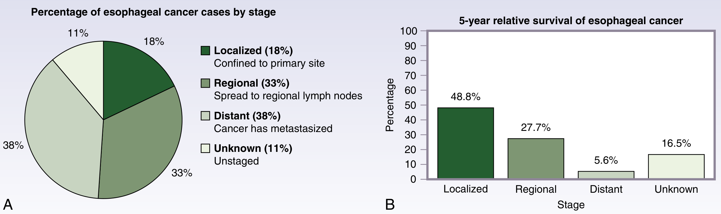
STEP 1 - Initial Assessment and Staging Workup
Before any treatment, you must stage the cancer to decide what treatment is appropriate.
A. History and Physical Examination
- Main symptom: progressive dysphagia (first solids, then liquids)
- Weight loss, odynophagia (pain on swallowing), regurgitation, hoarseness (suggests recurrent laryngeal nerve involvement)
- History of GERD / Barrett's esophagus (risk factor for adenocarcinoma)
- Tobacco and alcohol use (risk for SCC)
B. Endoscopy (Upper GI Endoscopy + Biopsy)
- First investigation
- Confirms diagnosis by tissue biopsy
- Assesses tumor location, length, and relation to GEJ
- Determines histologic type (adenocarcinoma vs SCC)
C. TNM Staging (AJCC 8th Edition - from Sabiston)
| Category | Criteria |
|---|---|
| T1a | Invades lamina propria or muscularis mucosa |
| T1b | Invades submucosa |
| T2 | Invades muscularis propria |
| T3 | Invades adventitia |
| T4a | Invades resectable adjacent structures (diaphragm, pleura, pericardium) |
| T4b | Invades unresectable structures (aorta, vertebra, trachea) |
| N0-N3 | 0 to ≥7 lymph nodes involved |
| M0/M1 | No distant / distant metastasis |
D. Staging Investigations (in order)
- CT scan of chest, abdomen, and pelvis - assesses local invasion and distant metastases (lung, liver, adrenals)
- FDG-PET/CT scan - detects occult distant metastases not seen on CT; changes management in ~20% of patients
- Endoscopic Ultrasound (EUS) - gold standard for T and N staging; determines depth of tumor invasion and regional lymph node involvement
- Diagnostic laparoscopy - for GEJ (junction) tumors to exclude peritoneal metastases
STEP 2 - Multidisciplinary Team (MDT) Discussion
All patients must be discussed in a multidisciplinary team meeting that includes:
- Surgeon (thoracic/upper GI)
- Medical oncologist
- Radiation oncologist
- Gastroenterologist
- Radiologist
- Pathologist
Treatment is then tailored to stage, fitness (performance status), and histology.
STEP 3 - Treatment Based on Stage
GROUP A: Very Early Disease (Tis, T1a - High-grade dysplasia or mucosal cancer)
These tumors are confined to the mucosa and have very low risk of lymph node spread.
Treatment: Endoscopic therapy (preferred over surgery)
- Endoscopic Mucosal Resection (EMR) - removes the full thickness of mucosa for lesions <2 cm
- Endoscopic Submucosal Dissection (ESD) - for larger lesions or when en bloc removal needed
- Radiofrequency Ablation (RFA) - burns away remaining Barrett's tissue after resection
- EMR successfully eradicates 91-98% of T1a cancer (Sabiston)
- After successful endoscopic therapy - regular endoscopic surveillance
Esophagectomy is reserved for patients with high-risk features (lymphovascular invasion, deep submucosal invasion >500 µm, or poor differentiation) or failed endoscopic therapy.
GROUP B: Locally Advanced Resectable Disease (T2-T3, N0-N1, M0)
This is the most common presentation for the lower third esophagus.
Treatment: Neoadjuvant therapy FIRST, then Surgery
Step 3B-1: Neoadjuvant (Pre-operative) Chemoradiotherapy (CRT) - STANDARD OF CARE
The landmark CROSS trial (2012) established this as standard:
- Regimen: Carboplatin + Paclitaxel given weekly, concurrent with 41.4 Gy radiation in 23 fractions
- Then surgery 4-6 weeks after completing CRT
- Results: Rate of complete (R0) resection = 92% vs 69% surgery alone
- Median overall survival: 48.6 months (CRT + surgery) vs 24 months (surgery alone)
- In adenocarcinoma patients: pathologic complete response (pCR) in ~23%
Alternative: Perioperative Chemotherapy (FLOT regimen)
- The FLOT4 trial showed perioperative FLOT (5-FU + Leucovorin + Oxaliplatin + Docetaxel) was superior to ECF/ECX regimens
- 4 cycles pre-op + 4 cycles post-op
- Median OS: 50 months (FLOT) vs 35 months (ECF)
- Preferred by many centers for lower esophageal/GEJ adenocarcinoma
Step 3B-2: Restaging Before Surgery
- FDG-PET/CT at 5-8 weeks after completing neoadjuvant therapy - mandatory to confirm no new distant disease before proceeding to surgery
Step 3B-3: Surgery - ESOPHAGECTOMY
Surgery for lower third esophageal cancer = Esophagectomy with gastric conduit reconstruction.
Types of Esophagectomy:
| Approach | Description | When Used |
|---|---|---|
| Ivor Lewis (transthoracic) | Laparotomy + right thoracotomy; anastomosis in chest | Most common for lower third/GEJ tumors |
| McKeown (three-hole) | Right thoracotomy + laparotomy + left neck incision; anastomosis in neck | When wider margin needed |
| Transhiatal Esophagectomy (THE) | No thoracotomy; esophagus removed through abdomen + neck incision | Reduces pulmonary complications; good for lower third |
| Minimally invasive esophagectomy (MIE) | Laparoscopic + thoracoscopic approach | Equivalent oncologic outcomes; less morbidity |
For lower third adenocarcinoma, Ivor Lewis is most commonly performed.
What the surgery involves:
- Mobilize and resect the esophagus with adequate proximal margin
- Remove regional lymph nodes (2-field lymphadenectomy - mediastinal + abdominal)
- Create a gastric conduit (stomach tubularized to replace the esophagus)
- Anastomose (join) conduit to remaining esophagus in the chest (Ivor Lewis)
Goal: R0 resection (no cancer at margins) + removal of at least 15 lymph nodes
Step 3B-4: Adjuvant (Post-operative) Therapy
- If pCR (no cancer left in surgical specimen): surveillance only
- If residual disease after neoadjuvant CRT: Adjuvant Nivolumab (immunotherapy, PD-1 inhibitor) for 1 year - shown to improve disease-free survival in the CheckMate-577 trial
- If perioperative chemotherapy (FLOT) used: complete remaining 4 cycles post-surgery
GROUP C: Unresectable or Patient Unfit for Surgery
For patients with T4b disease (invades aorta/trachea) or who are medically unfit for surgery:
Definitive Chemoradiotherapy (dCRT)
- Standard regimen: Cisplatin + 5-FU concurrent with 50.4 Gy radiation
- Aim: cure without surgery
- Complete response rates ~25-30%; long-term survival is achievable in responders
GROUP D: Metastatic / Stage IV Disease
Goal is palliative (comfort and symptom control), not cure.
Systemic Chemotherapy (First-line):
- HER2-positive adenocarcinoma: Trastuzumab (Herceptin) + chemotherapy (ToGA trial)
- HER2-negative / PD-L1 positive: Nivolumab + chemotherapy (CheckMate-649 trial) - first-line immunotherapy + chemo
- Standard chemo backbone: FOLFOX (5-FU + Oxaliplatin) or CF (Cisplatin + 5-FU)
Palliative Procedures for Dysphagia:
- Endoscopic stenting - self-expanding metal stent (SEMS) - fastest relief of dysphagia
- Palliative radiotherapy - helps shrink tumor to relieve obstruction
- Laser therapy / PDT (Photodynamic therapy) - recanalize the lumen
- Feeding jejunostomy or gastrostomy - nutritional support
STEP 4 - Surgical Complications to Know (Post-Esophagectomy)
| Complication | Notes |
|---|---|
| Anastomotic leak | Most serious; detected by water-soluble contrast swallow; treated conservatively or re-op |
| Pneumonia / ARDS | Major cause of mortality; early physiotherapy critical |
| Recurrent laryngeal nerve injury | Hoarseness, aspiration |
| Conduit necrosis | Rare but catastrophic |
| Dumping syndrome | Rapid emptying of gastric conduit |
| Stricture formation | Late complication; treated with dilatation |
STEP 5 - Follow-Up and Surveillance
After curative treatment:
- History, examination, and CT scan every 3-6 months for first 2 years, then annually
- Endoscopy if symptoms suggest recurrence
- PET-CT if recurrence suspected
- Nutritional support - dietitian involvement throughout
- Watch for and treat complications of surgery and treatment
Summary Flowchart (Simple)
Lower 1/3 Esophageal Cancer Confirmed
           |
           v
   STAGING (CT + PET + EUS)
           |
    ________|________
   |                 |
Tis/T1a           T2-T3/N+ (resectable)
   |                 |
Endoscopy          Neoadjuvant CRT (CROSS)
(EMR/ESD)          or Periop chemo (FLOT)
   |                 |
Surveillance    Esophagectomy (Ivor Lewis)
                     |
             Residual disease?
               YES -> Nivolumab
               NO  -> Surveillance
                     |
             Metastatic?
               -> Systemic chemo ± immunotherapy
               -> Palliative stenting/RT
Key Points to Remember
- Lower 1/3 cancer = mainly adenocarcinoma (from Barrett's/GERD)
- EUS is gold standard for T and N staging
- CROSS trial regimen (carboplatin/paclitaxel + 41.4 Gy) is standard neoadjuvant CRT
- FLOT is the preferred perioperative chemo for GEJ/lower esophageal adenocarcinoma
- Surgery = Ivor Lewis esophagectomy for lower third tumors
- Goal of surgery = R0 resection + ≥15 lymph nodes
- Nivolumab adjuvant therapy if residual disease after CRT + surgery (CheckMate-577)
- Metastatic disease: check HER2 status - trastuzumab if HER2-positive
- Palliative: SEMS stenting is fastest treatment for dysphagia in metastatic disease
Sources: Sabiston Textbook of Surgery 22nd Ed., Chapter 84 (Esophageal Cancer); Current Surgical Therapy 14e; Sleisenger & Fordtran's GI and Liver Disease
In brucelosis can fecal calprotectin increased or not
Reading File
This is a narrow, specific question about a lab value (fecal calprotectin) in the context of brucellosis. Let me search the textbook library and PubMed simultaneously - this is likely not covered in standard textbooks so PubMed will be key.
Searching sources...
Finding Sources
Searching PubMed
brucellosis fecal calprotectin
Finding Sources
There is exactly one published case report on this topic. Let me read its abstract and also search for brucellosis GI involvement context.
Reading Article
PMID: 32775548
Finding Sources
This is a great finding. The case report directly answers the question. Let me now compile the full answer with proper context.
Fecal Calprotectin in Brucellosis
Short Answer: It may NOT be elevated - even when GI involvement is present.
What the Evidence Says
There is only one published case in the entire medical literature that specifically reports fecal calprotectin levels in a patient with brucellosis and gastrointestinal involvement:
Rodrigues Dos Santos et al. (2020) - "A Case of Brucellosis with Possible Ileal Involvement" (GE Portuguese Journal of Gastroenterology, PMID: 32775548)
A 68-year-old woman presented with:
- 4 months of colicky abdominal pain, diarrhea, night sweats, anorexia
- Fever, splenomegaly, anaemia, leukopenia
- Abdominal CT showed diffuse thickening of the ileum and swollen locoregional lymph nodes - i.e., clear evidence of intestinal involvement
- Blood cultures and Brucella serology were positive (Rose-Bengal positive, ELISA IgM positive, Huddleson titer 1:320)
The key finding: "Fecal calprotectin was NOT elevated" - despite CT-confirmed ileal thickening and active systemic brucellosis with GI symptoms.
She was treated with rifampicin 600 mg/day + doxycycline 100 mg twice daily for 10 weeks with good recovery.
Why Is This Clinically Important?
Fecal calprotectin is a granulocyte-derived protein released when neutrophils migrate into the intestinal lumen due to mucosal inflammation. It is:
- 89-98% sensitive and 81-91% specific for IBD (Crohn's disease, ulcerative colitis)
- Elevated in colorectal cancer, intestinal infections with mucosal disruption (e.g., Clostridioides difficile, Campylobacter, Salmonella)
The fact that it was not elevated in brucellosis even with ileal wall thickening suggests that:
- Brucellosis causes a different type of intestinal inflammation - it is primarily a systemic reticuloendothelial infection (affecting liver, spleen, lymph nodes, bone marrow), not a primary mucosal/luminal intestinal disease
- The ileal thickening likely reflects reactive lymphoid hyperplasia and submucosal inflammation rather than mucosal granulocyte infiltration - which is what calprotectin measures
- Brucella species invade macrophages (intracellular pathogen) - the dominant immune response is monocyte/macrophage-mediated, not a neutrophilic mucosal response
- Normal calprotectin + CT ileal thickening in a patient from an endemic area = think brucellosis as a differential, not IBD
Practical Takeaway
| Scenario | Fecal Calprotectin |
|---|---|
| IBD (Crohn's/UC) | Significantly elevated |
| Bacterial enteritis (Salmonella, Campylobacter) | Elevated |
| Brucellosis with GI symptoms | Not elevated (based on current evidence) |
| IBS / functional diarrhea | Normal |
This distinction is diagnostically useful: if a patient from a brucellosis-endemic area presents with diarrhea, abdominal pain, fever, and splenomegaly but has a normal fecal calprotectin, brucellosis should be high on the differential - don't dismiss it just because calprotectin is normal.
Limitation
This conclusion is based on only one case report (the only published literature on this specific question). Standard surgery and gastroenterology textbooks do not address this topic. More studies are needed to confirm this pattern consistently.
Source: Rodrigues Dos Santos J et al., GE Port J Gastroenterol. 2020 Jul; PMID: 32775548
This is a shared conversation. Sign in to Orris to start your own chat.