Now I have enough material to compile a comprehensive viva answer. Let me put it all together.
🧠 INTRACRANIAL ANEURYSMS, BRAIN AVMs & DAVFs — Comprehensive Neurosurgery Viva Guide
Sources: Adams & Victor's Principles of Neurology 12e, Barash Clinical Anesthesia 9e, Harrison's Principles of Internal Medicine 22e, Robbins Pathologic Basis of Disease, Bradley & Daroff's Neurology in Clinical Practice
PART 1: INTRACRANIAL ANEURYSMS
1.1 CLASSIFICATION OF INTRACRANIAL ANEURYSMS
| Type | Key Features |
|---|
| Saccular (Berry) | Most common (90%). Thin-walled blister at vessel bifurcations. Congenital defect in media/elastica |
| Fusiform (Atherosclerotic) | Spindle-shaped, no neck. Usually basilar or vertebral artery. Associated with atherosclerosis |
| Mycotic (Infectious) | Bacterial emboli (endocarditis) → focal arterial wall destruction → peripheral vessel location |
| Traumatic (Pseudoaneurysm) | Post-traumatic disruption of vessel wall, often MCA branches |
| Dissecting | Tear in intimal layer; can thrombose or rupture |
| Giant | >25 mm diameter (by definition); >2.5 cm by some texts; behave as mass lesion |
By Size:
- Small: < 10 mm
- Large: 10–24 mm
- Giant: ≥ 25 mm
By Location:
- Anterior circulation (85–90%): ACoA (30%), PCoA (25%), MCA (20%), ICA (including ophthalmic, cavernous)
- Posterior circulation (10–15%): Basilar tip, PICA, vertebral artery
1.2 EPIDEMIOLOGY & DEMOGRAPHICS
| Parameter | Detail |
|---|
| Prevalence | ~2% autopsy incidence; ~400,000 Americans harbor unruptured aneurysms |
| Peak rupture age | 35–65 years |
| Sex | Slightly more common in females (3:2 for SAH) |
| Multiplicity | 20% of patients have multiple aneurysms |
| Annual SAH rate | ~26,000/year in USA |
| Associated conditions | ADPKD (5–10%), Ehlers-Danlos type IV, Marfan syndrome, fibromuscular dysplasia, moyamoya, coarctation of aorta, AVM (5% have associated aneurysm on feeding artery) |
| Familial | First-degree relatives harbor unsuspected aneurysm in ~4% |
⚠️ Viva point: Rupture in childhood is rare. Rupture risk markedly increases for aneurysms >6 mm; those ≥10 mm are especially high risk.
1.3 PATHOGENESIS (Saccular Aneurysm)
- Congenital theory: Developmental defect in tunica media and internal elastic lamina at bifurcations
- Acquired theory (Ferguson): Hemodynamic stress at apex of bifurcations → focal destruction of internal elastic membrane → intima bulges → adventitia-covered sac → gradually enlarges → rupture at dome
- Average size at rupture: 10 mm (but can rupture at smaller sizes)
- Site of rupture: Dome (may have secondary sacculations)
1.4 CLINICAL PRESENTATION
A. UNRUPTURED ANEURYSM (Incidental / Mass Effect)
| Symptom | Aneurysm |
|---|
| Painful 3rd nerve palsy (ptosis, mydriasis, down-and-out eye) | PCoA aneurysm |
| Visual field defect (bitemporal hemianopia) | Ophthalmic/carotid-ophthalmic aneurysm |
| Cavernous sinus syndrome | Cavernous ICA aneurysm |
| Brainstem compression signs | Giant basilar aneurysm |
| TIAs / distal emboli | Giant aneurysm with intraluminal clot |
B. RUPTURED ANEURYSM (SAH)
Classic Presentation:
- "Thunderclap headache" — "worst headache of my life," sudden onset, peaks in seconds (sentinel headache in 30–50% before major rupture)
- Meningism: nuchal rigidity, Kernig's sign, Brudzinski's sign
- Photophobia, phonophobia
- Nausea, vomiting
- Transient/prolonged loss of consciousness
- Focal neurological deficits (cranial nerve palsy)
- Subhyaloid hemorrhage (Terson syndrome — vitreous hemorrhage)
- Seizures (early)
Complications after rupture:
- Rebleeding — Highest risk day 1; 20–30% within 2 weeks without treatment (fatal in 78%)
- Vasospasm — Days 4–14 post-SAH (peak day 7); symptomatic in 30%; DCI (delayed cerebral ischemia)
- Hydrocephalus — Acute (obstructive) or chronic (communicating/normal pressure)
- Hyponatremia — SIADH or Cerebral Salt Wasting Syndrome (distinction critical!)
- Cardiac dysfunction — Neurogenic stunned myocardium, T-wave inversions, QT prolongation
- Neurogenic pulmonary edema
1.5 GRADING SCALES 📊
Hunt & Hess Scale (1968) — Clinical Severity
| Grade | Description |
|---|
| 0 | Unruptured aneurysm |
| I | Asymptomatic or minimal headache, slight nuchal rigidity |
| II | Moderate-severe headache, nuchal rigidity, CN palsy only |
| III | Drowsiness, confusion, mild focal deficit |
| IV | Stupor, moderate-severe hemiparesis, early decerebrate rigidity |
| V | Deep coma, decerebrate posturing, moribund |
Grade I/II → Operate early. Grade IV/V → Consider delayed surgery or medical stabilization first.
WFNS Scale (GCS-based)
| WFNS Grade | GCS | Motor Deficit |
|---|
| 1 | 15 | No |
| 2 | 13–14 | No |
| 3 | 13–14 | Yes |
| 4 | 7–12 | Yes or No |
| 5 | 3–6 | Yes or No |
Fisher Grade (CT — Predicts Vasospasm Risk)
| Grade | CT Finding |
|---|
| 1 | No blood detected |
| 2 | Diffuse SAH, no clot >1 mm thick |
| 3 | Localized clot or layer >1 mm thick ← Highest vasospasm risk |
| 4 | Intracerebral or intraventricular clot with diffuse SAH |
Modified Fisher Grade (more commonly used now): adds IVH as a separate variable.
1.6 INVESTIGATIONS
CT HEAD (Non-contrast) — FIRST LINE
- Indication: Sudden-onset severe headache ("thunderclap"), meningism, LOC
- Positive findings:
- Hyperdensity (white) in basal cisterns, Sylvian fissures, interhemispheric fissure = SAH
- "Star-shaped" pattern at Circle of Willis
- Sensitivity: >90% within 6 hours of bleeding; drops to ~85% at 24h, ~50% at 1 week
- Can show hydrocephalus, ICH, location of maximum clot (localizes the aneurysm)
CT Head showing Fisher Grade 3 SAH — thick hyperdense blood in the left Sylvian fissure
CT Head showing Fisher Grade 4 SAH with intraventricular hemorrhage (yellow arrows)
LUMBAR PUNCTURE — If CT negative but clinical suspicion high
- Indication: CT negative + symptoms >6 hours old (allows xanthochromia to develop)
- Wait ≥12 hours after headache onset (time for RBC lysis → oxyhaemoglobin → xanthochromia)
- Positive findings:
- Xanthochromia (yellow CSF) — gold standard for LP diagnosis
- RBCs not clearing across 3 tubes (vs. traumatic tap)
- Elevated opening pressure
- Spectrophotometry detects oxyhaemoglobin/bilirubin
CT ANGIOGRAPHY (CTA)
- Indication: SAH confirmed on CT → next step to identify aneurysm
- Positive findings: Saccular outpouching at vessel bifurcation, aneurysm neck/dome morphology
- Sensitivity >95% for aneurysms >3 mm
- Used for operative planning when DSA not immediately available
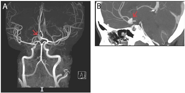
CTA/MRA showing 10mm saccular aneurysm at anterior communicating artery (ACoA)
DIGITAL SUBTRACTION ANGIOGRAPHY (DSA) — GOLD STANDARD
- Indication: CTA inconclusive; pre-operative planning; SAH with negative CTA (10% of SAH have negative initial CTA); follow-up after coiling
- Positive findings: Aneurysm neck, dome, feeding vessels, relationship to parent artery, multiplicity, vasospasm
- 4-vessel study (bilateral ICA + vertebral arteries)
- Repeat DSA at 2 weeks if initial is negative (angiographically negative SAH may be perimesencephalic or aneurysm missed due to spasm/thrombosis)
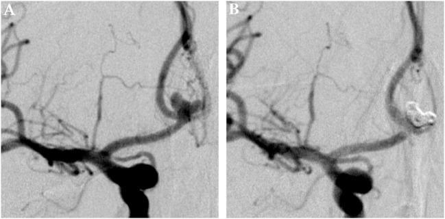
DSA: Pre-treatment multilobulated ACoA aneurysm (left) and complete exclusion after coil embolization (right)
MRI/MRA
- Indication: Incidental aneurysm screening; follow-up of coiled aneurysms; subacute SAH (FLAIR detects blood >24h)
- Positive findings: Flow-void; FLAIR hyperintensity in subarachnoid space; T2* GRE for chronic bleeds
1.7 MANAGEMENT
Medical Management (Acute SAH)
- Airway/breathing/circulation — Secure airway if GCS ≤8
- Bed rest in HDU/ICU
- Blood pressure control: Systolic ≤150 mmHg (until aneurysm secured); avoid hypotension
- Nimodipine — START IMMEDIATELY
💊 NIMODIPINE
- Drug class: Dihydropyridine calcium channel blocker (CCB)
- MOA: Blocks L-type voltage-gated Ca²⁺ channels in cerebral vascular smooth muscle → prevents Ca²⁺ influx → reduces sustained arterial spasm. Also has direct neuroprotective action via Ca²⁺-mediated neuronal death pathways
- Dose: 60 mg oral/NG every 4 hours × 21 days (or 0.5–2 mg/h IV in SIADH/unable to swallow)
- Special points:
- Only FDA-approved drug for reducing neurological deficits from vasospasm after SAH
- Does NOT decrease angiographic vasospasm but reduces DCI and improves neurological outcomes
- Must be given enterally (oral capsule or NG); IV preparation had risk of fatal errors if given as bolus — use IV preparation cautiously with infusion pump
- Reduces systemic BP — monitor for hypotension
- Statins (simvastatin, pravastatin) as adjunct — pleiotropic effects may reduce vasospasm (not first-line; evidence limited)
- Euvolemia — Maintain normal circulating blood volume (avoid hypovolemia → worsens vasospasm)
- Analgesia: Codeine, paracetamol (avoid NSAIDs)
- Antiepileptics: Short-term phenytoin or levetiracetam (prophylactic — controversial; not routine)
- DVT prophylaxis: Compression stockings; subcutaneous heparin after aneurysm secured
- Stool softeners (prevent Valsalva)
- Treat hyponatremia: If SIADH → fluid restriction; if CSWS → isotonic fluid replacement + fludrocortisone
- Hydrocephalus: EVD (External Ventricular Drain) if acute obstructive; VP shunt for chronic
1.8 SURGICAL & ENDOVASCULAR OPTIONS
A. SURGICAL CLIPPING
- Principle: Craniotomy + microsurgical placement of titanium clip across aneurysm neck, excluding it from circulation while preserving parent artery flow
- Indications (prefer clipping over coiling):
- Young patient with good grade (H&H I–III)
- Wide-necked aneurysm (dome:neck ratio <2, or neck >4 mm)
- MCA aneurysms (particularly good surgical access)
- Associated large ICH requiring evacuation
- Failed or recurrent after coiling
- Fusiform aneurysm
- Timing: Within 24–72 hours of SAH (early surgery to prevent rebleeding)
- Key surgical steps: Standard pterional craniotomy (most common), CSF drainage, sylvian fissure dissection, proximal control, clip application, intraoperative angiography/ICG to confirm complete exclusion
B. ENDOVASCULAR COIL EMBOLIZATION (GDC — Guglielmi Detachable Coils)
- Principle: Microcatheter navigated into aneurysm sac via femoral arterial access → platinum coils deployed → thrombosis fills the sac → excluded from circulation
- Indications (prefer coiling over clipping):
- Posterior circulation aneurysms (basilar tip, PICA) — hard surgical access
- Elderly or poor-grade patients (H&H IV–V)
- Narrow-necked aneurysm (dome:neck ratio >2)
- Medical comorbidities precluding craniotomy
- Patient preference
- ISAT Trial (2002): In patients suitable for both, coiling → better neurological outcome at 1 year vs. clipping (23.7% vs. 30.6% dependency/death); but higher rebleeding rate at 7 years
- BRAT Trial: Similar findings; favoured coiling for posterior circulation
Adjuncts to coiling:
- Balloon-assisted coiling — remodelling technique for wide-necked aneurysms
- Stent-assisted coiling — intracranial stent deployed across neck + coils
- Flow-diverter devices (Pipeline Embolization Device — PED): Redirects flow away from aneurysm into parent vessel → progressive thrombosis; used for large/giant fusiform aneurysms, wide-necked aneurysms (especially ICA)
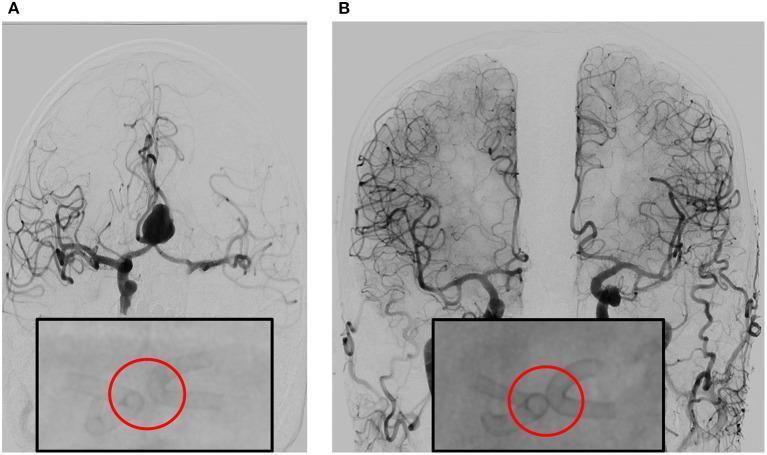
DSA showing bilateral flow-diverter stent deployment at ACoA — pre-treatment (A) and 6-month occlusion (B)
C. GIANT ANEURYSMS — Special Considerations
- Parent artery occlusion (PAO) with bypass (EC-IC bypass)
- Trapping (proximal + distal occlusion)
- Wrapping with muslin (palliative)
- Intraluminal stent stabilization (investigational)
D. MYCOTIC ANEURYSMS
- Treatment: Antibiotics (target organism for 4–6 weeks) first
- Surgery/coiling if: enlarging on serial angiography despite antibiotics, rupture, size >10 mm
- Serial DSA every 2 weeks while on antibiotics
1.9 UNRUPTURED INTRACRANIAL ANEURYSMS (UIA) — Management
ISUIA (International Study of Unruptured Intracranial Aneurysms) guidance:
- Aneurysms <7 mm with no prior SAH → very low annual rupture risk (~0.05%)
- Aneurysms ≥7 mm, posterior circulation location, or growth on serial imaging → treat
- Shared decision-making: Patient age, aneurysm size/location/morphology, comorbidities
Screening: MRA recommended for:
- First-degree relative of patient with SAH + aneurysm (especially ≥2 affected relatives)
- ADPKD patients (5–10% harbor aneurysms)
- Connective tissue disease
1.10 VASOSPASM MANAGEMENT (Triple-H Therapy — now modified)
Classic Triple-H: Hypertension, Hypervolemia, Hemodilution
Current evidence supports Induced Hypertension + Euvolemia (hypervolemia and hemodilution not proven beneficial)
- Target SBP 160–200 mmHg after aneurysm secured
- If refractory: Intra-arterial verapamil or nicardipine via cerebral angiography
- Transluminal balloon angioplasty for large vessel proximal vasospasm
- TCD (Transcranial Doppler) daily to monitor for vasospasm: MCA velocity >120 cm/s (mild), >200 cm/s (severe), Lindegaard ratio >3
PART 2: BRAIN ARTERIOVENOUS MALFORMATIONS (AVMs)
2.1 DEFINITION & PATHOLOGY
A brain AVM is a congenital vascular dysplasia consisting of:
- Abnormal cluster of arteries connected directly to veins without intervening capillary bed (nidus)
- High-pressure arteriovenous shunting
- Associated aneurysms in ~5% of cases (on main feeding artery)
- Somatic KRAS mutations in endothelial cells identified in ~50% of specimens
Genetics: Mostly sporadic; hereditary hemorrhagic telangiectasia (HHT/Osler-Weber-Rendu) is a recognized genetic association
2.2 EPIDEMIOLOGY
| Parameter | Detail |
|---|
| Prevalence | ~1/10th as common as saccular aneurysms |
| Sex | Males = Females |
| Age of presentation | Present from birth; symptoms most common age 10–30 years (occasionally up to age 50+) |
| Annual hemorrhage risk | ~3% per year (general); 6–9% in year after first hemorrhage |
| Rebleeding | 2–4% per year over decades |
2.3 CLINICAL PRESENTATION
| Mode of Presentation | Frequency |
|---|
| Intracerebral hemorrhage / SAH | ~45–50% (first manifestation) |
| Seizures (focal or generalized) | ~30% |
| Headache (migraine-type, especially parieto-occipital AVM) | ~20% |
| Progressive focal neurological deficit | ~10% |
| Bruit (systolic, over carotid/mastoid/eyeball) | <25% |
Features suggesting AVM:
- Young patient with ICH in unusual location (not basal ganglia/thalamus — which is hypertensive)
- No hypertension
- Recurrent hemorrhage in the same location
- Seizure disorder + unusual headaches
Additional signs:
- Pulsatile carotid in the neck (large AVM)
- Retinal vascular malformation (coextensive with optic nerve lesion)
- Vein of Galen enlargement → hydrocephalus in children
2.4 SPETZLER-MARTIN GRADING SCALE (Surgical Risk Stratification)
| Feature | Score |
|---|
| Nidus size | |
| < 3 cm | 1 |
| 3–6 cm | 2 |
| > 6 cm | 3 |
| Venous drainage | |
| Superficial only | 0 |
| Deep (includes internal cerebral veins, basal veins, or cerebellar veins) | 1 |
| Eloquence of adjacent brain | |
| Non-eloquent | 0 |
| Eloquent (sensorimotor, language, visual cortex, thalamus/hypothalamus, brainstem, cerebellar peduncles) | 1 |
Total score: 1–5
- Grade I–II: Low surgical risk → recommend surgery
- Grade III: Intermediate → individualized decision
- Grade IV–V: High surgical risk → consider SRS or embolization; surgery rarely recommended
Supplementary factors (Spetzler-Ponce "class" system):
- Age (<20, 20–40, >40), prior bleed, nidus compactness
2.5 INVESTIGATIONS
MRI Brain
- Indication: Screening, diagnosis, pre-operative mapping
- Positive findings:
- T2: Serpiginous flow voids (high-velocity arterial flow) = hallmark
- T1: Hemosiderin from old bleeds (dark rim)
- T2*/GRE/SWI: Microbleeds, old hemorrhage
- FLAIR: Perilesional gliosis (from chronic ischemia/"steal")
- Dilated feeding arteries and draining veins
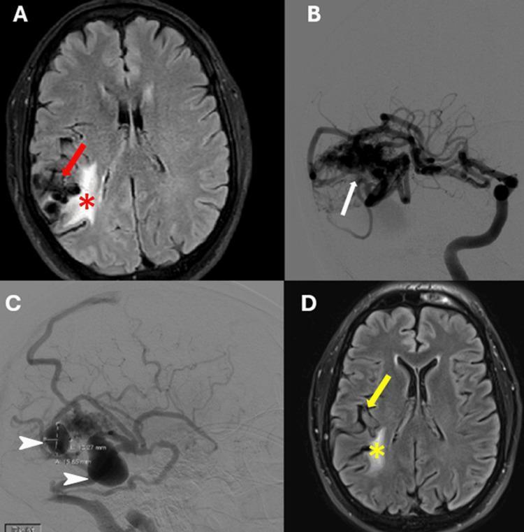
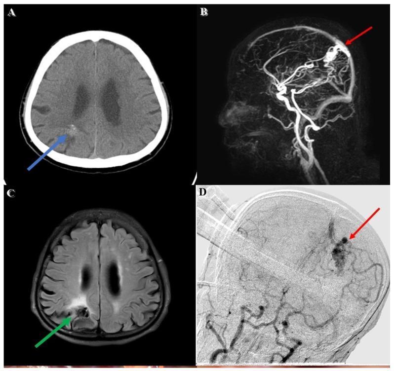
Spetzler-Martin Grade 3 AVM: FLAIR shows flow voids and gliosis (A); DSA reveals nidus from MCA feeders (B); venous pseudoaneurysms (C); post-radiosurgery improvement (D)
Grade I AVM: CT shows right parietal hemorrhage (A); MRA shows nidus (B); T2 shows flow voids (C); DSA confirms vascular architecture (D)
CT (non-contrast)
- Positive findings: ICH, calcifications within AVM (35%), hyperdense nidus
DSA (Gold Standard)
- Indication: Diagnosis confirmation; pre-treatment planning (surgical, embolization, SRS)
- Positive findings: Nidus (tangle of abnormal vessels), feeding arteries, draining veins (early venous filling = AV shunting), associated aneurysms
- 4D DSA (3D rotational) for pre-operative planning
2.6 MANAGEMENT OF BRAIN AVMs
Conservative (Observation)
- Small, deep, unruptured AVMs in elderly
- ARUBA Trial (2014): For unruptured AVMs, medical management alone may be superior to intervention in the short term — controversial, ongoing debate
A. MICROSURGICAL RESECTION
- Indications: Grade I–III AVMs; ruptured AVM in accessible location; lesion causing progressive deficits
- Steps: Craniotomy → identify and control feeding arteries first → circumferential dissection of nidus → coagulate feeding arteries → remove nidus en bloc → ligate draining veins last (ligate veins early → hemorrhagic infarction!)
- Intraoperative DSA to confirm complete resection
- Grade IV–V → rarely operated; morbidity very high
B. STEREOTACTIC RADIOSURGERY (SRS — Gamma Knife / CyberKnife / LINAC)
- Indications: Small AVMs (≤3 cm / <10 ml), deep/eloquent location (thalamus, brainstem), Grade III–IV, residual after embolization
- Mechanism: Ionizing radiation → endothelial injury → progressive thrombosis and fibrosis → obliteration over 2–3 years
- Obliteration rates: ~80% for small AVMs at 3 years
- Limitation: Latency period of 2–3 years during which AVM still bleeds; radiation necrosis (3–5%)
C. ENDOVASCULAR EMBOLIZATION (ONYX / NBCA)
- Indications: Rarely curative alone; used as adjunct to surgery (reduce blood loss) or SRS (reduce nidus volume)
- ONYX (Ethylene-vinyl alcohol copolymer): Liquid embolic agent; controlled injection; forms cast in nidus
- NBCA (n-butyl cyanoacrylate): Older liquid embolic; faster polymerization
- Pre-operative embolization 24–48 hours before surgery
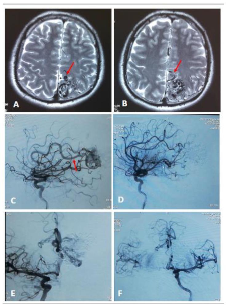
Grade 3 left parietal AVM: T2 flow voids (A,B); DSA pre-embolization (C,E); marked devascularization post-embolization (D,F)
D. MULTIMODALITY APPROACH
- Embolization → SRS (reduce nidus volume to <3 cm for SRS)
- Embolization → Surgery (reduce blood loss, easier resection)
PART 3: DURAL ARTERIOVENOUS FISTULAS (DAVFs)
3.1 DEFINITION
A dAVF is an acquired arteriovenous shunt within the dural leaflets, connecting dural arteries to dural venous sinuses or cortical veins. Unlike brain AVMs, these are NOT congenital.
Common locations: Transverse/sigmoid sinus (most common), cavernous sinus, superior sagittal sinus, anterior cranial fossa (ethmoidal DAVF — highest hemorrhage risk)
Etiology/Triggers:
- Dural sinus thrombosis (most important — angiogenesis-related recanalization)
- Trauma
- Prior surgery/craniotomy
- Infection
- Idiopathic
3.2 EPIDEMIOLOGY
| Parameter | Detail |
|---|
| Age | Adults; usually 40–60 years |
| Sex | Males > Females (for lateral sinus DAVFs); Females for cavernous sinus DAVFs |
| Proportion of all vascular malformations | ~10–15% |
3.3 CLASSIFICATION
COGNARD CLASSIFICATION (Venous Drainage Pattern — Hemorrhage Risk)
| Type | Venous Drainage | Hemorrhage Risk |
|---|
| I | Antegrade into sinus | Benign |
| IIa | Retrograde into sinus only | Low |
| IIb | Retrograde into cortical veins only | Moderate |
| IIa+b | Retrograde into both sinus and cortical veins | Moderate-High |
| III | Direct cortical venous drainage, no venous ectasia | High |
| IV | Direct cortical venous drainage + venous ectasia | Very High |
| V | Drainage to spinal perimedullary veins | Myelopathy |
BORDEN CLASSIFICATION (Simpler, 3 types)
| Type | Description | Risk |
|---|
| I | Drains into dural sinus (antegrade) | Benign |
| II | Drains into dural sinus + retrograde cortical veins | Moderate |
| III | Drains directly into cortical veins only | High hemorrhage risk |
Viva pearl: Borden III = Cognard III/IV = HIGH RISK → needs treatment
3.4 CLINICAL FEATURES
Based on location:
| Location | Symptoms |
|---|
| Cavernous sinus DAVF | Pulsatile proptosis, chemosis, conjunctival injection, bruit, diplopia (CN VI palsy most common), glaucoma |
| Transverse/Sigmoid sinus | Pulsatile tinnitus (most common presenting symptom), headache, bruit over mastoid |
| Superior sagittal sinus | Headache, intracranial hypertension, papilledema |
| Anterior cranial fossa (ethmoidal) | ICH (no benign form — all drain to cortical veins → high hemorrhage risk) |
| Tentorial | Severe symptoms, hemorrhage risk |
| Spinal DAV fistula (type V) | Progressive myelopathy, Foix-Alajouanine syndrome |
Symptoms of cortical venous hypertension (Borden II/III):
- ICH, SAH
- Focal neurological deficits
- Dementia (venous congestion)
- Seizures
- Papilledema
3.5 INVESTIGATIONS
MRI Brain
- Dilated cortical veins
- T2 hyperintensity in white matter (venous hypertension/congestion)
- Prominent superficial vessels on MRA
- GRE/SWI: Hemosiderin from old bleeds
CT/CTA
- Early venous filling
- Dilated cortical veins
- Venous ectasia (varix)
DSA (Gold Standard)
- Indication: Definitive diagnosis; pre-treatment planning; defines fistula point, feeding arteries (ECA/ICA dural branches), venous drainage
- Positive findings: Early arteriovenous shunting; retrograde cortical venous filling; venous varices; fistula point
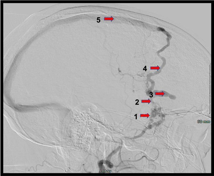
DSA lateral view: Borden III/Cognard IV dAVF — middle meningeal artery (1) feeds fistula point (2) → venous ectasia (3) → arterialized Sylvian vein (4) → superior sagittal sinus (5)
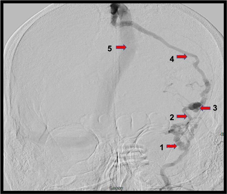
AP DSA: same Borden III/Cognard IV dAVF demonstrating retrograde cortical venous drainage — high hemorrhage risk
3.6 MANAGEMENT OF DAVFs
Conservative Management
- Borden I (antegrade sinus drainage only) — low risk; observe or treat symptoms
- Cavernous sinus DAVF: 50% spontaneous closure; manual carotid compression (patient compresses ipsilateral carotid with contralateral hand × 10 min 3× daily for 4 weeks — Halbach technique) — effective in some CCF/DAVF
A. ENDOVASCULAR EMBOLIZATION (PREFERRED for most)
Transarterial embolization:
- ONYX injected via microcatheter into feeding arteries → occludes fistula point
- Used for anterior cranial fossa DAVF, transverse/sigmoid DAVF
Transvenous embolization:
- Microcatheter navigated via venous route into sinus/vein near fistula → coils/ONYX → occlude fistula
- Particularly effective for cavernous sinus DAVF (via inferior petrosal sinus)
- Transvenous coiling of involved sinus segment for sigmoid/transverse sinus DAVF
B. SURGICAL TREATMENT
- Indications: Failed endovascular; anterior cranial fossa DAVF (ethmoidal — surgical disconnection preferred due to proximity to ophthalmic artery); accessible Borden III DAVFs
- Technique: Craniotomy → identify and interrupt cortical draining vein at fistula point → sinus skeletonization → coagulation/clip of fistulous connection
C. STEREOTACTIC RADIOSURGERY
- Adjunct or alternative for small DAVFs; slower obliteration
- Suitable for deep/inaccessible locations
Cavernous-Carotid Fistula (CCF) — Special Type of DAVF
| Type | Description | Cause | Treatment |
|---|
| Type A (Direct CCF) | High-flow; ICA directly into cavernous sinus | Trauma, ruptured ICA aneurysm | Transarterial/transvenous coiling; detachable balloon (historical) |
| Type B | ICA meningeal branches → cavernous sinus | Dural (spontaneous) | Conservative/embolization |
| Type C | ECA meningeal branches → cavernous sinus | Dural | Embolization |
| Type D | Both ICA + ECA branches → cavernous sinus | Dural | Embolization |
PART 4: FOLLOW-UP PROTOCOLS
Aneurysm Follow-Up
| Scenario | Modality | Frequency |
|---|
| After surgical clipping | CT angio or DSA at 1 year; MRA at 5 years | Annual initially |
| After coil embolization | DSA at 3–6 months; then MRA at 1, 3, 5 years | More frequent (coil compaction/recanalization risk 20–30%) |
| Unruptured, untreated | MRA or CTA every 6–12 months × 2 years; then annually if stable | Annual |
| Flow diverter (PED) | DSA at 3–6 months; MRA at 1–3 years | Per protocol |
AVM Follow-Up
| Scenario | Modality | Frequency |
|---|
| Post-surgical resection | Immediate post-op DSA (same day or day 1); MRI at 3–6 months | Annual MRI × 3 years; then as needed |
| Post-SRS | MRI 6 months, 1 year, 2 years; DSA at 3 years (to confirm obliteration) | Annual MRI until obliteration confirmed on DSA |
| Conservative management | MRI ± MRA every 1–2 years | Annual |
DAVF Follow-Up
| Scenario | Modality | Frequency |
|---|
| Post-embolization | DSA at 3–6 months | As needed; repeat if residual |
| Borden I (conservative) | MRI ± MRA at 1 year | Annual |
| Post-surgical | DSA at 3 months | Annual MRI thereafter |
QUICK VIVA SUMMARY TABLE
| Feature | Saccular Aneurysm (SAH) | Brain AVM | DAVF |
|---|
| Age | 35–65 years | 10–30 years | 40–60 years |
| Congenital vs Acquired | Congenital weakness | Congenital | Acquired |
| Sex | F > M (SAH) | M = F | M > F (lateral); F > M (cavernous) |
| Presentation | Thunderclap headache, SAH | ICH, seizures, headache | Tinnitus, proptosis, ICH (high-grade) |
| Key Scale | Hunt & Hess, WFNS, Fisher | Spetzler-Martin | Cognard, Borden |
| Gold standard Ix | DSA | DSA | DSA |
| First-line treatment | Coiling (ISAT) or Clipping | Surgery (SM I–III), SRS | Endovascular embolization |
| Key drug | Nimodipine (60 mg q4h × 21 days) | None specific | None specific |
| Hallmark imaging | CT: hyperdense cisterns | MRI: flow voids | DSA: retrograde cortical veins |
Key Viva Tips:
- Nimodipine does NOT prevent vasospasm angiographically — it prevents the neurological consequences (DCI)
- Ligate AVM veins LAST during surgery — ligating early causes hemorrhagic infarction
- Anterior cranial fossa DAVF has no benign form — all drain to cortical veins → always treat surgically
- ISAT Trial favoured coiling; BRAT Trial confirmed coiling for posterior circulation
- Spetzler-Martin Grade ≥ IV → surgery carries unacceptably high morbidity; consider SRS or observation
- Cerebral salt wasting vs SIADH — both cause hyponatremia post-SAH; CSWS requires volume replacement (not restriction!)
- 20% of aneurysm patients have multiple aneurysms — identify the ruptured one by the clot location on CT
Based on: Adams & Victor's Principles of Neurology 12e | Barash Clinical Anesthesia 9e | Harrison's 22e | Bradley & Daroff's Neurology in Clinical PracticeHere is your complete neurosurgery viva guide covering all three entities. Here's a quick orientation of what's covered:
Structure of the guide:
Part 1 — Intracranial Aneurysms
- Full classification (saccular/fusiform/mycotic/traumatic/giant) with size categories
- Demographics: peak age 35–65, F>M, 20% multiple
- Pathogenesis: congenital defect in media + hemodynamic theory (Ferguson)
- Clinical: thunderclap headache, CN III palsy patterns, mass effect
- 3 Grading Scales: Hunt & Hess (clinical), WFNS (GCS-based), Fisher (CT/vasospasm risk)
- Investigations: CT → LP (xanthochromia) → CTA → DSA with indications and positive findings
- Management: Nimodipine (MOA + dose + special points), general ICU care
- Surgery: Clipping vs. Coiling with ISAT/BRAT evidence, flow diverters, indications for each
- Vasospasm management (Triple-H → evolved to induced HTN + euvolemia)
- Unruptured aneurysm (ISUIA criteria)
Part 2 — Brain AVMs
- Epidemiology: age 10–30, 3%/year bleed risk
- Spetzler-Martin Scale (full table with scoring)
- MRI findings (flow voids), DSA planning
- Surgical resection, SRS (Gamma Knife), ONYX embolization with clear indications
- ARUBA Trial mention
Part 3 — DAVFs
- Acquired (sinus thrombosis) vs. congenital AVM
- Cognard Classification (I–V) + Borden Classification (I–III) with hemorrhage risk
- Location-specific symptoms (cavernous → proptosis; transverse → tinnitus; anterior fossa → ICH)
- Endovascular (transarterial/transvenous ONYX), surgical, SRS options
- Carotid-Cavernous Fistula (Barrow A–D types)
Part 4 — Follow-up protocols for each entity with modality and frequency