
ECG finding
I'd be happy to help with ECG findings! Let me clarify what you're looking for.
Asking for Preferences
What ECG topic would you like to explore?
Got it! You want 10 diseases with their characteristic ECG findings and images. Let me pull that from the medical library now.
Searching sources...
Reading File
Finding Sources
Finding Sources
Finding Sources
Finding Sources
Finding Sources
Finding Sources
Reading File
Reading File
Searching the Web
ECG findings 10 cardiac diseases STEMI atrial fibrillation heart block WPW hyperkalemia pericarditis images
I now have great textbook content and ECG images. Let me fetch the key ECG images and read more sections for a comprehensive response covering 10 diseases.
Loading Image
Loading Image
Loading Image
Finding Sources
Reading File
Reading File
Loading Image
Reading File
Loading Image
Finding Sources
Finding Sources
Reading File
Reading File
I now have excellent textbook content. Let me fetch a few more key ECG images and compile the comprehensive answer.
Loading Image
Searching the Web
ECG pericarditis hyperkalemia WPW long QT Brugada ventricular tachycardia images site:litfl.com OR site:ecgwaves.com
Here is your comprehensive guide to 10 diseases and their characteristic ECG findings, with textbook images.
ECG Findings in 10 Diseases
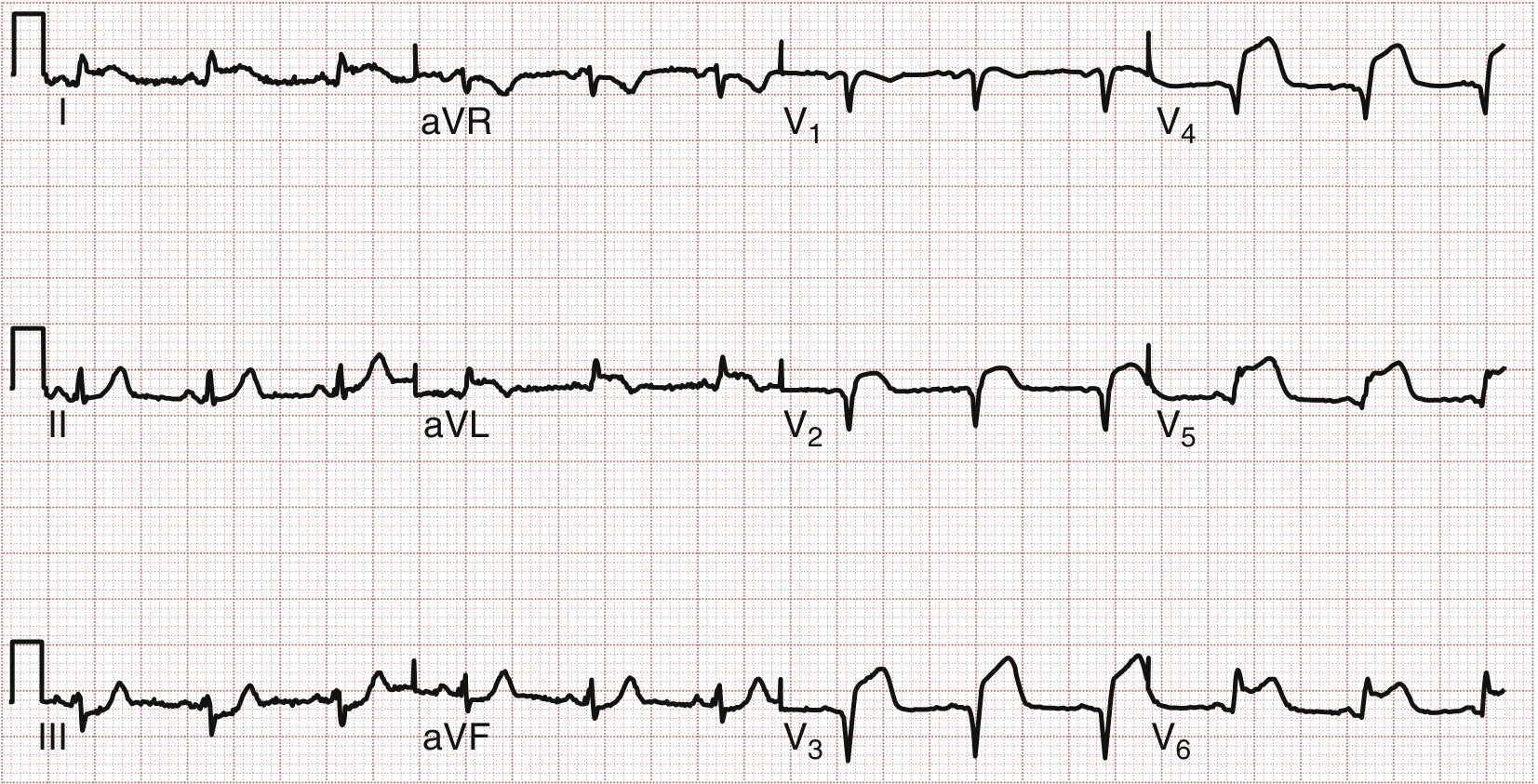
1. Anterolateral ST-Elevation Myocardial Infarction (STEMI)
ECG Features:
- ST elevation in leads V1-V4 (anterior) and I, aVL, V5-V6 (lateral)
- Reciprocal ST depression in inferior leads (III, aVF)
- Hyperacute T waves early; Q waves develop as infarct evolves
- Proximal LAD occlusion produces the widest distribution of changes
2. Inferior STEMI
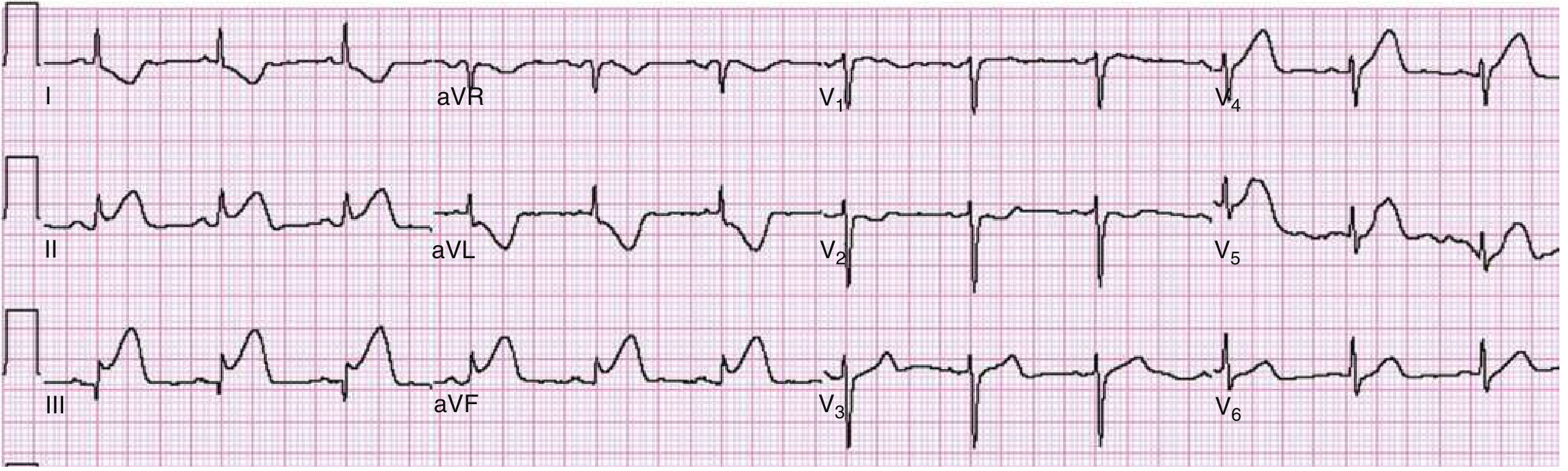
ECG Features:
- ST elevation in leads II, III, aVF
- Reciprocal ST depression in I and aVL
- If ST elevation is greater in lead III than lead II: suggests RCA occlusion (90% sensitive, 71% specific)
- ST elevation in V1 with inferior MI suggests right ventricular infarction
- Posterior extension: ST depression + tall R waves in V1-V3
3. High Lateral STEMI
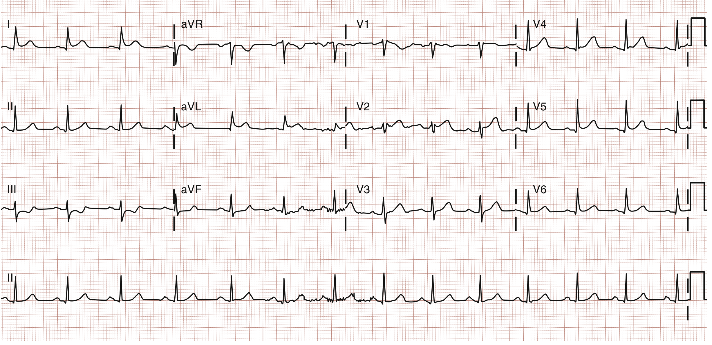
ECG Features:
- ST elevation in leads I and aVL (first diagonal or left circumflex occlusion)
- Reciprocal ST depression in leads III, aVF, and V1
- May have subtle ST elevation in V2
4. Extensive Inferior + Posterior + RV MI (15-Lead ECG)
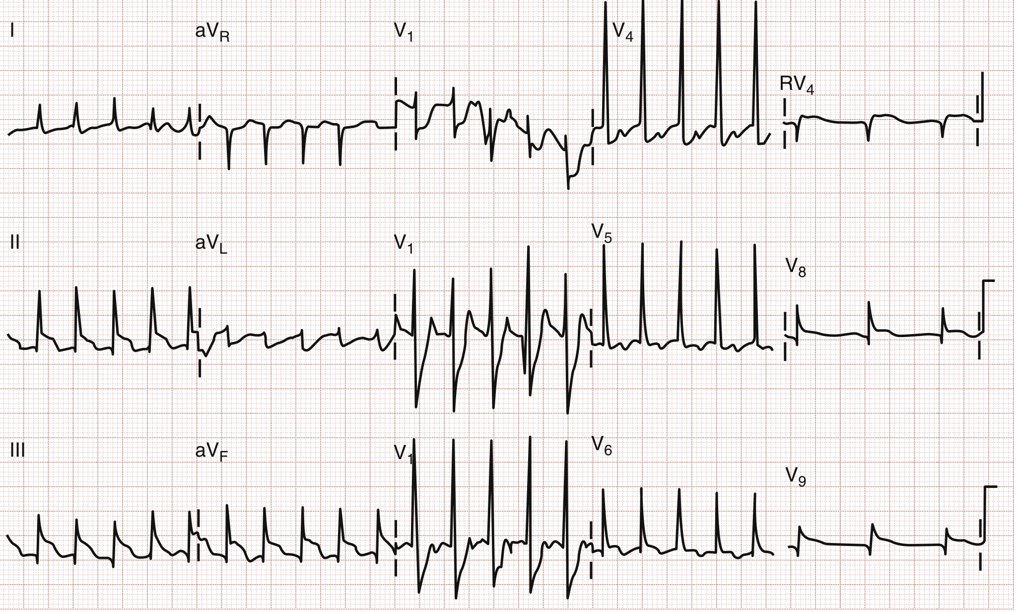
ECG Features:
- ST elevation II, III, aVF (inferior)
- ST depression V1-V3 with prominent R wave = posterior MI (reciprocal)
- ST elevation in posterior leads V8-V9
- ST elevation in right-sided leads (RVq) = right ventricular MI
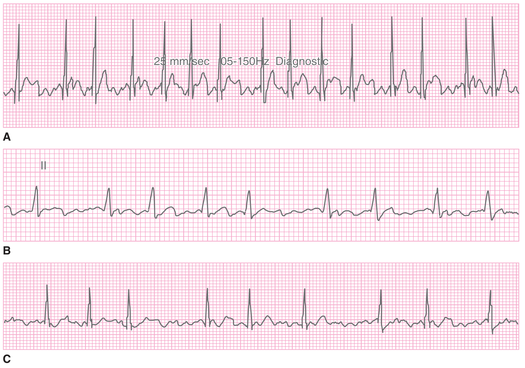
5. Atrial Fibrillation (AF)
ECG Features:
- No discernible P waves - replaced by chaotic/fibrillatory baseline
- Irregularly irregular ventricular response
- Narrow QRS complexes (unless bundle branch block/preexcitation coexists)
- Atrial rate >600 bpm; ventricular rate typically 120-170 bpm when AV node unaffected
- A very rapid ventricular response (>200 bpm) suggests an accessory pathway (WPW)
6. Atrial Flutter
ECG Features:
- Regular "sawtooth" flutter waves at ~300 bpm - best seen in II, III, aVF, V1
- Usually regular ventricular rate due to fixed AV block ratio (2:1 most common = ~150 bpm)
- Regular narrow-complex tachycardia at exactly ~150 bpm should always raise suspicion for flutter with 2:1 block
- P waves (flutter waves) are single morphology, downward-deflecting
7. Ventricular Tachycardia (VT)
ECG Features:
- Wide-complex tachycardia (QRS >120 ms, often >160 ms)
- AV dissociation (P waves and QRS complexes independent) - most reliable sign of VT
- Fusion beats and capture beats confirm AV dissociation
- Concordance: all precordial leads positive (positive concordance) or all negative (negative concordance) - strongly suggests VT
- No RS complex in any precordial lead favors VT
- Initial R wave in aVR favors VT
Key rule: Any wide-complex tachycardia should be treated as VT until proven otherwise, especially in patients with structural heart disease or prior MI.
8. Third-Degree (Complete) AV Block
ECG Features:
- Complete dissociation of P waves and QRS complexes
- Atrial rate is faster than ventricular rate
- P waves march through at their own rate; QRS complexes at a slow escape rate (20-60 bpm)
- Wide QRS escape rhythm = block below AV node (His-Purkinje); narrow QRS = junctional escape
- Causes: acute MI (especially inferior), Lyme disease, autoimmune, infiltrative cardiomyopathy, drug toxicity
Key finding: Regular P waves at ~80 bpm + slow regular wide QRS at ~35 bpm, completely independent of each other.
9. Wolff-Parkinson-White (WPW) Syndrome
ECG Features:
- Short PR interval (<120 ms)
- Delta wave - slurred upstroke at start of QRS due to ventricular preexcitation
- Widened QRS (>120 ms)
- Pseudo-ST and T changes (secondary repolarization abnormalities)
- Risk: AF with rapid conduction through accessory pathway can precipitate VF
Classic triad: Short PR + delta wave + widened QRS = WPW pattern.
10. Hyperkalemia
ECG Features (progressive with rising K+):
| K+ Level | ECG Change |
|---|---|
| 5.5-6.0 mEq/L | Tall, peaked (tented) T waves - narrow base, symmetric |
| 6.0-7.0 mEq/L | PR prolongation, P wave flattening |
| 7.0-8.0 mEq/L | Widening of QRS |
| >8.0 mEq/L | Sine wave pattern (QRS merges with T wave) |
| Critical | Ventricular fibrillation / asystole |
Peaked T waves in hyperkalemia are narrow-based and symmetric, distinguishing them from the broad asymmetric T waves in ischemia.
Summary Table
| # | Disease | Key ECG Finding |
|---|---|---|
| 1 | Anterolateral STEMI | ST elevation V1-V4, I, aVL |
| 2 | Inferior STEMI | ST elevation II, III, aVF + reciprocal changes |
| 3 | High Lateral STEMI | ST elevation I, aVL |
| 4 | Posterior/RV MI | ST depression V1-V3 + tall R, ST elevation V8-V9/RV leads |
| 5 | Atrial Fibrillation | No P waves, irregularly irregular QRS |
| 6 | Atrial Flutter | Sawtooth flutter waves ~300 bpm, regular ventricular rate ~150 |
| 7 | Ventricular Tachycardia | Wide QRS, AV dissociation, fusion beats |
| 8 | Complete Heart Block | P-QRS dissociation, atrial rate > ventricular rate |
| 9 | WPW Syndrome | Short PR, delta wave, wide QRS |
| 10 | Hyperkalemia | Peaked T waves → wide QRS → sine wave pattern |
*Sources: ROSEN's Emergency Medicine (9th Ed.), Tintinalli's Emergency Medicine, Braunwald's Heart Disease, Goldman-Cecil Medicine, Harrison's Principles of Internal Medicine 22E (2025)*This covers 10 important conditions with their hallmark ECG patterns and real textbook ECG images. Here are a few high-yield mnemonics to remember:
- STEMI localization: ST elevation in II/III/aVF = inferior; V1-V4 = anterior; I/aVL = lateral; V1-V3 depression + tall R = posterior
- AF vs Flutter: AF = irregularly irregular + no P waves; Flutter = regular sawtooth at 300 bpm with 2:1 block giving rate ~150
- VT vs SVT: Wide complex + AV dissociation + fusion beats = VT until proven otherwise
- WPW triad: Short PR + delta wave + wide QRS
- Hyperkalemia progression: Peaked T → flat P → wide QRS → sine wave → VF
Let me know if you'd like to go deeper on any specific condition!
ECG finding with images and ECG reading with all disease.
Pulling ECG images and readings for all major diseases now - running parallel searches across the medical library.
Searching sources...
Finding Sources
Finding Sources
Finding Sources
Finding Sources
Reading File
Reading File
Reading File
Reading File
Excellent - found images for pericarditis, Brugada, LBBB/RBBB. Let me fetch all these images in parallel now.
Loading Image
Loading Image
Loading Image
Reading File
Loading Image
Loading Image
Finding Sources
Finding Sources
Reading File
Reading File
Finding Sources
Loading Image
Reading File
Finding Sources
Reading File
Finding Sources
Loading Image
Reading File
Loading Image
Finding Sources
I now have an excellent collection of textbook ECG images. Let me compile the full, comprehensive illustrated guide.---
Complete ECG Guide: Findings & Readings Across All Major Diseases
HOW TO READ ANY ECG - Systematic Approach
Before reading individual diseases, always follow this order:
- Rate - Count R waves in 6-second strip x 10, or 300/large squares between R-R
- Rhythm - Regular? Irregular? Identify P waves
- P waves - Present? Normal shape? One before each QRS?
- PR interval - Normal: 120-200 ms (3-5 small squares)
- QRS duration - Normal: <120 ms (3 small squares)
- QRS axis - Normal: -30° to +90°
- ST segments - Elevation? Depression? Horizontal/upsloping/downsloping?
- T waves - Upright? Inverted? Peaked?
- QT interval - QTc normal: <440 ms (male), <460 ms (female)
- Ancillary - Q waves, bundle branch blocks, hypertrophy
SECTION A: ISCHEMIC HEART DISEASE
1. Anterior STEMI (LAD Occlusion)
ECG Reading:
| Feature | Finding |
|---|---|
| Rate | Usually normal or tachycardic |
| Rhythm | Sinus rhythm (may have PVCs) |
| P waves | Normal |
| PR interval | Normal |
| QRS | May develop pathological Q waves in V1-V4 |
| ST segments | Elevation in V1-V4 (and I, aVL if anterolateral) |
| T waves | Hyperacute upright early; inversion later |
| Reciprocal | ST depression in II, III, aVF |
Key ECG findings:
- ST elevation ≥1 mm in 2 contiguous leads
- "Tombstone" ST elevations in severe LAD occlusion
- Loss of R-wave progression (R waves become smaller V1→V4)
- Pathological Q waves (>1 small sq wide, >¼ R-wave height) = completed infarct
2. Inferior STEMI (RCA or LCx Occlusion)
ECG Reading:
| Feature | Finding |
|---|---|
| ST Elevation | II, III, aVF |
| Reciprocal | ST depression in I, aVL |
| Key clue | If STE greater in III than II → RCA occlusion (90% sens.) |
| RV involvement | STE in V1 with inferior MI = right ventricular MI |
| Posterior extension | ST depression + tall R in V1-V3 |
How to distinguish RCA vs LCx inferior STEMI:
- RCA: STE III > II, ST depression in aVL and I
- LCx: STE II ≥ III, aVL isoelectric or elevated
3. High Lateral STEMI (Diagonal / LCx Occlusion)
ECG Reading:
| Feature | Finding |
|---|---|
| ST Elevation | I, aVL (sometimes V2) |
| Reciprocal | ST depression in III, aVF, V1 |
| Culprit | First diagonal (D1) or left circumflex |
4. Extensive Inferior + Posterior + Right Ventricular MI
ECG Reading (15-lead):
| Territory | Leads | Finding |
|---|---|---|
| Inferior | II, III, aVF | ST elevation |
| Lateral | I, aVL, V5-V6 | ST elevation |
| Posterior | V1-V3 (standard) | ST depression + tall R wave (reciprocal) |
| Posterior (direct) | V7-V9 | ST elevation (smaller magnitude) |
| Right ventricle | RV4 | ST elevation |
Reading tip: Posterior MI is the "mirror image" - flip V1-V3 upside down mentally and you see STEMI pattern.
SECTION B: ARRHYTHMIAS
5. Atrial Fibrillation (AF)
ECG Reading:
| Feature | Finding |
|---|---|
| Rate | Ventricular: 120-170 bpm (uncontrolled); slower if controlled/AV node disease |
| Rhythm | Irregularly IRREGULAR - hallmark |
| P waves | ABSENT - replaced by chaotic fibrillatory baseline |
| PR interval | Not measurable |
| QRS | Narrow (unless BBB or WPW coexists) |
| Baseline | Fine fibrillatory waves, most prominent in V1 |
Key rule: If you see NO P waves + irregularly irregular QRS = AF until proven otherwise.
- AF with rate >200 bpm = suspect WPW accessory pathway (medical emergency - avoid AV nodal blockers!)
6. Atrial Flutter
ECG Reading:
| Feature | Finding |
|---|---|
| Rate | Atrial: ~300 bpm; Ventricular: ~150 bpm (2:1 block) |
| Rhythm | Usually regular (if fixed AV block ratio) |
| P waves | Sawtooth flutter waves - no isoelectric baseline between them |
| Best leads | II, III, aVF, V1 for flutter waves |
| AV ratio | Usually 2:1 (rate ~150), can be 3:1 or 4:1 |
Bedside clue: Regular narrow-complex tachycardia at exactly ~150 bpm = atrial flutter with 2:1 block until proven otherwise. Apply vagal maneuver to slow rate and reveal flutter waves.
7. Ventricular Tachycardia (VT)
ECG Reading:
| Feature | Finding |
|---|---|
| Rate | 100-250 bpm |
| Rhythm | Usually regular |
| QRS | Wide (>120 ms), often >160 ms |
| AV dissociation | P waves and QRS independent - most specific sign |
| Fusion beats | Hybrid QRS (sinus + VT) = confirms AV dissociation |
| Capture beats | Narrow QRS "captured" in a run of wide QRS = confirms VT |
| Concordance | All precordial QRS all positive (positive) or all negative (negative) = strongly suggests VT |
| aVR sign | Initial R wave in aVR, or notching of initial downstroke = VT |
Critical rule: Any wide-complex tachycardia in a patient with prior MI or cardiomyopathy = VT until proven otherwise. Never give verapamil to wide-complex tachycardia.
8. Supraventricular Tachycardia (SVT/AVNRT)
ECG Reading:
| Feature | Finding |
|---|---|
| Rate | 150-250 bpm |
| Rhythm | Perfectly regular |
| QRS | Narrow (<120 ms) unless aberrant conduction |
| P waves | Hidden in or just after QRS (retrograde P waves) |
| RP interval | Very short (<70 ms in AVNRT) |
| Response | Terminates abruptly with vagal maneuver or adenosine |
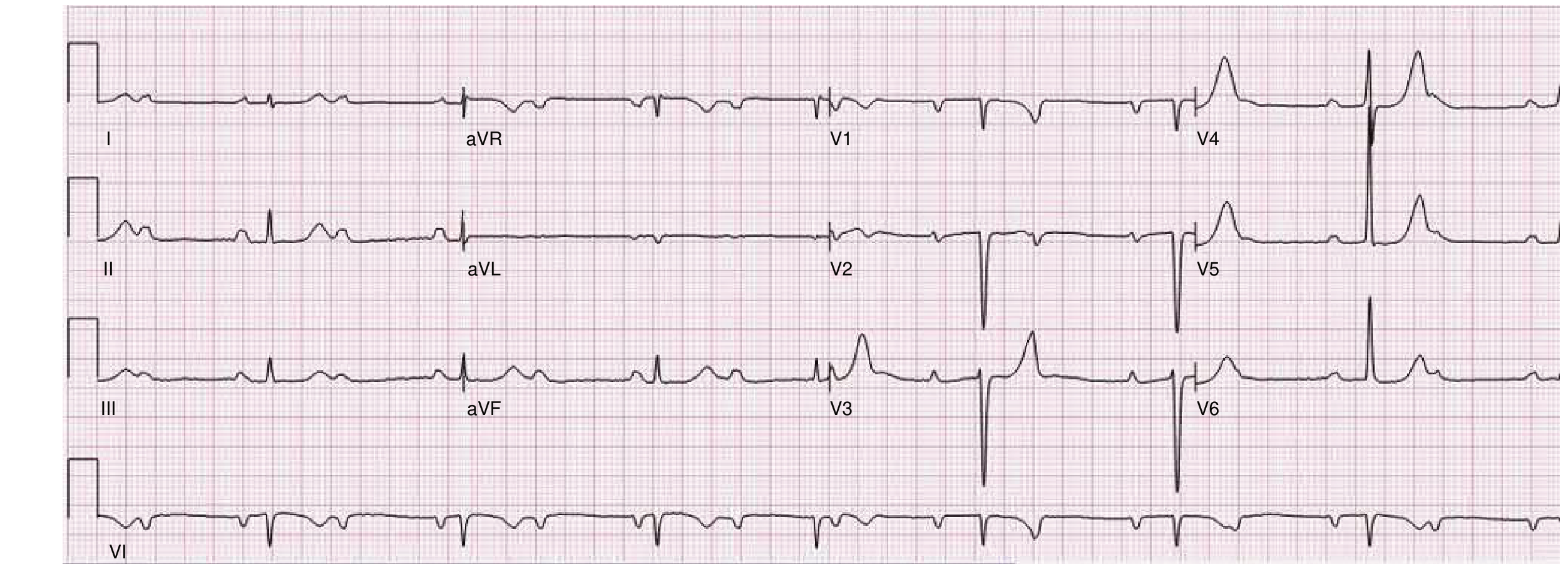
9. Complete (Third-Degree) AV Heart Block
ECG Reading:
| Feature | Finding |
|---|---|
| P waves | Present, regular at own rate (~60-100 bpm) |
| QRS | Present, regular at slow escape rate (20-60 bpm) |
| Key finding | P waves and QRS are COMPLETELY INDEPENDENT |
| Atrial rate | Always FASTER than ventricular rate |
| QRS morphology | Narrow = junctional escape; Wide = ventricular escape (worse) |
How to read: Measure P-P interval and R-R interval separately. They march out at their own rates with NO relationship to each other.
SECTION C: CONDUCTION DISORDERS
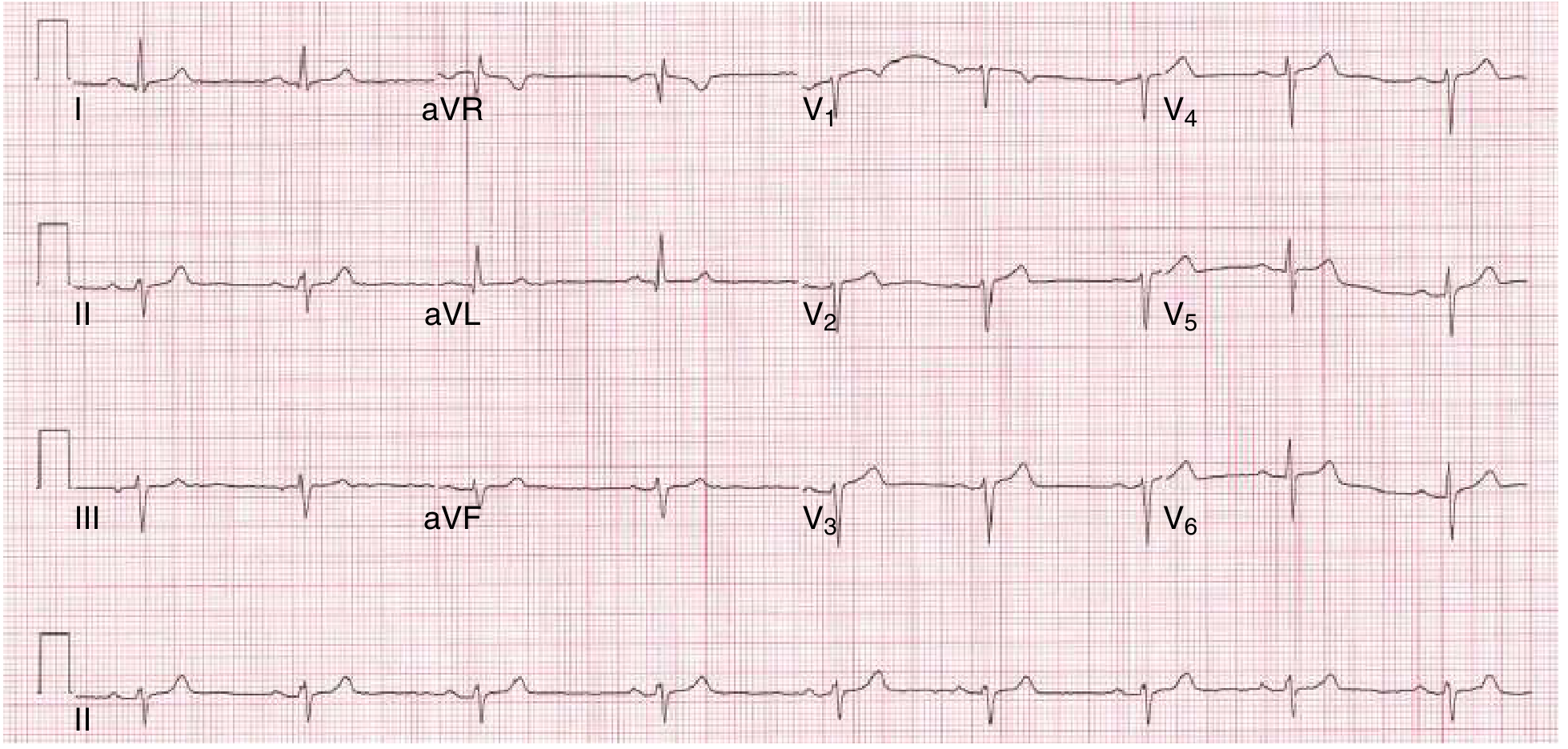
10. Right Bundle Branch Block (RBBB)
ECG Reading:
| Feature | Finding |
|---|---|
| QRS duration | ≥120 ms (wide) |
| V1 pattern | rsR' or rSR' ("rabbit ears") pattern |
| Wide S wave | Wide terminal S in V5-V6 and lead I |
| ST/T | Discordant ST/T in V1-V2 (opposite to QRS) |
| Axis | Usually normal |
Memory aid: "WiLLiaM MaRRoW" for LBBB vs RBBB - in V1: LBBB = W shape, RBBB = M shape. In V6: LBBB = M, RBBB = W.
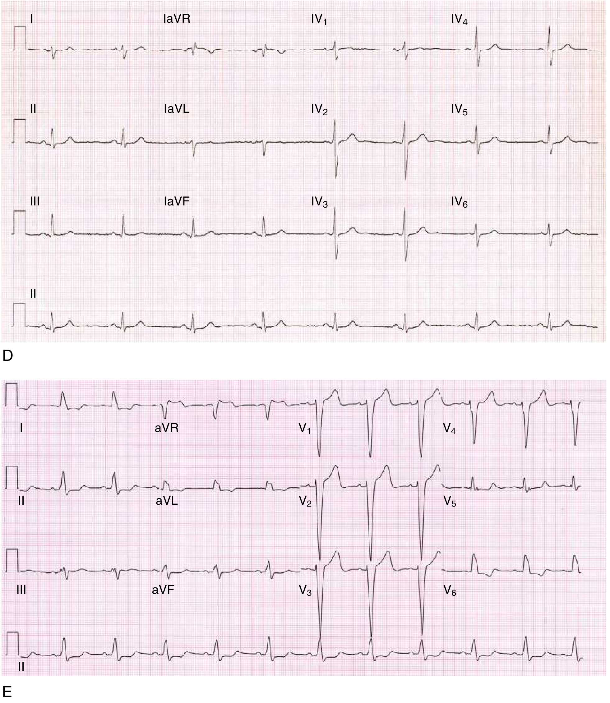
11. Left Bundle Branch Block (LBBB)
ECG Reading:
| Feature | Finding |
|---|---|
| QRS duration | ≥120 ms |
| V1 pattern | QS or rS pattern (deep S, no R) |
| V5-V6 pattern | Broad notched R wave without Q |
| Lead I/aVL | Broad notched R wave |
| ST/T | Discordant throughout (ST/T opposite to QRS) |
| Clinical significance | New LBBB with chest pain = treat as STEMI equivalent! |
Reading LBBB: In V1 you see a deep wide S (or QS); in V6 and I you see a tall notched R. The ST-T changes are EXPECTED (secondary) and should NOT be over-interpreted unless using Sgarbossa criteria.
12. Left Anterior Fascicular Block (LAFB)
ECG Reading:
| Feature | Finding |
|---|---|
| QRS duration | <120 ms (normal width) |
| Axis | Left axis deviation: -45° to -90° |
| Lead I, aVL | qR pattern (small Q, tall R) |
| Lead II, III, aVF | rS pattern (small R, deep S) |
| Clinical | Often benign; may indicate LAD disease if new |
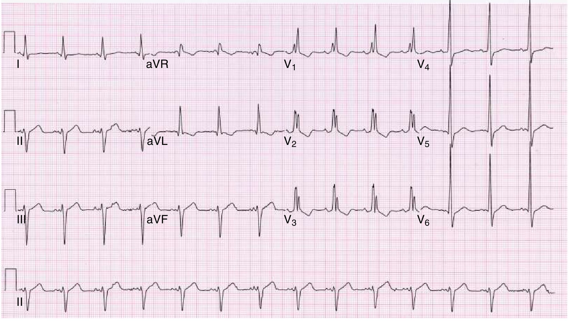
13. RBBB + LAFB (Bifascicular Block)
ECG Reading:
| Feature | Finding |
|---|---|
| QRS duration | ≥120 ms |
| V1 | rsR' (RBBB pattern) |
| Axis | Left axis deviation (-45° to -90°) |
| Clinical risk | May progress to complete heart block; consider pacing |
SECTION D: CHANNELOPATHIES & GENETIC SYNDROMES
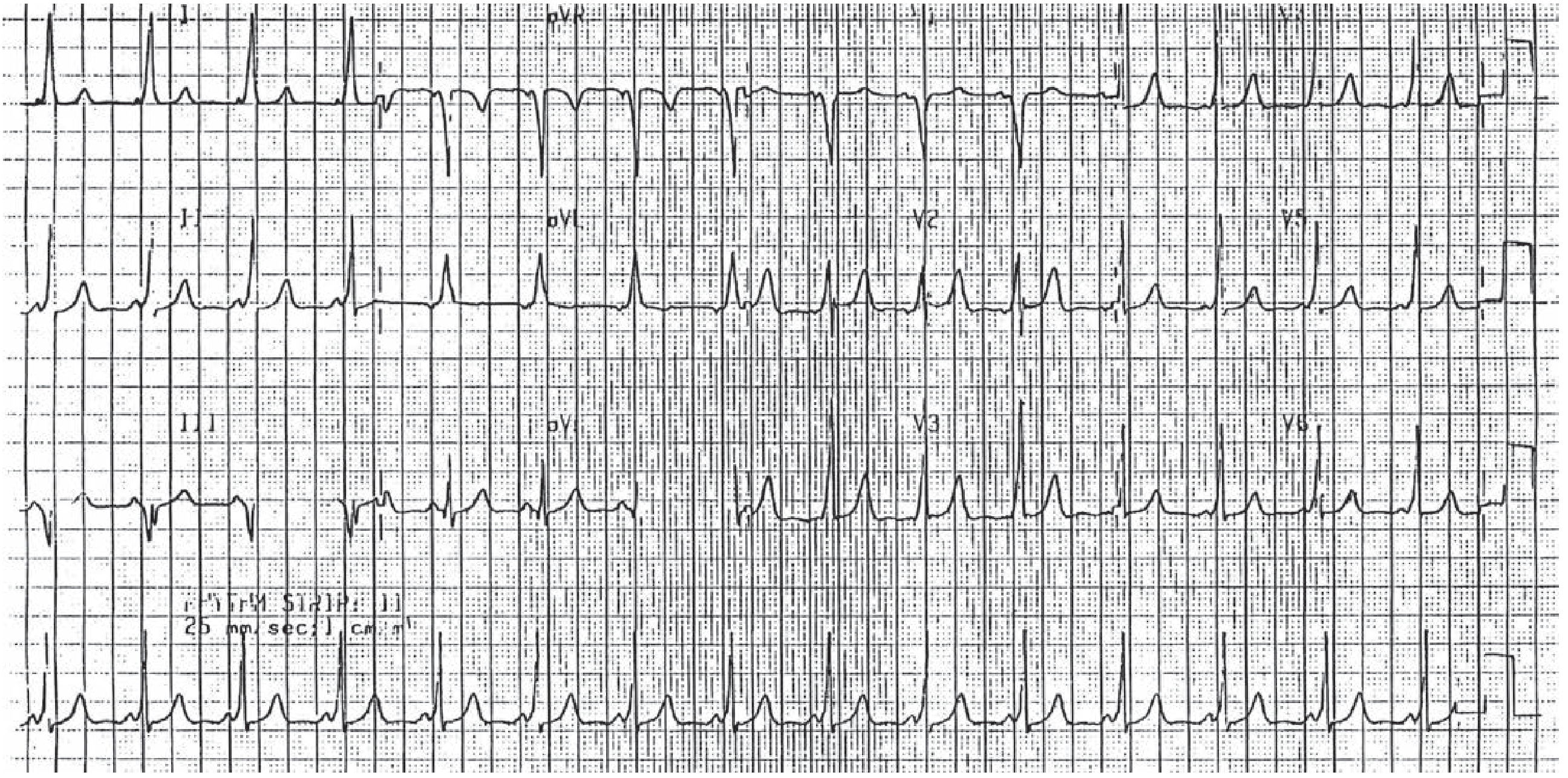
14. Wolff-Parkinson-White (WPW) Syndrome
ECG Reading (Sinus Rhythm):
| Feature | Finding |
|---|---|
| PR interval | Short (<120 ms) |
| Delta wave | Slurred upstroke at start of QRS |
| QRS | Widened (>120 ms) |
| ST/T | Pseudo-changes (secondary to pre-excitation) |
| Risk | AF with rapid accessory pathway conduction → VF |
Classic ECG triad: Short PR + Delta wave + Wide QRS
During WPW tachycardias:
- Orthodromic AVRT (65%): Regular, narrow complex, rate 160-220 bpm
- Antidromic AVRT (5-10%): Wide complex, looks like VT
- AF with WPW: Irregular, wide-complex, rates >200 bpm (most dangerous!)
Never give: Adenosine, verapamil, digoxin, or beta-blockers in AF + WPW (can cause VF)
15. Long QT Syndrome
ECG Reading:
| Feature | Finding |
|---|---|
| QTc | >440 ms (male), >460 ms (female) - corrected by Bazett: QTc = QT/√RR |
| T waves | Broad, notched, or "humped" T waves |
| Arrhythmia | Torsades de pointes (twisting QRS axis around baseline) |
| Risk triggers | Hypokalemia, hypomagnesemia, QT-prolonging drugs, exercise |
Bazett formula: QTc = QTm ÷ √(R-R interval in seconds)
- Normal QT at 60 bpm: 350-440 ms
- QTc >500 ms = very high risk for torsades
Causes of acquired long QT:
- Drugs: antipsychotics, macrolides, fluoroquinolones, antifungals, sotalol, amiodarone
- Electrolytes: hypokalemia, hypomagnesemia, hypocalcemia
- Ischemia, CNS events (stroke, SAH)
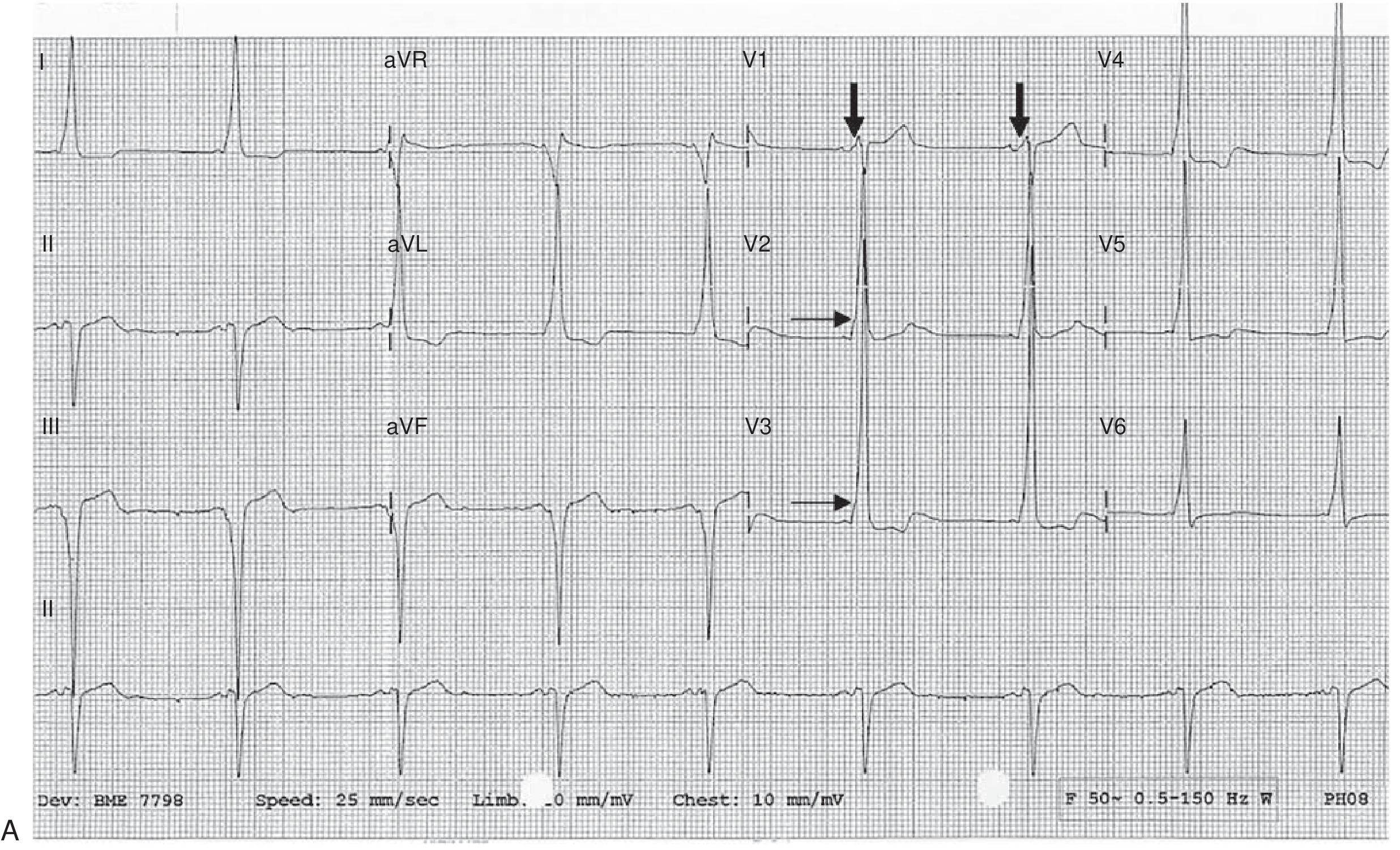
16. Brugada Syndrome
ECG Reading:
| Feature | Finding |
|---|---|
| Key leads | V1-V2 (and V3) |
| Type 1 (diagnostic) | Coved ST elevation ≥2 mm with downsloping to negative T wave |
| Type 2 | Saddleback pattern (>2 mm ST elevation, positive or biphasic T) |
| QRS | May resemble RBBB |
| Provocation | ST changes worsen with fever, Na+ channel blockers, drugs |
Reading Brugada: In V1, look for a "shark fin" or "coved" pattern - the ST goes up, curves over, and descends to a negative T wave. This is NOT normal RBBB.
Clinical: Predominantly affects young males. Risk of polymorphic VT and VF during sleep or fever. Associated with SCN5A loss-of-function mutations. Treatment: ICD.
SECTION E: ELECTROLYTE DISORDERS
17. Hyperkalemia
ECG Reading (Progressive Changes):
| K+ Level | ECG Change |
|---|---|
| 5.5-6.0 mEq/L | Tall, peaked (tented) T waves - narrow base, symmetric, pointy |
| 6.0-7.0 mEq/L | PR prolongation, P wave flattening/disappearance |
| 7.0-8.0 mEq/L | Progressive QRS widening |
| >8.0 mEq/L | Sine wave pattern (QRS merges with T wave) |
| Critical | VF or asystole |
Peaked T waves in hyperkalemia vs ischemia:
- Hyperkalemia: Narrow base, symmetric, tall, pointy ("tented")
- Ischemia (hyperacute): Broad base, asymmetric, usually in one territory
Hyperkalemia can also mimic:
- Brugada pattern (pseudo-RBBB + coved ST in V1-V2)
- STEMI pattern (especially when QRS widens)
18. Hypokalemia
ECG Reading:
| Feature | Finding |
|---|---|
| T waves | Flattened or inverted T waves |
| U waves | Prominent U waves (after T wave, especially V2-V3) |
| QT interval | Apparent QT prolongation (actually QU prolongation) |
| ST changes | ST depression |
| Risk | Torsades de pointes |
Key sign: Prominent U wave (positive deflection after T wave) larger than T wave in same lead = hypokalemia until proven otherwise.
SECTION F: PERICARDIAL & MYOCARDIAL DISEASE
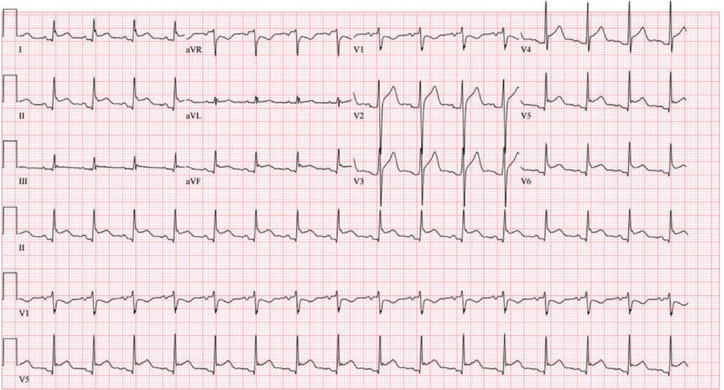
19. Acute Pericarditis
ECG Reading (4 Stages):
Stage 1 (Hours to days - diagnostic stage):
| Feature | Finding |
|---|---|
| ST elevation | Diffuse, concave (saddle-shaped), in MULTIPLE territories |
| PR depression | Diffuse PR depression (most leads) |
| aVR | ST depression + PR ELEVATION ("knuckle sign") |
| Key distinction | NO reciprocal ST depression (unlike STEMI) |
Stage 2 (Days): ST and PR normalize
Stage 3 (1-3 weeks): Diffuse T-wave inversions
Stage 4 (Weeks-months): ECG returns to normal
Pericarditis vs STEMI:
| Feature | Pericarditis | STEMI |
|---|---|---|
| ST distribution | Diffuse (multiple territories) | Localized (one territory) |
| ST shape | Concave (saddle-shaped) | Convex (domed/tombstone) |
| PR depression | YES (hallmark) | NO |
| Reciprocal changes | ABSENT | PRESENT |
| Q waves | NO | YES (infarct) |
20. Hypertrophic Cardiomyopathy (HCM)
ECG Reading:
| Feature | Finding |
|---|---|
| LVH voltage | Tall R in V5-V6 (>26 mm), tall R in aVL (>13 mm) |
| Pathological Q waves | Deep, narrow Q waves in I, aVL, V5-V6 (septal hypertrophy) |
| T waves | Deep T-wave inversions (especially lateral) |
| ST changes | ST depression in lateral leads |
| Apical HCM | Giant T-wave inversions (deeply negative, "giant negative T waves") in V4-V6 |
SECTION G: PULMONARY DISEASE
21. Pulmonary Embolism (PE)
ECG Reading:
| Feature | Finding |
|---|---|
| Most common | Sinus tachycardia (most frequent finding) |
| S1Q3T3 | S wave in I + Q wave in III + T-wave inversion in III |
| Right heart strain | T-wave inversions V1-V4 (right precordial) |
| RBBB | New incomplete or complete RBBB |
| Right axis deviation | QRS axis shifts rightward |
| P pulmonale | Tall peaked P waves in II (right atrial enlargement) |
Caution: S1Q3T3 is specific but not sensitive (seen in <20% of PE). Most PE ECGs show only sinus tachycardia or are normal. The most specific finding for massive PE is new RBBB + right precordial T-wave inversions.
22. Chronic Obstructive Pulmonary Disease (COPD) / Cor Pulmonale
ECG Reading:
| Feature | Finding |
|---|---|
| Rate/rhythm | Sinus tachycardia; Multifocal atrial tachycardia (MAT) in COPD exacerbations |
| P pulmonale | Tall peaked P waves in II, III, aVF (>2.5 mm) |
| Right axis deviation | QRS axis +90° to +180° |
| Poor R progression | Small R waves across precordium (low voltage) |
| RVH pattern | R > S in V1, deep S in V5-V6 |
| Low voltage | QRS <5 mm limb leads (air trapping/barrel chest) |
MAT (Multifocal Atrial Tachycardia): Rate >100 bpm, ≥3 different P-wave morphologies, irregular rhythm - classic in COPD/respiratory failure.
SECTION H: STRUCTURAL HEART DISEASE
23. Left Ventricular Hypertrophy (LVH)
ECG Reading:
| Feature | Finding |
|---|---|
| Cornell criteria | S in V3 + R in aVL >2.8 mV (men), >2.0 mV (women) |
| Sokolow-Lyon | S in V1 + R in V5 or V6 >35 mm |
| "Strain pattern" | ST depression + T inversion in I, aVL, V5-V6 |
| Left axis deviation | Mild LAD common |
24. Right Ventricular Hypertrophy (RVH)
ECG Reading:
| Feature | Finding |
|---|---|
| R > S in V1 | Dominant R in V1 (R wave taller than S wave) |
| Right axis deviation | Axis >+90° |
| Deep S in V5-V6 | Persistence of S waves laterally |
| ST/T changes | Strain pattern: ST depression + T inversion in V1-V3 |
SECTION I: OTHER IMPORTANT ECG PATTERNS
25. Digoxin Effect vs Digoxin Toxicity
Digoxin Effect (therapeutic):
| Feature | Finding |
|---|---|
| ST changes | "Scooped" or reversed tick ST depression - characteristic downsloping |
| QT | Shortened |
| PR | Mildly prolonged |
| T waves | Flattened or inverted |
Digoxin Toxicity:
| Feature | Finding |
|---|---|
| Bradycardia | SA or AV node dysfunction |
| Heart blocks | 1st, 2nd, or 3rd degree AV block |
| Atrial tachycardia with block | Classic: PAT with 2:1 block |
| Bidirectional VT | Alternating QRS axis - pathognomonic |
| Ventricular arrhythmias | PVCs, bigeminy, VT, VF |
26. Hypercalcemia vs Hypocalcemia
| Condition | ECG Finding |
|---|---|
| Hypercalcemia | Shortened QT interval (shortened ST segment) |
| Hypocalcemia | Prolonged QT interval (prolonged ST segment, QT >500 ms) |
27. Early Repolarization (Benign)
ECG Reading:
| Feature | Finding |
|---|---|
| J point | Notched or slurred J point at QRS-ST junction |
| ST shape | Upsloping/concave ST elevation |
| Distribution | Mid-lateral precordial leads, can be inferior |
| aVR | Reciprocal ST depression in aVR |
| Common in | Young males, athletes (up to 100% in endurance athletes) |
Distinguish from pericarditis: Early repolarization has no PR depression, no evolution through stages, and changes normalize with exercise/tachycardia.
28. Hypothermia
ECG Reading:
| Feature | Finding |
|---|---|
| Rate | Bradycardia (profound) |
| Osborn (J) wave | Positive deflection at J point - pathognomonic for hypothermia |
| QRS | Widening |
| QT | Prolonged |
| Arrhythmia | AF, VF at <28°C core temperature |
| Muscle tremor | Fine artifact simulating AF |
Summary Quick-Reference Table
| # | Disease | Rate | Rhythm | Key ECG Finding |
|---|---|---|---|---|
| 1 | Anterior STEMI | Normal/↑ | Sinus | STE V1-V4, Q waves developing |
| 2 | Inferior STEMI | Normal/↑ | Sinus | STE II, III, aVF + reciprocal STD I, aVL |
| 3 | High Lateral STEMI | Normal/↑ | Sinus | STE I, aVL + reciprocal STD III, aVF |
| 4 | Posterior MI | Normal | Sinus | STD V1-V3 + tall R wave, STE V7-V9 |
| 5 | Atrial Fibrillation | Variable | Irregularly irregular | No P waves, chaotic baseline |
| 6 | Atrial Flutter | ~150 | Regular | Sawtooth flutter waves at 300 bpm |
| 7 | Ventricular Tachycardia | 100-250 | Regular | Wide QRS + AV dissociation |
| 8 | SVT/AVNRT | 150-250 | Regular | Narrow QRS, P hidden in QRS |
| 9 | Complete Heart Block | Slow escape <40 | AV dissociated | Independent P waves and QRS |
| 10 | RBBB | Normal | Sinus | rSR' in V1, wide S in V5-V6 |
| 11 | LBBB | Normal | Sinus | Broad notched R in I, V5-V6; QS in V1 |
| 12 | LAFB | Normal | Sinus | Left axis deviation, qR in aVL |
| 13 | WPW | Variable | Variable | Short PR + delta wave + wide QRS |
| 14 | Long QT | Normal | Sinus (risk of TdP) | QTc >440-460 ms, broad T waves |
| 15 | Brugada | Normal | Sinus (risk of VF) | Coved ST elevation V1-V2, negative T |
| 16 | Hyperkalemia | Slow | Sinus → sinusoidal | Peaked T → flat P → wide QRS → sine wave |
| 17 | Hypokalemia | Normal | Sinus (risk of TdP) | Flat T, prominent U wave, long QU |
| 18 | Acute Pericarditis | Normal/↑ | Sinus | Diffuse STE concave + PR depression |
| 19 | PE | ↑ (tachycardia) | Sinus | S1Q3T3, T inversion V1-V4, RBBB |
| 20 | COPD/Cor Pulmonale | ↑ | MAT or sinus | P pulmonale, RAD, poor R progression |
| 21 | LVH | Normal | Sinus | Voltage criteria + strain pattern |
| 22 | Digoxin toxicity | Slow | Various | PAT with block, bidirectional VT |
| 23 | Hypothermia | Slow | AF/VF at <28°C | Osborn (J) wave, bradycardia |
| 24 | Hypocalcemia | Normal | Sinus (risk of TdP) | Long QT (prolonged ST) |
*Sources: ROSEN's Emergency Medicine (9th Ed.), Tintinalli's Emergency Medicine, Braunwald's Heart Disease, Fuster & Hurst's The Heart (15th Ed.), Goldman-Cecil Medicine, Harrison's Principles of Internal Medicine 22E (2025)*This is a full illustrated ECG reference guide covering 24 diseases across 8 clinical categories, with real textbook ECG images. Here's a recap of what's covered:
Sections:
- A - Ischemic Heart Disease: Anterior STEMI, Inferior STEMI, High Lateral STEMI, Posterior/RV MI
- B - Arrhythmias: AF, Atrial Flutter, VT, SVT, Complete Heart Block
- C - Conduction Disorders: RBBB, LBBB, LAFB, Bifascicular block
- D - Channelopathies: WPW, Long QT, Brugada syndrome
- E - Electrolytes: Hyperkalemia, Hypokalemia
- F - Pericardial/Myocardial: Acute Pericarditis, HCM
- G - Pulmonary: PE (S1Q3T3), COPD/Cor pulmonale/MAT
- H - Structural + Other: LVH, RVH, Digoxin toxicity, Hypothermia (Osborn waves), Hypocalcemia
High-yield memory pearls:
- PR depression = pericarditis (no other cause does this diffusely)
- Short PR + delta wave = WPW
- Peaked T → sine wave = hyperkalemia progression
- S1Q3T3 + sinus tach = think PE
- Wide QRS + AV dissociation = VT
- Irregularly irregular + no P waves = AF
This is a shared conversation. Sign in to Orris to start your own chat.