
Teach me everything about sudden cardiac death/arrest for an emergency resident from basic physiology, deep pathology, complete pharmacology and resuscitation guidelines according to latest AHA guidelines.
Reading File
Now launching parallel searches across the medical library, PubMed, and web simultaneously.
Finding Sources
Finding Sources
Searching PubMed
"cardiac arrest" AND "resuscitation" AND "guidelines"
Searching the Web
AHA 2024 2025 cardiac arrest ACLS guidelines updates sudden cardiac death
Excellent - found the 2025 AHA guidelines (published Oct 2025). Now reading the key textbook sections in parallel.
Reading File
Reading File
Reading File
Reading File
Reading Article
PMID: 41122884
Reading Article
PMID: 41122894
Now reading the Tintinalli's chapter on SCD in detail plus searching for pharmacology, channelopathies, and getting the AHA 2025 highlights.
Reading File
Finding Sources
Finding Sources
Reading Web Page
https://cpr.heart.org/-/media/CPR-Files/2025-documents-for-c…
Now reading the Tintinalli's ACLS pharmacology section and the Harrison's post-arrest management in parallel, plus the channelopathy and post-arrest care content.
Reading File
Reading File
Finding Sources
Finding Sources
Excellent data gathered. Now reading the channelopathy section and the Tintinalli ACLS pharmacology in detail.
Reading File
Reading File
Reading File
Reading Web Page
https://ronlitman.substack.com/p/adult-advanced-life-support…
Now reading the Tintinalli pharmacology detail and fetching the ACLS algorithm image, plus reading post-arrest care content.
Reading File
Reading File
Loading Image
Finding Sources
I now have comprehensive material. Let me fetch the Harrison's VF/VT algorithm image and do a final read of the H's and T's / post-arrest section.
Loading Image
Loading Image
Reading File
I now have all the information needed to write a comprehensive teaching document. Let me update the todos and compose the full response.---
Sudden Cardiac Death & Arrest: A Complete Guide for the Emergency Resident
Sources: Robbins & Kumar Pathology (10e), Tintinalli's Emergency Medicine, Harrison's Principles (22e), Rosen's Emergency Medicine, Barash's Clinical Anesthesia, Miller's Anesthesia, and the 2025 AHA Guidelines for CPR & ECC (Circulation, October 2025, PMID 41122884, 41122893, 41122894).
PART 1 - DEFINITIONS AND EPIDEMIOLOGY
Sudden Cardiac Death (SCD): Unexpected death from cardiac causes within 1 hour of symptom onset, or within 24 hours of last being seen alive and well.
Sudden Cardiac Arrest (SCA): The same event, but the patient is successfully resuscitated. SCD = SCA + failure to resuscitate.
Scale of the problem:
- ~341,000-450,000 out-of-hospital cardiac arrests (OHCA) per year in the US
- Estimated overall OHCA survival: 5.4-10.8% (EMS-treated cases only)
- 60% male; peak incidence age >45-50 years
- Only 23% of all cardiac arrests present with VF; the rest are non-shockable
- Survival from VF/pVT is vastly better than from asystole/PEA
- Strong circadian pattern: highest incidence in first hours after awakening (peak sympathetic tone)
- SCD is 30-80% more common in lowest vs highest socioeconomic quartile
- Tintinalli's Emergency Medicine, Ch. 11; Robbins & Kumar Basic Pathology 10e, p. 362
PART 2 - BASIC PHYSIOLOGY OF CARDIAC ARREST
Normal Cardiac Electrophysiology Briefly
The cardiac action potential has 5 phases:
- Phase 0: Rapid Na+ influx (fast depolarization) - targeted by Class I antiarrhythmics
- Phase 1: Transient K+ efflux
- Phase 2: Plateau - Ca2+ influx (L-type) balanced by K+ efflux - targeted by Class IV (CCBs)
- Phase 3: Rapid K+ efflux (repolarization) - targeted by Class III antiarrhythmics
- Phase 4: Resting potential; automaticity in pacemaker cells via If (funny current)
Sinoatrial (SA) node fires at 60-100 bpm; AV node at 40-60 bpm; Purkinje/ventricular cells at 20-40 bpm.
Why the Heart Arrests
Three fundamental arrhythmia mechanisms lead to SCA:
| Mechanism | Description | Example |
|---|---|---|
| Re-entry | Unidirectional block + slow conduction creates circular wavefronts | VF, VT, torsades |
| Abnormal automaticity | Enhanced phase 4 depolarization in non-pacemaker cells | Ischemic foci triggering VT |
| Triggered activity | Early (EAD) or delayed (DAD) afterdepolarizations | Torsades (EAD), digitalis toxicity (DAD) |
Re-entry is the dominant mechanism in SCA. Ischemia creates islands of tissue that depolarize and repolarize at different rates (inhomogeneous refractoriness). Dilated ventricles further promote dispersion of repolarization. These heterogeneous circuits sustain VF.
- Tintinalli's Emergency Medicine, p. 95
Hemodynamics of Cardiac Arrest
When the heart arrests, cardiac output drops to zero. With high-quality CPR, you generate at most 30% of normal cardiac output. This means:
- Coronary perfusion pressure (CPP) = Aortic relaxation pressure - Right atrial pressure. CPP must be >15 mmHg for ROSC to occur; epinephrine increases this by peripheral vasoconstriction.
- Cerebral blood flow is critically dependent on uninterrupted CPR; each pause in compressions takes 10-15 seconds to rebuild.
- After 4-6 minutes of no-flow time, cerebral neurons begin irreversible injury.
Three phases of cardiac arrest (Weisfeldt model):
- Electrical phase (0-4 min): Immediate defibrillation is most effective
- Circulatory phase (4-10 min): CPR first to replenish cardiac energy stores, THEN shock
- Metabolic phase (>10 min): Metabolic derangement limits survival; novel therapies (ECMO) may help
- Miller's Anesthesia 10e, Ch. 31
PART 3 - PATHOLOGY: CAUSES AND STRUCTURAL SUBSTRATES
Etiologies by Frequency
| Category | Cause | % of SCD |
|---|---|---|
| Coronary artery disease | Atherosclerosis, plaque rupture, chronic ischemia | 65-80% |
| Cardiomyopathies | DCM, HCM, ARVC | 10-15% |
| Channelopathies/primary arrhythmia | Long QT, Brugada, CPVT | 5-10% |
| Structural (non-coronary) | MVP, congenital anomalies, myocarditis | 5% |
| Non-cardiac | PE, tension PTX, electrolyte crisis | ~5% |
- Robbins & Kumar Basic Pathology 10e, p. 362; Tintinalli's, Ch. 11
Coronary Artery Disease (CAD) - The Leading Cause
- Coronary atherosclerosis on autopsy in 80% of SCD victims
- Importantly, only 10-20% have acute plaque rupture - most have chronic severe stenosis
- 80-90% of resuscitated SCA patients show NO enzymatic/ECG evidence of acute MI - the arrhythmia precedes necrosis
- Healed remote MIs found in ~40%; subendocardial vacuolization indicating chronic ischemia is common
- Mechanism: chronic ischemia creates areas of fibrosis and electrically unstable periinfarct zones that serve as re-entry substrates
Cardiomyopathies
Dilated Cardiomyopathy (DCM):
- Dilated ventricles cause dispersion of depolarization/repolarization
- Creates "islands" firing at different rates - sustains re-entry VT/VF
- EF ≤35% is the single best predictor of SCD risk (primary ICD indication)
- NYHA class II-III carries higher SCD risk; class IV patients more often die from pump failure
Hypertrophic Cardiomyopathy (HCM):
- Most common cardiovascular cause of SCD in young athletes (~1/3 of exercise-related SCA)
- Risk ~1% per year
- Mechanism: myocyte disarray, fibrosis, LVOTO creating dynamic outflow obstruction and arrhythmia substrate
- ICD indications: prior VF/VT, family history of SCD, syncope, LV wall thickness ≥30 mm, abnormal BP response to exercise, massive LVH in children
Arrhythmogenic Right Ventricular Cardiomyopathy (ARVC):
- Hereditary; right-sided failure + VT with LBBB morphology (right ventricular origin)
- ECG: epsilon wave in V1-V3; T-wave inversions in right precordial leads
- Fibrofatty replacement of RV myocardium disrupts conduction
- Tintinalli's Emergency Medicine, p. 95-96
Channelopathies - The "Electrically Silent" Hearts
These cause SCA in structurally normal hearts, most common in the young:
| Syndrome | Mechanism | ECG | Trigger | Treatment |
|---|---|---|---|---|
| Long QT Syndrome (10+ types) | Reduced repolarizing K+ currents (LQT1,2) or gain-of-function Na+ channels (LQT3) | QTc >460 ms (F), >440 ms (M) | Exercise (LQT1), loud noise (LQT2), sleep (LQT3) | Beta-blockers, ICD; avoid QT-prolonging drugs |
| Brugada Syndrome | SCN5A mutation (Na+ channel); partial RBBB + ST elevation V1-V3 | Coved ST elevation V1-V3 | Fever, sodium channel blockers, vagotonia | ICD; quinidine, isoproterenol for electrical storm |
| CPVT (Catecholaminergic Polymorphic VT) | RYR2/CASQ2 mutations; Ca2+ overload → DADs | Normal at rest; bidirectional VT on exercise | Exercise, emotional stress | Beta-blockers, flecainide, ICD |
| Short QT Syndrome | Gain-of-function K+ channels | QTc <340 ms | - | ICD |
Clinical presentation: syncope, near-syncope, palpitations, or SCD. Strong family history is a diagnostic clue. All require admission to monitored settings and cardiology referral for ICD evaluation. - Rosen's Emergency Medicine, Ch. 68
Myocarditis
- Histologically defined by mononuclear cell infiltrates
- Leading viral causes: Coxsackie B, SARS-CoV-2, parvovirus B19, HHV-6
- Can cause SCA through direct myocyte injury + acute arrhythmia substrate
- Treat underlying cause; arrhythmias managed per standard ACLS
PART 4 - CARDIAC ARREST RHYTHMS AND THEIR SIGNIFICANCE
The Four Rhythms
| Rhythm | Description | Shockable? | Survival |
|---|---|---|---|
| VF | Chaotic, disorganized, no effective contraction | YES | Best (with early defib) |
| Pulseless VT (pVT) | Organized but no pulse | YES | Good |
| PEA | Organized ECG, no pulse | NO | Moderate if reversible cause found |
| Asystole | Flatline | NO | Very poor |
Practical point: Coarse VF responds better to defibrillation than fine VF (more myocardial energy reserve). VF within the first minutes of witnessed arrest is associated with an acute coronary syndrome in the majority.
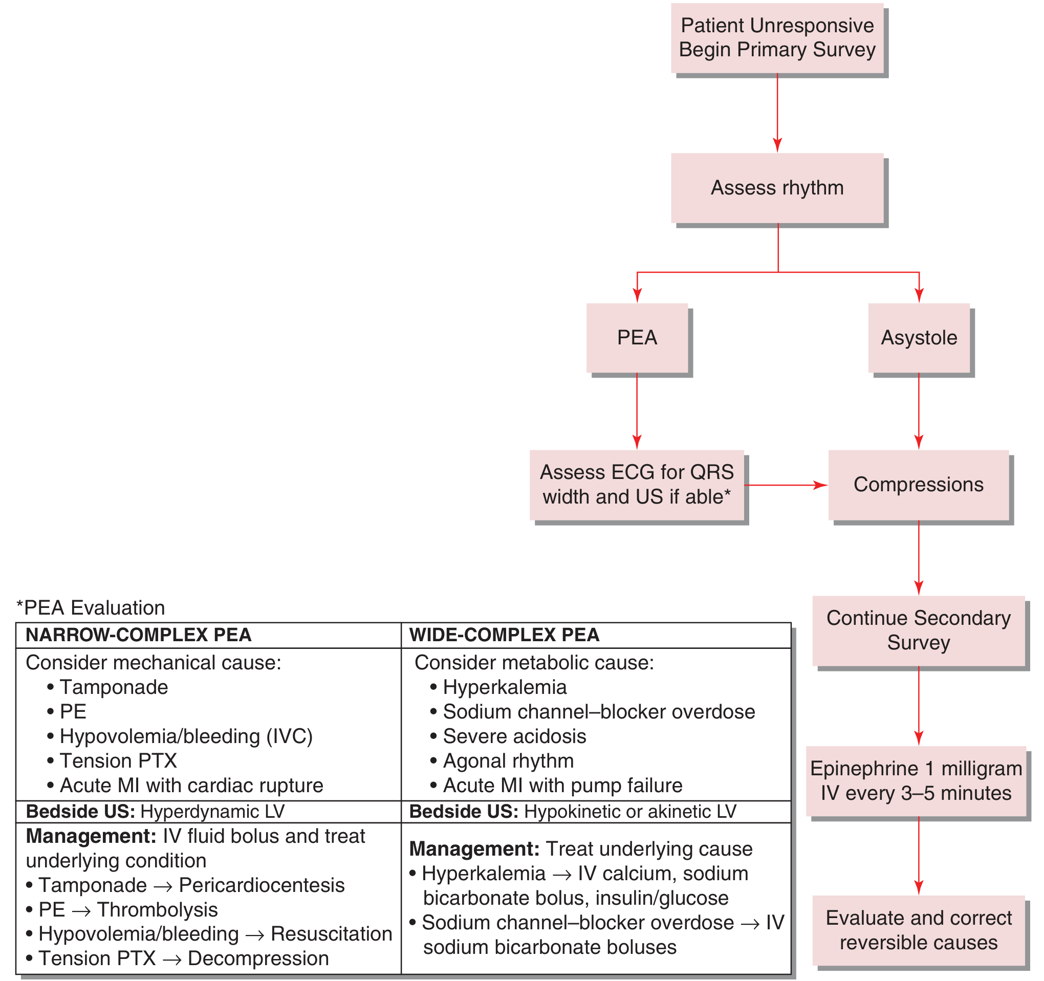
PEA: narrow vs. wide complex matters:
- Narrow-complex PEA = mechanical cause (tamponade, PE, tension PTX, hypovolemia, acute MI with rupture) - bedside US shows hyperdynamic LV
- Wide-complex PEA = metabolic cause (hyperkalemia, Na-channel blocker toxicity, severe acidosis, agonal rhythm) - bedside US shows hypokinetic/akinetic LV
- Tintinalli's Emergency Medicine, Ch. 24
PART 5 - COMPLETE PHARMACOLOGY OF CARDIAC ARREST
Vasopressors
Epinephrine (Adrenaline) - First-Line for All Rhythms
Mechanism: Endogenous catecholamine. Alpha-1 agonism causes intense peripheral vasoconstriction, increasing aortic diastolic pressure and therefore coronary perfusion pressure and cerebral perfusion. Beta-1 effects increase heart rate and contractility (but also myocardial O2 demand - a trade-off).
Dose: 1 mg IV/IO bolus (10 mL of 1:10,000) every 3-5 minutes. No maximum dose but benefit diminishes beyond 15-20 minutes.
2025 AHA recommendation: Administer as early as possible for non-shockable rhythms (PEA/asystole). For shockable rhythms (VF/pVT), give after the first failed defibrillation attempt. Vasopressin offers no advantage as a substitute or combination. Higher initial doses are NOT recommended.
Evidence caveat (important!): Epinephrine improves ROSC and short-term survival, but the landmark 2018 PARAMEDIC2 trial showed no improvement in survival to hospital discharge, and survivors had higher rates of severe neurologic disability. Nevertheless, consensus recommendations retain its use. Benefit is most evident in the first 15-20 minutes; beyond 20 minutes, it may worsen myocardial ischemia and cause ventricular ectopy.
- Tintinalli's Emergency Medicine, p. 199-200
Vasopressin
- Not recommended as substitute or addition to epinephrine (2025 AHA: no advantage)
Antiarrhythmics for Shockable Rhythms (VF/pVT)
Amiodarone - Preferred Agent for Refractory VF/pVT
Class: III (predominantly), but has activity across all 4 Vaughan-Williams classes (Na+ channel block, K+ channel block, beta-block, Ca2+ channel block).
Indication: Persistent VT/VF after defibrillation AND epinephrine.
Dose:
- Cardiac arrest: 300 mg IV bolus (in 20 mL D5W) → may repeat 150 mg once
- Stable VT: 150 mg IV over 10-15 min, then 1 mg/min x6h, then 0.5 mg/min x18h
- Infusions >2h: use glass or polyolefin containers (precipitates in PVC)
Adverse effects: Hypotension, bradycardia (slow infusion), phlebitis with peripheral IV. Prolonged oral use: pulmonary toxicity, thyroid dysfunction, corneal deposits, hepatotoxicity, photosensitivity, blue-gray skin.
Evidence: The 2016 ROC ALPS trial (Kudenchuk et al.) showed no difference in survival to hospital discharge for amiodarone vs. lidocaine vs. placebo for shock-refractory VF. However, amiodarone and lidocaine both showed improved rates of survival to hospital admission. Guidelines retain amiodarone as first-line antiarrhythmic.
- Tintinalli's Emergency Medicine, p. 200; Barash Clinical Anesthesia 9e
Lidocaine - Acceptable Alternative
Class: Ib sodium channel blocker. Decreases automaticity and phase 4 depolarization.
Indication: VF/pVT refractory to defib + epi + amiodarone, OR when amiodarone unavailable.
Dose: 1-1.5 mg/kg IV bolus; repeat 0.5-0.75 mg/kg every 5-10 min if needed (max 3 mg/kg). Maintenance infusion: 1-4 mg/min.
Side effects: CNS toxicity (perioral numbness, tinnitus, seizures, altered consciousness) - especially in elderly and liver disease.
Magnesium Sulfate
Specific indication: Torsades de Pointes (polymorphic VT with QT prolongation), or hypomagnesemia-driven arrhythmias.
Dose: 1-2 g IV over 5-20 minutes.
Mechanism: Blocks Ca2+ influx, stabilizes membrane potential, terminates EAD-driven torsades.
NOT recommended for routine VF without documented hypomagnesemia or torsades.
Drugs for Non-Shockable Rhythms
Atropine
- No longer recommended in cardiac arrest for PEA/asystole (2010 guidelines removed it from the algorithm)
- Still indicated for symptomatic bradycardia with a pulse: 0.5 mg IV every 3-5 min (max 3 mg)
- Mechanism: muscarinic antagonist, blocks vagal slowing of SA/AV nodes
Sodium Bicarbonate
- NOT routine in cardiac arrest
- Use for: severe pre-existing metabolic acidosis, hyperkalemia, tricyclic antidepressant overdose, sodium channel blocker toxicity
- Dose: 1 mEq/kg IV
- Risks: hypernatremia, paradoxical intracellular acidosis, hyperosmolarity; only give if good ventilation is established
Calcium
- Indication: Hyperkalemia, hypocalcemia, calcium channel blocker overdose, hypermagnesemia
- Dose: Calcium chloride 1 g (10 mL of 10% solution) IV - preferred in cardiac arrest; calcium gluconate 3x less potent per mL
- Do NOT mix with sodium bicarbonate (precipitates)
Glucose + Insulin
- For hyperkalemia (drives K+ intracellularly)
- Not part of standard cardiac arrest protocol unless hyperkalemia confirmed
Naloxone
- 2025 AHA Update: Expanded role - administer for suspected opioid-associated respiratory/cardiac arrest
- Dose: 0.4-2 mg IV/IO/IM/IN; may repeat
- BLS and HCP algorithms now include opioid overdose response with naloxone
Specific Antidotes in Special Situations
| Cause | Antidote/Treatment |
|---|---|
| TCA overdose (wide QRS, hypotension) | NaHCO3 boluses (1-2 mEq/kg) |
| Digoxin toxicity (Brady/VF) | Digoxin-specific Fab antibodies |
| Beta-blocker overdose (refractory) | IV Glucagon, high-dose insulin-euglycemia, lipid emulsion |
| Ca-channel blocker overdose | IV Calcium, glucagon, high-dose insulin-euglycemia |
| Local anesthetic toxicity (Bupivacaine) | Intralipid 20% (1.5 mL/kg IV bolus) |
| Hyperkalemia | Calcium, bicarb, insulin/dextrose, albuterol, furosemide |
| Torsades de Pointes | Magnesium 2g IV; overdrive pacing; isoproterenol |
PART 6 - HIGH-QUALITY CPR
The Non-Negotiables (2025 AHA)
| Parameter | Target |
|---|---|
| Rate | 100-120 compressions/min |
| Depth | ≥2 inches (5 cm); not >2.4 inches (6 cm) |
| Chest recoil | Full recoil between compressions - do not lean |
| Interruptions | Minimize - pre-charge defibrillator during compressions |
| Compression fraction | >80% of resuscitation time |
| Ventilation (with advanced airway) | 1 breath every 6 seconds (10/min); do NOT hyperventilate |
| Ventilation (without advanced airway) | 30:2 ratio |
2025 New/Updated Points:
- Hand and body position optimization improves compression quality
- CPR should be performed on a firm surface whenever possible
- Head-up CPR (Trendelenburg reversal): emerging evidence that 15-30° head elevation during CPR may reduce intracranial pressure and improve cerebral perfusion - 2025 guidelines include this as an option
- Breaths with compressions are now recommended for both HCP and lay rescuers when willing and capable (not compression-only only)
Airway Management During Arrest
- Bag-mask ventilation (BMV) is acceptable and may be preferred over intubation in early arrest
- Supraglottic airways (LMA, i-gel) are reasonable alternatives to ETT in many settings
- ETT remains standard if an experienced provider is available and compressions not interrupted
- Capnography (waveform ETCO2) is MANDATORY for confirming ETT and monitoring resuscitation:
- ETCO2 <10 mmHg after 20 min = very low ROSC probability
- ETCO2 ≥20 mmHg = good CPR quality
- Sudden spike in ETCO2 = first sign of ROSC
- Do NOT use ETCO2 alone to stop resuscitation
Vascular Access
- IV (peripheral) > IO > central for drug delivery speed in cardiac arrest
- IO is equivalent to IV for drug dosing - same doses
- Central line: longer drug circulation time; reserve for monitoring post-ROSC
- Endotracheal drug delivery is no longer recommended (unreliable absorption in pulmonary edema)
- Flush all IV drugs with 20 mL NS bolus; continue CPR 30-60 seconds before shocking
PART 7 - RESUSCITATION ALGORITHMS (2025 AHA)
Algorithm 1: VF / Pulseless VT (Shockable)
Unresponsive, no breathing, no pulse
         ↓
Activate emergency response, get AED/defibrillator
         ↓
START HIGH-QUALITY CPR (100-120/min)
         ↓
Rhythm check → VF/pVT confirmed
         ↓
SHOCK (biphasic 200 J or manufacturer-recommended)
Immediately resume CPR x 2 min
         ↓
Rhythm check → Still VF/pVT?
         ↓
SHOCK again (max energy)
Resume CPR x 2 min
Establish IV/IO access
EPINEPHRINE 1 mg IV/IO → repeat q3-5 min
         ↓
Rhythm check → Still VF/pVT?
         ↓
SHOCK
Resume CPR x 2 min
AMIODARONE 300 mg IV/IO bolus
(may repeat 150 mg once)
         ↓
Rhythm check → Still VF/pVT?
         ↓
Continue cycles: shock → CPR x 2 min → epi q3-5 min
Consider LIDOCAINE if amiodarone unavailable or failed
Consider reversible causes (H's and T's)
Consider double sequential defibrillation for refractory VF
Key timing principle: Epinephrine for VF/pVT is given AFTER the first failed shock (not at the start). For PEA/asystole, give epinephrine as early as possible.
VF/VT Algorithm from Harrison's Principles of Internal Medicine 22e:
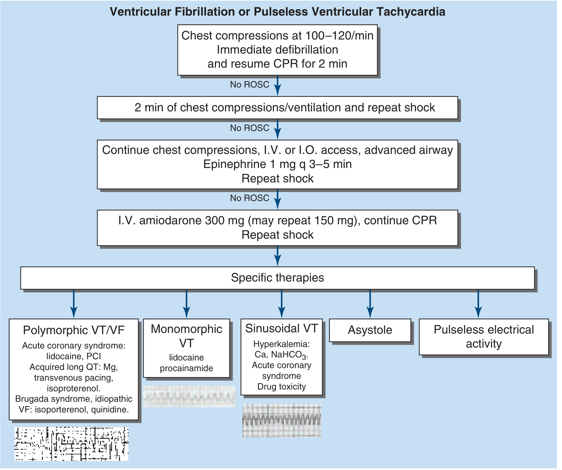
Algorithm 2: PEA / Asystole (Non-Shockable)
Unresponsive, no pulse
         ↓
Confirm rhythm (asystole confirmed in ≥2 leads)
START HIGH-QUALITY CPR
         ↓
EPINEPHRINE 1 mg IV/IO (give ASAP)
Repeat q3-5 min
         ↓
Identify and treat H's and T's throughout
         ↓
Rhythm check q2 min
         ↓
If shockable rhythm develops → switch to VF algorithm
If ROSC → post-arrest care
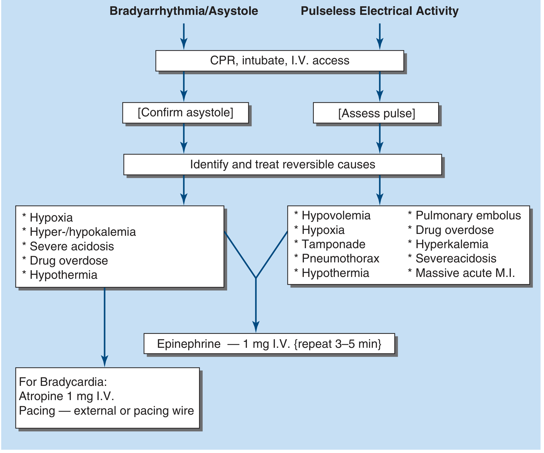
PEA Management (Tintinalli's Tintinalli algorithm showing QRS analysis):
The H's and T's - Reversible Causes
Always run through these at EVERY resuscitation:
| H's | T's |
|---|---|
| Hypovolemia/Hemorrhage | Tamponade (cardiac) |
| Hypoxia | Tension pneumothorax |
| Hydrogen ion (acidosis) | Thrombosis - coronary (ACS) |
| Hypo/Hyperkalemia | Thrombosis - pulmonary (PE) |
| Hypothermia | Tablets/Toxins (drug OD) |
Bedside POCUS during arrest: Can identify tamponade, RV strain (PE), hypovolemia (IVC collapse), pneumothorax, wall motion abnormalities. Do NOT interrupt compressions - use pulse check windows (≤10 sec). Point-of-care US may guide decision-making for PEA but does NOT improve survival by itself.
Defibrillation - Technical Points
- Biphasic waveform: start at 120-200 J (device-specific; manufacturer-recommended) or 200 J if unknown
- Resume compressions IMMEDIATELY after shock - do not wait to assess rhythm
- Pre-charge defibrillator while compressions are ongoing to minimize pause
2025 New Recommendations on Advanced Defibrillation:
-
Double Sequential External Defibrillation (DSED): Two defibrillators placed on different vector axes, both fired within seconds for shock-refractory VF (≥3 failed shocks). The 2022 DOSE VF trial showed superiority over standard defibrillation. 2025 guidelines: may be considered for refractory VF (updated - evidence now supports it), though routine use is not yet established.
-
Vector Change Defibrillation: Repositioning pad placement to change the electrical vector for refractory VF. 2025 (New): usefulness not yet established - insufficient evidence to recommend for or against.
PART 8 - SPECIAL CONSIDERATIONS IN CARDIAC ARREST
Peri-Arrest Conditions (2025 AHA new section)
The 2025 guidelines explicitly address peri-arrest - conditions that can cause arrest or complicate management:
- Atrial fibrillation/flutter with RVR: Electrical cardioversion (synchronized, 120-200 J biphasic for AF; 50-100 J for flutter). Rate control with diltiazem, metoprolol, or amiodarone if hemodynamically stable.
- New 2025: Updated bradycardia algorithm - transvenous pacing is now explicitly recommended for persistent hemodynamically unstable bradycardia refractory to medications (atropine, dopamine/epinephrine infusion).
ECMO-CPR (eCPR)
- Extracorporeal cardiopulmonary resuscitation - VA-ECMO during cardiac arrest
- Considered for refractory arrest in select patients: witnessed arrest, short no-flow time, reversible etiology (PE, hypothermia, ACS), age <65-75, no DNAR
- Requires institutional capability; emerging evidence supports improved survival in select centers
- 2025 AHA: recommends consideration for refractory arrest at ECMO-capable centers
Hypothermia-Induced Arrest
- "Not dead until warm and dead" - continue resuscitation until core temperature ≥30-32°C
- Use warm IV fluids, forced air warming, ECMO rewarming in severe cases
- Hypothermia protects the brain; excellent neurologic outcomes possible even after prolonged arrest
Pregnancy
- Displace uterus leftward (manual displacement or left lateral tilt ≥15°) to relieve aortocaval compression
- Peri-mortem C-section (PMCD) within 5 minutes of maternal cardiac arrest if no ROSC
- PMCD improves maternal ROSC by relieving aortocaval compression
Opioid Overdose (2025 AHA Update)
- Naloxone 0.4-2 mg IV/IO/IM/IN for respiratory arrest/arrest suspected due to opioids
- Updated BLS algorithm now explicitly includes opioid antagonist step
- If no response to naloxone and no pulse: proceed to standard CPR algorithm
- Public access opioid emergency kits (expanded emphasis in 2025 guidelines)
PART 9 - POST-CARDIAC ARREST CARE (PCAC)
This is the "fourth link in the chain" and critically determines neurological outcomes.
Immediate Stabilization Post-ROSC
Airway/Breathing:
- Intubate if not already done for patients not fully awake
- Titrate FiO2 to SpO2 94-99% - avoid hyperoxia (O2 free radicals worsen reperfusion injury)
- Avoid hypoxia (SpO2 <94%) - equally harmful
- Target PaCO2 35-45 mmHg - avoid hypocapnia (<30 mmHg), which causes cerebral vasoconstriction
- Ventilator rate to maintain normocarbia
Circulation:
- Target MAP 65-100 mmHg (many centers target ≥65-70 mmHg minimum; 90-100 mmHg if needed for cerebral autoregulation)
- Treat hypotension aggressively: IV fluids + vasopressors (norepinephrine is first-line pressor post-ROSC)
- 12-lead ECG immediately to assess for STEMI
Glucose:
- Target glucose 140-180 mg/dL (120-180 mg/dL per some guidelines)
- Avoid both hypoglycemia AND hyperglycemia - both worsen neurological outcome
- Check glucose every 30-60 min
Coronary Angiography Post-ROSC
- STEMI on post-ROSC ECG: Immediate emergent PCI (Class I recommendation - unchanged)
- No STEMI but hemodynamic instability, electrical instability, or evidence of ischemia: Consider emergent angiography
- No STEMI, hemodynamically stable: 2023 TOMAHAWK/COACT trials changed the approach - delayed or selective angiography is now acceptable; emergent angiography does NOT improve outcomes in non-STEMI post-arrest patients
- 2025 AHA: Emergent angiography is reasonable for STEMI; for non-STEMI OHCA, delayed angiography (within 24-72h) is acceptable unless clinical instability dictates earlier intervention
Targeted Temperature Management (TTM) - 2025 Update
The evidence evolution:
- Original 2002 HACA trial and Bernard et al.: hypothermia (32-34°C) improved neurological outcomes in shockable OHCA
- 2013 TTM trial (Nielsen et al.): 33°C vs. 36°C - no difference
- 2021 TTM2 trial (Dankiewicz et al.): Normothermia (37.5°C) with aggressive fever prevention vs. hypothermia (33°C) - no difference in mortality or neurological outcome
2025 AHA Current Recommendation:
- Fever prevention is mandatory: Keep temperature ≤37.5°C for at least 72 hours post-ROSC in comatose patients - aggressive fever treatment even if not actively cooling
- Active cooling to 32-36°C may still be reasonable for select comatose patients, but is no longer universally mandated
- DO NOT use cold saline prehospital boluses (increases pulmonary edema, no benefit)
- Shivering must be treated: analgesia + sedation; NMB if refractory
- Miller's Anesthesia 10e; Harrison's Principles 22e; 2025 AHA Post-Cardiac Arrest Guidelines PMID 41122894
Neuroprognostication
Do NOT prognosticate early. Mandatory waiting periods:
- Without TTM: ≥72 hours post-ROSC before declaring poor prognosis
- With TTM (32-34°C): ≥72 hours after rewarming (total ~120 hours from arrest)
Multimodal approach required:
| Tool | Poor Prognosis Marker |
|---|---|
| Clinical exam | Absent pupillary light reflex bilaterally at 72h; absent corneal reflexes |
| EEG | Burst suppression, status epilepticus, absence of reactivity |
| SSEPs (somatosensory evoked potentials) | Bilateral absent N20 response (most reliable single predictor) |
| CT brain | Loss of gray-white differentiation; global cerebral edema |
| MRI brain | Diffuse diffusion restriction |
| Biomarkers | NSE (neuron-specific enolase) >60-80 ug/L at 48-72h; GFAP |
2025 New: Guidelines now differentiate prognostication for favorable vs. unfavorable outcome (not just poor outcome). No single test is sufficient - always use multimodal assessment.
Seizures Post-Arrest
- Seizures (including non-convulsive status epilepticus - NCSE) occur in 10-30% of post-arrest comatose patients
- NCSE is particularly dangerous and clinically silent
- Continuous EEG monitoring is indicated for all comatose post-arrest patients (at least 24-48h)
- Treat clinical seizures and NCSE promptly: Benzodiazepines → Levetiracetam/Valproate → Keppra → anaesthetic doses
- 2025 Update: Myoclonus definitions and treatment options now explicitly included - differentiate Lance-Adams syndrome (good prognosis) from malignant myoclonus
Mechanical Circulatory Support (MCS)
- Intra-aortic balloon pump (IABP): improves coronary perfusion diastolically; use in cardiogenic shock
- Impella/LVAD: for refractory cardiogenic shock post-arrest
- VA-ECMO: bridge to recovery, transplant, or device; most benefit in reversible causes
Survivorship and Rehabilitation (2025 AHA New Section)
- Structured follow-up for patients, caregivers, and rescuers
- Screen for PTSD, anxiety, cognitive impairment in arrest survivors
- Genetic testing and family screening for all SCA survivors without clear cause (especially for channelopathies, HCM)
- ICD evaluation for all appropriate survivors
PART 10 - ICD THERAPY AND PREVENTION
Primary Prevention (ICD before first SCA)
Evidence-based indications (Class I):
- EF ≤35% + symptomatic HF (NYHA II-III) + optimal medical therapy x3 months (MADIT-II, SCD-HeFT trials)
- Prior MI (>40 days) + EF ≤30% + NYHA I symptoms
- HCM with risk factors (wall ≥30 mm, syncope, family SCD, abnormal BP response, prior VT/VF)
- Confirmed channelopathy (Long QT, Brugada, CPVT) with symptoms or high-risk features
- ARVC with VT/VF or high-risk features
Secondary Prevention (ICD after SCA or VT/VF)
- All survivors of SCA without clearly reversible cause (Class I)
- Sustained VT causing hemodynamic compromise or syncope (Class I)
Pharmacologic Prevention
| Drug | Indication | Benefit |
|---|---|---|
| Beta-blockers | Post-MI, HF, HCM, channelopathies | Reduce SCD significantly; CAST trial showed Class I drugs are harmful |
| Amiodarone | Adjunct to ICD; prevents shocks; not primary prevention monotherapy | Reduces VT burden but NOT mortality benefit over ICD alone |
| Sotalol | Class III + II; VT suppression | Proarrhythmic (torsades); requires QT monitoring |
| CAST warning | Class I drugs (flecainide, encainide, moricizine) INCREASE SCD in post-MI patients (CAST trial) | AVOID in structural heart disease |
| Flecainide | Safe in CPVT (reduces VT) and structurally normal heart AF | Do NOT use in structural heart disease |
- Tintinalli's Emergency Medicine, Ch. 11 (SCD Antiarrhythmic Drug section)
PART 11 - TERMINATION OF RESUSCITATION (2025 AHA Updated)
Out-of-Hospital Cardiac Arrest (OHCA) - BLS TOR Rule
All 3 criteria must be present to terminate:
- Arrest NOT witnessed by EMS
- No ROSC at any time
- No shock delivered (AED)
If ANY criterion is absent → transport.
Advanced Provider TOR Rule
All criteria must be present:
- Arrest not witnessed by bystander or EMS
- No bystander CPR provided
- No ROSC after 3 full ALS rounds (ALS on scene ≥20 min)
- Not presenting rhythm VF/pVT
2025 Update: TOR algorithm was updated to include both BLS and universal TOR rules. Rhythm (VF/pVT vs. non-shockable) is now explicitly part of the universal TOR decision.
Always consider: Hypothermia, drug overdose, drowning, and pediatric arrests - lower threshold to transport.
PART 12 - CHAIN OF SURVIVAL (2025 AHA Update)
2025 Change: A single unified Chain of Survival now applies across all ages and settings (previously separate adult/pediatric chains). The links are:
- Recognition and activation - Call 911; recognize arrest
- Immediate high-quality CPR - Bystander CPR with compressions + breaths (when capable)
- Rapid defibrillation - AED use as soon as available
- Advanced resuscitation - EMS/ACLS team with drugs, airway, monitoring
- Post-cardiac arrest care - ICU management, TTM, coronary angio, neuroprognostication
- Recovery - Survivorship support, rehabilitation, ICD, genetic counseling
Community survival strategies: Public Access Defibrillation (PAD) programs can double survival compared to CPR alone (Public Access Defibrillation trial). Dispatcher-assisted CPR instructions improve bystander CPR rates.
KEY CLINICAL PEARLS FOR THE EMERGENCY RESIDENT
-
The first 4 minutes are everything. Every minute of VF without defibrillation drops survival by ~7-10%. If an AED is present, use it before calling for more help.
-
CPR quality is the intervention that saves lives. Drugs are adjuncts. Maximize compression fraction, rate, and depth. No interruptions.
-
Amiodarone and lidocaine don't improve hospital discharge survival - they help get the patient to the hospital, which is still valuable.
-
PEA demands you think harder, not just push harder. Every PEA patient has a reason. Narrow QRS = mechanical problem; use POCUS. Wide QRS = metabolic/toxic problem; treat with specific antidotes.
-
ETCO2 is your friend. If it's >20 mmHg, your CPR is adequate. If it suddenly jumps 10+ mmHg, ROSC has occurred - check pulse before next cycle.
-
Post-arrest fever kills neurons. Keep temp ≤37.5°C aggressively. That is now the minimum standard regardless of whether you use active cooling.
-
Don't prognosticate too early. Many patients declared to have "poor prognosis" make meaningful recoveries if given 72+ hours. The 2025 guidelines strongly reinforce multimodal, time-appropriate assessment.
-
STEMI + cardiac arrest = cath lab. No debate. For non-STEMI post-arrest patients with hemodynamic stability, it is now acceptable to wait.
-
Think about ICD for every SCA survivor you resuscitate. If they survive to discharge without a clearly reversible cause, they need an ICD - this is a Class I recommendation.
Sources: Robbins & Kumar Basic Pathology 10e; Robbins Cotran & Kumar Pathologic Basis of Disease; Tintinalli's Emergency Medicine (Comprehensive Study Guide); Harrison's Principles of Internal Medicine 22e; Rosen's Emergency Medicine; Barash Clinical Anesthesia 9e; Miller's Anesthesia 10e; 2025 AHA Guidelines for CPR & ECC (Circulation 2025;152:suppl 2) — PMID 41122884, 41122893, 41122894; ERC/ESICM 2025 Post-Resuscitation Care Guidelines PMID 41123621.
This is a shared conversation. Sign in to Orris to start your own chat.