
Give me a flashcard. Showing USG images of cogwheel sign, beats on spring appearance, and waist sign. In Pelvic Inflammatory Disease.
cogwheel sign ultrasound pelvic inflammatory disease fallopian tube
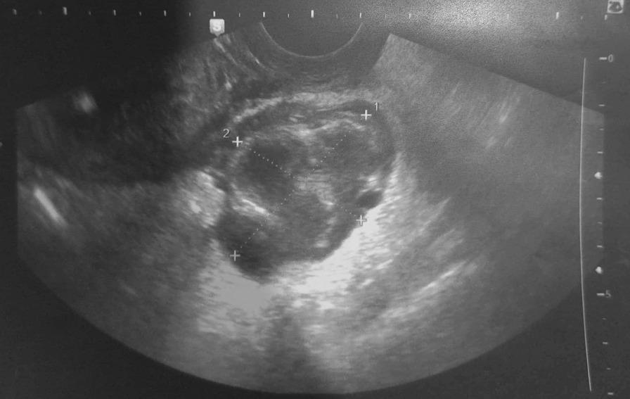
A transvaginal grey-scale ultrasound image demonstrating a complex adnexal mass, approximately 6 cm in diameter, located in the juxta-uterine region. The mass is primarily cystic with irregular, thickened walls and heterogeneous internal echogenicity, suggesting the presence of debris, pus, or complex fluid. A characteristic 'cogwheel sign' is visible, represented by thickened endosalpingeal folds that project into the lumen of the dilated fallopian tube, which is a classic sonographic indicator of pyosalpinx. The morphology is tubular and tortuous rather than purely spherical, typical of pelvic inflammatory disease progression. Caliper markers are placed to measure the dimensions of the collection. The surrounding stroma shows increased echogenicity, consistent with inflammatory changes in the adnexa. This diagnostic image illustrates the sonographic features used to differentiate pyosalpinx from other adnexal masses like ovarian cysts or ectopic pregnancies.
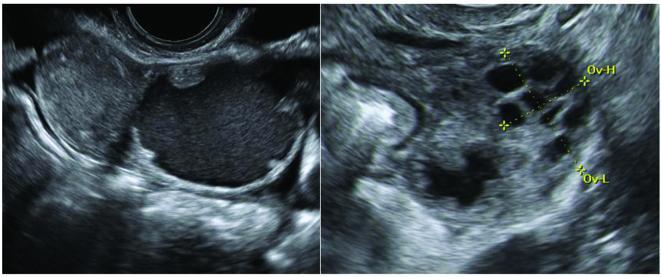
This diagnostic ultrasound image demonstrates classic sonographic features of acute salpingitis in two planes. The left panel shows a longitudinal view of a distended fallopian tube appearing as a pear-shaped unilocular mass with low-level internal echogenicity and thickened walls exceeding 5 mm. The right panel displays a transverse section illustrating the 'cogwheel sign,' characterized by multiple small, round, hypoechoic to anechoic structures arranged in a circular or serrated pattern. These protrusions represent cross-sections of thickened, edematous endosalpingeal folds projecting into the tubal lumen. The structures are bounded by incomplete septae and are adjacent to the ovary, which is marked with calipers (Ov-H and Ov-L). These findings are pathognomonic for inflammatory processes of the fallopian tube, such as pelvic inflammatory disease (PID), and help distinguish tubal pathology from primary ovarian masses.
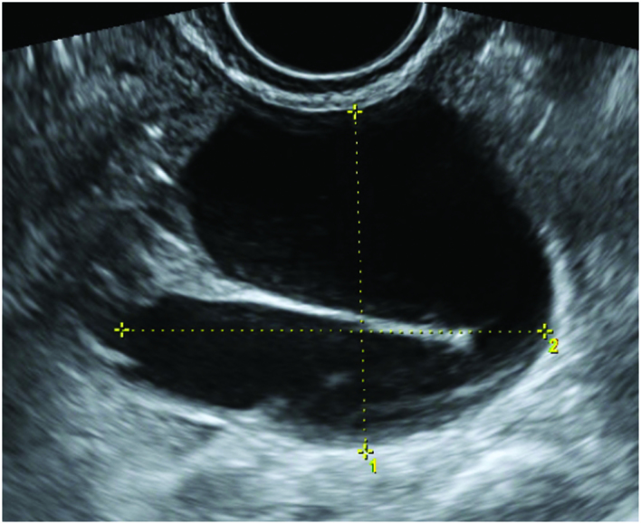
**Modality:** Transvaginal Ultrasound (TVUS), B-mode. **Anatomic Region:** Adnexa / Fallopian tube. **Description:** The image displays a cross-sectional view of a hydrosalpinx, appearing as an elongated, fusiform, and well-circumscribed cystic structure with anechoic internal fluid. A prominent, hyperechoic thin linear band is visible extending into the lumen, representing an incomplete septum or "cogwheel" sign. This feature is a classic sonographic hallmark of a dilated, folded fallopian tube, distinguishing it from an ovarian cyst or para-ovarian mass. **Visual Features:** - **Fluid Collection:** Anechoic, simple fluid without internal debris or vascularity. - **Wall Structure:** Smooth, thin walls with no solid papillary projections or thickened mural nodules. - **Septation:** A pathognomonic incomplete septum (cogwheel sign/waist sign) characteristic of chronic tubal dilatation. - **Measurements:** Orthogonal electronic calipers (marked 1 and 2) are positioned to quantify the maximum diameter of the distended lumen. **Diagnostic Significance:** These features are highly suggestive of hydrosalpinx, typically secondary to pelvic inflammatory disease (PID) or previous tubal pathology, aiding in the differentiation of adnexal masses.
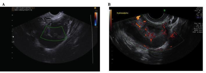
Two-panel transvaginal sonography (TVS) image demonstrating the features of acute salpingitis. Panel A displays a color Doppler ultrasound of a dilated fallopian tube, appearing as a pear-shaped, unilocular adnexal mass. The mass contains low-level echogenic fluid and is characterized by thickened walls (exceeding 5 mm) and multiple incomplete septae extending into the lumen. Panel B shows a power Doppler ultrasound of the same structure, highlighting significant hypervascularity concentrated within the incomplete septae and the thickened tubal wall. These internal projections and the increased vascular signal are classic sonographic indicators of an active inflammatory process, such as acute pelvic inflammatory disease (PID). The morphology of the tube with internal protrusions may also suggest a 'cogwheel sign' when viewed in transverse section, distinguishing acute salpingitis from simple hydrosalpinx.
beads on string appearance ultrasound PID tubo-ovarian
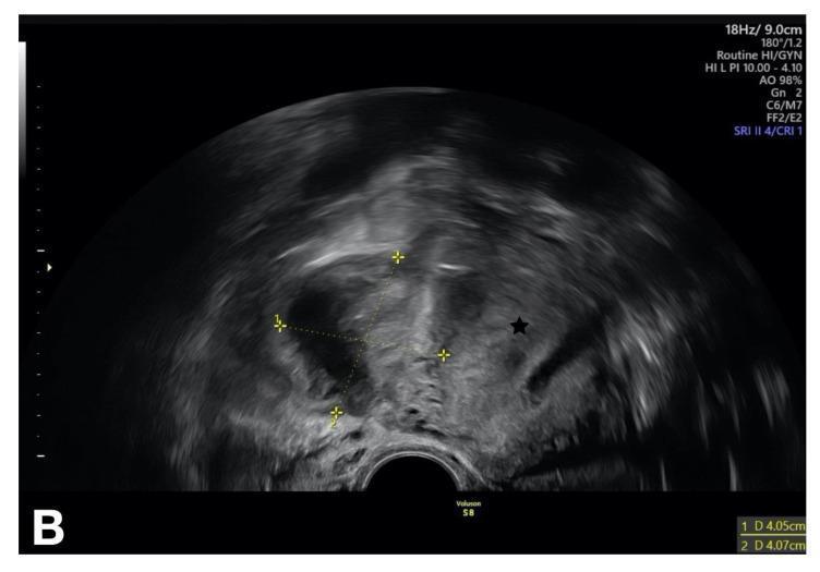
This diagnostic image is a transvaginal ultrasound (TVUS) in the transverse plane, illustrating a complex adnexal mass consistent with a tubo-ovarian abscess (TOA) secondary to pelvic inflammatory disease (PID). The mass is located in the right adnexa and is characterized by heterogeneous echogenicity, featuring mixed solid (hyperechoic) and cystic/fluid-filled (hypoechoic) components. The internal architecture appears disorganized with thick, irregular walls and ill-defined borders, indicating an inflammatory process. Electronic calipers are seen measuring the lesion, and a black star identifies the adjacent uterus, which appears mildly displaced. The surrounding tissue demonstrates typical sonographic signs of pelvic inflammation, including obscured anatomical planes and potential fluid in the Pouch of Douglas. This image serves as an educational example of advanced pelvic infection, emphasizing the transformation of normal ovarian and tubal anatomy into a unified inflammatory complex. It is intended for medical students and radiology residents to recognize the classic 'ground-glass' or multi-loculated appearance of a pelvic abscess.
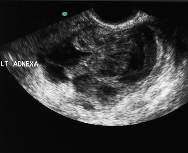
**Imaging Modality:** Transvaginal Ultrasound (TVUS) **Anatomical Region:** Left adnexa, coronal plane. **Observed Pathology:** Large, complex tubo-ovarian mass consistent with a Tubo-Ovarian Abscess (TOA). **Characteristic Visual Features:** The image demonstrates a poorly defined, multi-loculated heterogeneous mass involving the left adnexal region. Key features include: * **Echo Texture:** A mixture of hypoechoic and solid-appearing components with internal debris, characteristic of an inflammatory or purulent collection. * **Architecture:** The mass shows thick, irregular walls and internal septations. There is a lack of clear demarcation between the ovary and the fallopian tube, forming a "tubo-ovarian complex." * **Fluid Components:** Presence of low-level internal echoes within cystic areas, suggesting complex fluid or pus. **Key Diagnostic Features:** The primary diagnostic cue is the complete loss of normal adnexal architecture, replaced by a disorganized, heterogeneous mass. The "cogwheel" or "beaded" appearance often associated with salpingitis is obscured by the expansive, organized abscess formation. These sonographic findings are highly suggestive of Pelvic Inflammatory Disease (PID) progressing to a tubo-ovarian abscess.
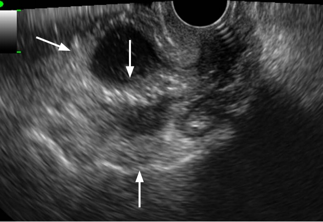
**Imaging Modality:** Transvaginal Ultrasound (TVUS) **Anatomical Region:** Pelvis (Adnexa) **Observed Pathology:** Tubo-ovarian abscess (TOA) **Characteristic Visual Features:** The image displays a complex, multilocular cystic adnexal mass. Key sonographic features include: * **Wall Morphology:** Distinct, regular thickening of the outer capsule and internal septations (indicated by arrows). * **Internal Content:** The cystic spaces contain low-level internal echogenic debris/fluid, consistent with purulent material or complex fluid. * **Architecture:** The mass lacks solid papillary projections but exhibits a "cogwheel" or "beaded" appearance characteristic of inflammatory adnexal processes involving the fallopian tube and ovary. **Diagnostic Differentiating Features:** The presence of thick, regular walls and septa combined with internal echogenicity helps differentiate this inflammatory complex from simple ovarian cysts or hydrosalpinx. The lack of irregular solid components or high-vascularity nodules (not visible here) helps distinguish it from malignant epithelial ovarian tumors. This presentation is highly suggestive of Pelvic Inflammatory Disease (PID) progressing to a tubo-ovarian abscess.

**Imaging Modality:** Transvaginal Ultrasound (TVUS), B-mode. **Anatomical Region:** Female pelvis, right adnexa. **Observed Pathology:** Hydrosalpinx. **Characteristic Visual Features:** The image demonstrates a prominent, elongated, tubular cystic structure in the adnexa. The mass is primarily anechoic to hypoechoic, indicating fluid collection within a dilated fallopian tube. Distinctive "waist signs" or indentations along the opposite walls of the tube are visible, creating a multiloculated appearance often referred to as a "cogwheel" or "beads-on-a-string" sign in cross-section. **Diagnostic Features:** The structure is separate from the ovary (not visualized in this plane) and lacks the thick, vascularized walls characteristic of a tubo-ovarian abscess or pyosalpinx. Electronic calipers measure the maximal dimensions of the dilated segment (approximately 2.66 cm x 1.76 cm). The presence of incomplete septa (indentations) is a key differentiating feature that distinguishes a dilated fallopian tube from other pelvic cystic adnexal masses such as para-ovarian cysts. **Clinical Context:** Findings are diagnostic of tubal dilation and fluid accumulation, consistent with chronic tubal occlusion and hydrosalpinx in a reproductive-age patient.
waist sign ultrasound PID tubo-ovarian complex
This diagnostic image is a transvaginal ultrasound (TVUS) in the transverse plane, illustrating a complex adnexal mass consistent with a tubo-ovarian abscess (TOA) secondary to pelvic inflammatory disease (PID). The mass is located in the right adnexa and is characterized by heterogeneous echogenicity, featuring mixed solid (hyperechoic) and cystic/fluid-filled (hypoechoic) components. The internal architecture appears disorganized with thick, irregular walls and ill-defined borders, indicating an inflammatory process. Electronic calipers are seen measuring the lesion, and a black star identifies the adjacent uterus, which appears mildly displaced. The surrounding tissue demonstrates typical sonographic signs of pelvic inflammation, including obscured anatomical planes and potential fluid in the Pouch of Douglas. This image serves as an educational example of advanced pelvic infection, emphasizing the transformation of normal ovarian and tubal anatomy into a unified inflammatory complex. It is intended for medical students and radiology residents to recognize the classic 'ground-glass' or multi-loculated appearance of a pelvic abscess.
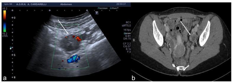
This dual-panel image demonstrates a tubo-ovarian complex involving a left adnexal abscess, illustrated through two diagnostic imaging modalities. Panel (a) is an axial color Doppler transabdominal ultrasound (TSA-US) scan. It reveals a complex cystic mass in the left adnexa characterized by a predominantly anechoic central region, consistent with fluid or purulent material. The color Doppler overlay shows peripheral vascularization (arrow) indicated by red and blue signals around the margins of the mass, reflecting active blood flow in the surrounding inflammatory tissue. Panel (b) is a contrast-enhanced axial CT image of the pelvis. It shows a corresponding left adnexal abscess with a distinctively thickened and hyper-enhancing wall (arrows). The enhancement indicates increased vascularity and inflammation characteristic of an infectious process. These findings are critical for diagnosing pelvic inflammatory disease (PID) and its complications, such as tubo-ovarian abscesses (TOA). This comparison illustrates how ultrasound serves as a primary tool for evaluating vascular compromise and cystic morphology, while CT provides superior detail regarding wall thickening and regional inflammatory extent.
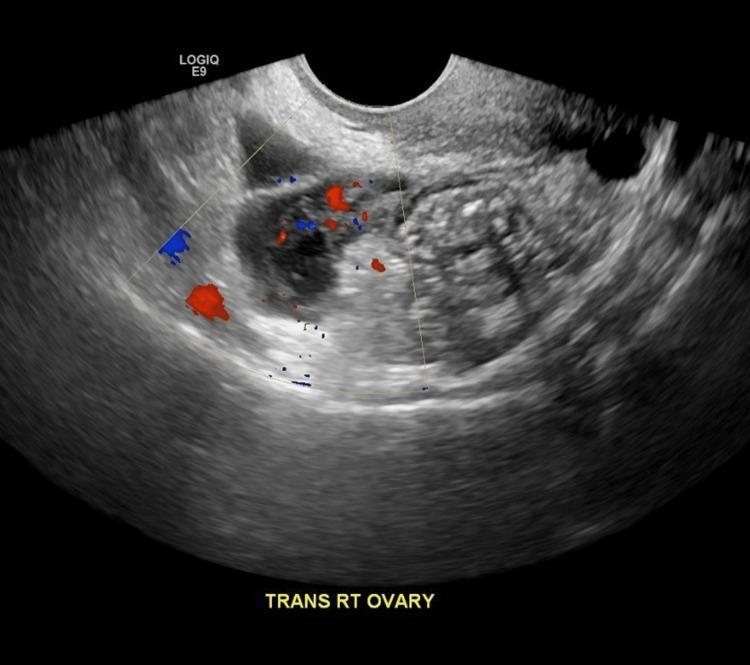
This diagnostic image is a transabdominal ultrasound of the right adnexa in the transverse plane, demonstrating a complex right tubo-ovarian abscess (TOA) measuring approximately 6.2 x 3 x 2.5 cm. The mass exhibits a heterogeneous echotexture with mixed cystic and solid components, characterized by internal low-level echoes and irregular thick walls. The borders of the mass are poorly defined and irregular, blending with adjacent pelvic structures, which is indicative of localized inflammation and pelvic inflammatory disease (PID). Color Doppler overlay reveals internal and peripheral vascularity, showing punctate red and blue signals that represent blood flow within the inflammatory tissue. There is no clear evidence of posterior acoustic shadowing or enhancement. This visual finding is a key diagnostic indicator for advanced pelvic infection and is used in clinical education to distinguish complex adnexal masses from simple ovarian cysts or endometriomas.
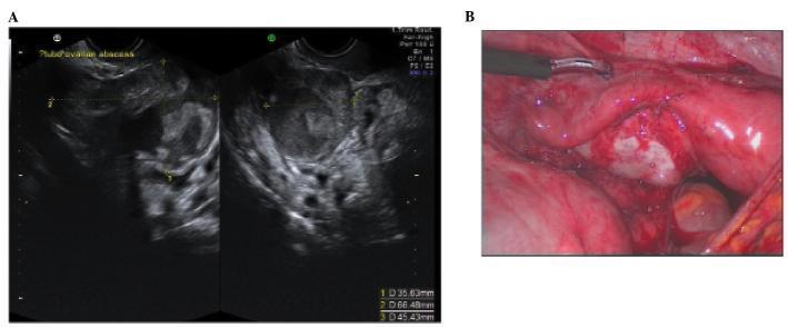
This dual-panel image provides a side-by-side comparison of a tubo-ovarian complex (TOC) using two imaging modalities. Panel A is a transvaginal ultrasound (TVUS) showing a large, heterogeneous, complex adnexal mass. The sonogram reveals mixed echogenicity with ill-defined borders, internal septations, and cystic components, indicating the involvement of both the fallopian tube and the ovary in an inflammatory process. Quantitative calipers measure the dimensions of the mass as approximately 35.6 mm x 66.5 mm x 45.4 mm. Panel B is a corresponding laparoscopic photograph of the same case, providing direct visualization of the pelvic cavity. It shows significant erythema and edema of the adnexal structures, with thick inflammatory adhesions and a whitish-yellow purulent-appearing exudate characteristic of a tubo-ovarian abscess. The laparoscopic view clarifies the anatomical distortion and tissue friability suggested by the ultrasound. This comparison illustrates the diagnostic pathway for pelvic inflammatory disease (PID) progressing to abscess formation, highlighting the correlation between sonographic heterogeneity and gross surgical inflammation.
ls /tmp/workspace/ 2>/dev/null || mkdir -p /tmp/workspace/pid-flashcard
mkdir -p /tmp/workspace/pid-flashcard && cd /tmp/workspace/pid-flashcard && python3 - <<'EOF' import urllib.request images = { "cogwheel.jpg": "https://cdn.orris.care/cdss_images/pmc_clinical_VQA_02e423c587589b4eded1f9dc96a2b0177fb9296c4cf12f5a01fa73457ac0ceb2.jpg", "beads.png": "https://cdn.orris.care/cdss_images/roco_radiology_ROCO_03616_1766647544617.png", "waist.png": "https://cdn.orris.care/cdss_images/roco_radiology_ROCO_78750_1766650452670.png", } headers = {'User-Agent': 'Mozilla/5.0'} for fname, url in images.items(): req = urllib.request.Request(url, headers=headers) with urllib.request.urlopen(req, timeout=20) as r: data = r.read() with open(fname, 'wb') as f: f.write(data) print(f"Downloaded {fname}: {len(data)} bytes") EOF
cd /tmp/workspace/pid-flashcard && python3 - <<'PYEOF' from PIL import Image, ImageDraw, ImageFont import textwrap, os # ── Canvas ───────────────────────────────────────────────────────────────── W, H = 1500, 980 card = Image.new("RGB", (W, H), "#0d1b2a") draw = ImageDraw.Draw(card) # ── Gradient-like top bar ─────────────────────────────────────────────────── for y in range(80): t = y / 80 r = int(30 + (10 - 30) * t) g = int(80 + (50 - 80) * t) b = int(160 + (120 - 160) * t) draw.line([(0, y), (W, y)], fill=(r, g, b)) # ── Fonts ────────────────────────────────────────────────────────────────── try: font_title = ImageFont.truetype("/usr/share/fonts/truetype/dejavu/DejaVuSans-Bold.ttf", 38) font_sub = ImageFont.truetype("/usr/share/fonts/truetype/dejavu/DejaVuSans-Bold.ttf", 22) font_label = ImageFont.truetype("/usr/share/fonts/truetype/dejavu/DejaVuSans-Bold.ttf", 20) font_desc = ImageFont.truetype("/usr/share/fonts/truetype/dejavu/DejaVuSans.ttf", 17) font_tag = ImageFont.truetype("/usr/share/fonts/truetype/dejavu/DejaVuSans-Bold.ttf", 15) except: font_title = font_sub = font_label = font_desc = font_tag = ImageFont.load_default() # ── Title bar text ───────────────────────────────────────────────────────── title = "USG Signs in Pelvic Inflammatory Disease (PID)" bbox = draw.textbbox((0,0), title, font=font_title) tw = bbox[2] - bbox[0] draw.text(((W - tw)//2, 18), title, font=font_title, fill="#ffffff") # ── Helper: rounded rectangle ────────────────────────────────────────────── def rounded_rect(draw, xy, radius, fill, outline=None, width=2): x0,y0,x1,y1 = xy draw.rounded_rectangle([x0,y0,x1,y1], radius=radius, fill=fill, outline=outline, width=width) # ── Panel data ───────────────────────────────────────────────────────────── panels = [ { "img": "cogwheel.jpg", "sign": "Cogwheel Sign", "emoji": "⚙️", "color": "#e63946", "light": "#ffd6d8", "what": "Thickened endosalpingeal folds seen in TRANSVERSE section of the inflamed tube", "where": "Acute salpingitis / Pyosalpinx", "note": "Serrated wheel-like projections into tubal lumen", }, { "img": "beads.png", "sign": "Beads-on-a-String Sign", "emoji": "🔵", "color": "#2a9d8f", "light": "#c8f5ef", "what": "Multiple small cystic nodules strung along a dilated tube in LONGITUDINAL section", "where": "Hydrosalpinx / Chronic PID", "note": "Incomplete septa create bead-like locules", }, { "img": "waist.png", "sign": "Waist Sign", "emoji": "⏳", "color": "#f4a261", "light": "#fdebd0", "what": "Opposing indentations on walls of dilated tube producing an hourglass narrowing", "where": "Hydrosalpinx / Chronic PID", "note": "Pathognomonic for tortuous fallopian tube dilation", }, ] NCOLS = 3 PAD = 22 # outer padding GAP = 18 # gap between panels TOP_BAR = 85 # title bar height IMG_H = 320 # USG image display height col_w = (W - 2*PAD - (NCOLS-1)*GAP) // NCOLS panel_h = H - TOP_BAR - PAD - 10 py0 = TOP_BAR + 10 for i, p in enumerate(panels): px0 = PAD + i*(col_w + GAP) px1 = px0 + col_w py1 = py0 + panel_h # Panel background rounded_rect(draw, [px0, py0, px1, py1], radius=16, fill="#111e2e", outline=p["color"], width=3) # Coloured sign-name header strip rounded_rect(draw, [px0+3, py0+3, px1-3, py0+50], radius=14, fill=p["color"]) label = p["emoji"] + " " + p["sign"] lb = draw.textbbox((0,0), label, font=font_sub) lw = lb[2]-lb[0] draw.text((px0 + (col_w-lw)//2, py0+13), label, font=font_sub, fill="#ffffff") # ── Paste USG image ──────────────────────────────────────────────────── img_path = p["img"] try: usg = Image.open(img_path).convert("RGB") # crop to centre-square then resize iw, ih = usg.size side = min(iw, ih) left = (iw - side)//2 top = (ih - side)//2 usg = usg.crop((left, top, left+side, top+side)) usg = usg.resize((col_w - 20, IMG_H), Image.LANCZOS) card.paste(usg, (px0+10, py0+58)) # thin border over image draw.rectangle([px0+10, py0+58, px0+10+col_w-20, py0+58+IMG_H], outline=p["color"], width=2) except Exception as e: draw.rectangle([px0+10, py0+58, px1-10, py0+58+IMG_H], fill="#1a2a3a") draw.text((px0+20, py0+80), f"[image error: {e}]", font=font_desc, fill="#aaaaaa") # ── Info rows ────────────────────────────────────────────────────────── info_y = py0 + 58 + IMG_H + 14 def wrap_draw(text, x, y, w, fnt, color, line_h=24): for line in textwrap.wrap(text, width=int(w/10)): draw.text((x, y), line, font=fnt, fill=color) y += line_h return y # WHERE draw.text((px0+14, info_y), "📍 Structure:", font=font_label, fill=p["color"]) info_y = wrap_draw(p["where"], px0+14, info_y+24, col_w-28, font_desc, "#e0e8f0", 22) info_y += 8 # WHAT draw.text((px0+14, info_y), "🔍 Finding:", font=font_label, fill=p["color"]) info_y = wrap_draw(p["what"], px0+14, info_y+24, col_w-28, font_desc, "#e0e8f0", 22) info_y += 8 # KEY NOTE pill pill_x0, pill_y0 = px0+14, info_y+2 pill_text = "⚡ " + p["note"] pb = draw.textbbox((0,0), pill_text, font=font_tag) pw = pb[2]-pb[0]+16 ph = pb[3]-pb[1]+10 rounded_rect(draw, [pill_x0, pill_y0, pill_x0+pw, pill_y0+ph], radius=8, fill=p["light"]) draw.text((pill_x0+8, pill_y0+5), pill_text, font=font_tag, fill="#1a1a2e") # ── Footer ───────────────────────────────────────────────────────────────── footer = "All signs seen on Transvaginal Ultrasound (TVUS) • PID Spectrum: Salpingitis → Pyosalpinx → Hydrosalpinx → Tubo-Ovarian Abscess" fb = draw.textbbox((0,0), footer, font=font_tag) fw = fb[2]-fb[0] draw.text(((W-fw)//2, H-22), footer, font=font_tag, fill="#6a8fa8") # ── Save ─────────────────────────────────────────────────────────────────── out = "/tmp/workspace/pid-flashcard/PID_USG_Signs_Flashcard.png" card.save(out, "PNG", dpi=(150,150)) print(f"Saved: {out} ({os.path.getsize(out)//1024} KB)") PYEOF
🩻 PID USG Signs - Flashcard
PID USG Signs Flashcard
Image · PNG
⚙️ Cogwheel Sign
- Structure: Acute salpingitis / Pyosalpinx
- Finding: Thickened endosalpingeal folds seen in transverse section of the inflamed fallopian tube - they project inward like gear teeth around the lumen
- Key: Pathognomonic for acute PID-related tubal inflammation
🔵 Beads-on-a-String Sign
- Structure: Hydrosalpinx / Chronic PID
- Finding: Multiple small cystic nodules strung along a dilated tube in longitudinal section - incomplete septa create bead-like locules separated by partial walls
- Key: Represents chronic tubal occlusion with folded, fluid-filled segments
⏳ Waist Sign
- Structure: Hydrosalpinx / Chronic PID
- Finding: Opposing indentations on the walls of a dilated tube creating an hourglass narrowing - the "waist" - where the tube folds back on itself
- Key: Pathognomonic for tortuous fallopian tube dilation; distinguishes hydrosalpinx from a simple ovarian cyst
Create a PDF of this flashcard for offline use
python3 - <<'PYEOF' from reportlab.lib.pagesizes import A4, landscape from reportlab.lib.utils import ImageReader from reportlab.pdfgen import canvas from reportlab.lib import colors import os src_img = "/tmp/workspace/pid-flashcard/PID_USG_Signs_Flashcard.png" out_pdf = "/tmp/workspace/pid-flashcard/PID_USG_Signs_Flashcard.pdf" # Use landscape A4 so the wide flashcard fits perfectly page_w, page_h = landscape(A4) # 841.89 x 595.28 pts c = canvas.Canvas(out_pdf, pagesize=landscape(A4)) # Metadata c.setTitle("PID USG Signs Flashcard") c.setSubject("Cogwheel Sign, Beads-on-a-String Sign, Waist Sign – Pelvic Inflammatory Disease") c.setAuthor("Orris Medical AI") c.setKeywords("PID, ultrasound, cogwheel sign, beads on string, waist sign, salpingitis") # Dark background to match the card c.setFillColorRGB(0.051, 0.106, 0.165) # #0d1b2a c.rect(0, 0, page_w, page_h, fill=1, stroke=0) # Draw the PNG stretched to fill the page with small margin margin = 8 img = ImageReader(src_img) iw, ih = img.getSize() aspect = iw / ih avail_w = page_w - 2*margin avail_h = page_h - 2*margin if avail_w / aspect <= avail_h: draw_w = avail_w draw_h = avail_w / aspect else: draw_h = avail_h draw_w = avail_h * aspect x = (page_w - draw_w) / 2 y = (page_h - draw_h) / 2 c.drawImage(img, x, y, width=draw_w, height=draw_h, preserveAspectRatio=True, mask='auto') c.save() size_kb = os.path.getsize(out_pdf) // 1024 print(f"PDF saved: {out_pdf} ({size_kb} KB)") PYEOF
PID USG Signs Flashcard
PDF Document · PDF
- Format: Landscape A4 - optimised to display the full three-panel flashcard without cropping
- Size: ~983 KB
- Metadata embedded: Title, subject, author, and keywords (PID, cogwheel sign, beads-on-a-string, waist sign) - so it's searchable in PDF viewers
- Print-ready: The dark background and panel layout print well on colour printers, or you can view it on any device offline
Can you make me? A flashcard. Show me the new updates in Samagra Shishu Bal Swasthya Karyakram before our new updates in a single flashcard.
What are the 'new updates' you want shown on the flashcard? Please list the updated items (e.g. new conditions added, screening changes, age groups, etc.) so I can show BEFORE vs AFTER accurately.
Samagra Shishu Bal Swasthya Karyakram SSPBSK new updates 2024 2025 latest notification
RBSK Rashtriya Bal Swasthya Karyakram renamed Samagra Shishu changes updated conditions 2024
- RBSK (old) = 0-18 yrs, 4Ds, 30 conditions, school + AWC mobile teams
- SSBSK (new, launched June 29, 2026) = integrates HBNC + HBYC, 0-36 months, risk-stratified, new digital tracking, maternal mental health, Shishu Shivirs
mkdir -p /tmp/workspace/ssbsk-flashcard && python3 - <<'PYEOF' from PIL import Image, ImageDraw, ImageFont import textwrap, os # ── Canvas ────────────────────────────────────────────────────────────────── W, H = 1600, 1020 card = Image.new("RGB", (W, H), "#0a0f1e") draw = ImageDraw.Draw(card) # ── Fonts ─────────────────────────────────────────────────────────────────── def load_font(path, size): try: return ImageFont.truetype(path, size) except: return ImageFont.load_default() BOLD = "/usr/share/fonts/truetype/dejavu/DejaVuSans-Bold.ttf" REG = "/usr/share/fonts/truetype/dejavu/DejaVuSans.ttf" OBLIQ = "/usr/share/fonts/truetype/dejavu/DejaVuSans-Oblique.ttf" f_title = load_font(BOLD, 34) f_head = load_font(BOLD, 20) f_sub = load_font(BOLD, 17) f_body = load_font(REG, 15) f_tag = load_font(BOLD, 13) f_small = load_font(OBLIQ, 13) f_footer = load_font(REG, 12) f_arrow = load_font(BOLD, 52) # ── Helper ────────────────────────────────────────────────────────────────── def rr(d, xy, r, fill=None, outline=None, w=2): d.rounded_rectangle(xy, radius=r, fill=fill, outline=outline, width=w) def tw(text, font): bb = draw.textbbox((0,0), text, font=font) return bb[2]-bb[0], bb[3]-bb[1] def draw_text_wrapped(text, x, y, max_w, font, color, line_h=22): chars_per_line = max(1, int(max_w / (tw("A", font)[0] or 8))) for line in textwrap.wrap(text, width=chars_per_line): draw.text((x, y), line, font=font, fill=color) y += line_h return y def bullet_block(items, x, y, max_w, font, color, bullet="•", line_h=22, gap=6): bw = tw(bullet + " ", font)[0] for item in items: draw.text((x, y), bullet, font=font, fill=color) y = draw_text_wrapped(item, x + bw, y, max_w - bw, font, color, line_h) + gap return y # ═══════════════════════════════════════════════════════════════════════════ # TOP HEADER GRADIENT BAR # ═══════════════════════════════════════════════════════════════════════════ for yi in range(90): t = yi / 90 r = int(15 + (40 - 15)*t) g = int(30 + (10 - 30)*t) b = int(80 + (50 - 80)*t) draw.line([(0, yi), (W, yi)], fill=(r, g, b)) title_text = "RBSK → SSBSK : What's New?" ttw, _ = tw(title_text, f_title) draw.text(((W-ttw)//2, 22), title_text, font=f_title, fill="#ffffff") subtitle = "Rashtriya Bal Swasthya Karyakram → Samagra Shishu Bal Swasthya Karyakram (Launched: 29 June 2026)" stw, _ = tw(subtitle, f_small) draw.text(((W-stw)//2, 62), subtitle, font=f_small, fill="#aac8e8") # ═══════════════════════════════════════════════════════════════════════════ # TWO COLUMN LAYOUT # ═══════════════════════════════════════════════════════════════════════════ PAD = 20 GAP = 18 TOP = 100 BOT = H - 30 COL_W = (W - 2*PAD - GAP) // 2 # Centre arrow strip ARROW_W = 50 COL_W2 = (W - 2*PAD - ARROW_W - 2*GAP) // 2 L_X0 = PAD L_X1 = PAD + COL_W2 A_X0 = L_X1 + GAP A_X1 = A_X0 + ARROW_W R_X0 = A_X1 + GAP R_X1 = R_X0 + COL_W2 # ── LEFT panel (RBSK - OLD) ────────────────────────────────────────────── rr(draw, [L_X0, TOP, L_X1, BOT], r=18, fill="#12203a", outline="#e63946", w=3) # header rr(draw, [L_X0+3, TOP+3, L_X1-3, TOP+52], r=16, fill="#e63946") old_h = "❌ RBSK (Old Programme)" ohw, _ = tw(old_h, f_head) draw.text((L_X0 + (COL_W2-ohw)//2, TOP+15), old_h, font=f_head, fill="#ffffff") cy = TOP + 65 # Tag rr(draw, [L_X0+12, cy, L_X0+160, cy+26], r=8, fill="#6b1a1a") draw.text((L_X0+18, cy+5), "Rashtriya Bal Swasthya Karyakram", font=load_font(BOLD,12), fill="#ffaaaa") cy += 38 sections_old = [ ("📅 Age Group", "#ffaaaa", ["Birth to 18 years"]), ("🧩 Framework", "#ffaaaa", ["4 Ds: Defects at Birth, Diseases, Deficiencies, Developmental Delays"]), ("🔢 Conditions Screened", "#ffaaaa", ["30 conditions (32 with optional TB, Leprosy, Sickle cell)"]), ("👥 Coverage", "#ffaaaa", ["Children 0-6 wks (AWC), 6wks-6yrs (AWC), 6-18yrs (Schools)"]), ("🚗 Delivery Mode", "#ffaaaa", ["Mobile Health Teams (MHT) at AWCs & schools"]), ("🏥 Referral", "#ffaaaa", ["DEIC (District Early Intervention Centre) → DHs/Tertiary"]), ("💊 Treatment", "#ffaaaa", ["Free treatment incl. surgeries via empanelled hospitals"]), ("📊 Monitoring", "#ffaaaa", ["Paper-based + partial digital; no unified tracker"]), ("🧠 Mental Health", "#ffaaaa", ["Limited — only Behaviour Disorder (Autism), ADHD, Learning Disorder"]), ("🌱 Home Visits", "#ffaaaa", ["HBNC (ASHAs) and HBYC (ASHAs + AWWs) — separate programmes"]), ("📱 Digital", "#ffaaaa", ["No integrated national digital platform"]), ] for icon_title, col, items in sections_old: draw.text((L_X0+14, cy), icon_title, font=f_sub, fill=col) cy += 22 cy = bullet_block(items, L_X0+22, cy, COL_W2-36, f_body, "#dce8f0", line_h=20, gap=4) cy += 4 # ── ARROW strip ────────────────────────────────────────────────────────── arrow_cx = (A_X0 + A_X1) // 2 arrow_cy = (TOP + BOT) // 2 for yi in range(TOP+5, BOT-5): t = (yi - TOP) / (BOT - TOP) r = int(220 + (255-220)*t) g = int(100 + (180-100)*t) b = int(0) draw.line([(arrow_cx, yi), (arrow_cx, yi)], fill=(r,g,b)) # draw thick arrow body draw.rectangle([arrow_cx-4, TOP+20, arrow_cx+4, BOT-50], fill="#f4a261") # arrowhead aw = 20 for di in range(aw): draw.line([(arrow_cx-aw+di, BOT-50-di), (arrow_cx+aw-di, BOT-50-di)], fill="#f4a261", width=3) # ── RIGHT panel (SSBSK - NEW) ──────────────────────────────────────────── rr(draw, [R_X0, TOP, R_X1, BOT], r=18, fill="#0d2a1a", outline="#2dc653", w=3) # header rr(draw, [R_X0+3, TOP+3, R_X1-3, TOP+52], r=16, fill="#1a8a3a") new_h = "✅ SSBSK (New Programme)" nhw, _ = tw(new_h, f_head) draw.text((R_X0 + (COL_W2-nhw)//2, TOP+15), new_h, font=f_head, fill="#ffffff") cy2 = TOP + 65 # Tag rr(draw, [R_X0+12, cy2, R_X0+190, cy2+26], r=8, fill="#0a3d1a") draw.text((R_X0+18, cy2+5), "Samagra Shishu Bal Swasthya Karyakram", font=load_font(BOLD,12), fill="#88ffbb") cy2 += 38 NEW_GREEN = "#88ffcc" NEW_GOLD = "#ffd700" sections_new = [ ("📅 Age Group", NEW_GREEN, ["Birth to 36 months (0–3 years) — focused on first 1000 days"]), ("🧩 Framework", NEW_GREEN, ["Integrates HBNC + HBYC into ONE unified programme"]), ("⭐ Risk-Stratified Care [NEW]", NEW_GOLD, [ "At-risk newborns: up to 9 home visits in first 42 days", "At-risk children: up to 8 home visits up to 36 months", "At-risk criteria: LBW, prematurity, late BF start, SNCU discharge, SAM, recurrent illness, dev. delay", ]), ("👥 Joint Home Visits [NEW]", NEW_GOLD, ["ASHAs + ANMs + CHOs + AWWs visit together (coordinated care)"]), ("🏫 Community Platforms [NEW]", NEW_GOLD, [ "Well-Baby Sessions at every VHSND", "Monthly Shishu Shivirs at Ayushman Arogya Mandirs", ]), ("🧠 Mental Health [NEW]", NEW_GOLD, ["Post-partum maternal mental health screening (first time)"]), ("🌱 Early Childhood Dev [NEW]", NEW_GOLD, ["Nurturing care: play, learning, child safety, family engagement mainstreamed"]), ("📱 Digital Integration [NEW]", NEW_GOLD, [ "Decision-Support Systems (DSS) + child-wise digital tracking", "Integrated with: JANANI, U-WIN, RBSK 2.0, POSHAN Tracker", "ABHA & Baal-ABHA IDs for every child", ]), ("📺 Screen Time Advisory [NEW]", NEW_GOLD, ["Guidelines on age-appropriate play; adverse impact of screen time addressed"]), ] for icon_title, col, items in sections_new: draw.text((R_X0+14, cy2), icon_title, font=f_sub, fill=col) cy2 += 22 cy2 = bullet_block(items, R_X0+22, cy2, COL_W2-36, f_body, "#c8f0d8", line_h=20, gap=4) cy2 += 4 # ═══════════════════════════════════════════════════════════════════════════ # FOOTER # ═══════════════════════════════════════════════════════════════════════════ footer_y = H - 22 footer = "Source: MoHFW PIB Release (June 28, 2026) • RBSK 2.0 Guidelines (May 2026) • NHM.gov.in | ⭐ = New in SSBSK 🟡 = Major change" ftw, _ = tw(footer, f_footer) draw.text(((W-ftw)//2, footer_y), footer, font=f_footer, fill="#556688") # ═══════════════════════════════════════════════════════════════════════════ # Save # ═══════════════════════════════════════════════════════════════════════════ out = "/tmp/workspace/ssbsk-flashcard/RBSK_vs_SSBSK_Flashcard.png" card.save(out, "PNG", dpi=(150,150)) print(f"Saved: {out} size={os.path.getsize(out)//1024} KB dims={card.size}") PYEOF
python3 - <<'PYEOF' from reportlab.lib.pagesizes import landscape, A4 from reportlab.lib.utils import ImageReader from reportlab.pdfgen import canvas import os src = "/tmp/workspace/ssbsk-flashcard/RBSK_vs_SSBSK_Flashcard.png" out = "/tmp/workspace/ssbsk-flashcard/RBSK_vs_SSBSK_Flashcard.pdf" pw, ph = landscape(A4) c = canvas.Canvas(out, pagesize=landscape(A4)) c.setTitle("RBSK vs SSBSK Comparison Flashcard") c.setSubject("Samagra Shishu Bal Swasthya Karyakram – New Updates vs RBSK") c.setAuthor("Orris Medical AI") c.setKeywords("RBSK SSBSK HBNC HBYC child health India 2026") # dark bg c.setFillColorRGB(0.039, 0.059, 0.118) c.rect(0, 0, pw, ph, fill=1, stroke=0) margin = 6 img = ImageReader(src) iw, ih = img.getSize() aspect = iw / ih avail_w = pw - 2*margin avail_h = ph - 2*margin if avail_w / aspect <= avail_h: dw = avail_w; dh = avail_w / aspect else: dh = avail_h; dw = avail_h * aspect c.drawImage(img, (pw-dw)/2, (ph-dh)/2, width=dw, height=dh, preserveAspectRatio=True, mask='auto') c.save() print(f"PDF: {out} {os.path.getsize(out)//1024} KB") PYEOF
RBSK vs SSBSK Flashcard
Image · PNG
RBSK vs SSBSK Flashcard
PDF Document · PDF
What Changed: RBSK → SSBSK
| Feature | RBSK (Old) | SSBSK (New - June 2026) |
|---|---|---|
| Age group | 0–18 years | 0–36 months (first 3 years) |
| Framework | 4 Ds, 30 conditions | HBNC + HBYC merged into one |
| Home visits | Separate HBNC/HBYC programmes | Risk-stratified: up to 9 visits (newborns), 8 visits (children) |
| Home visit team | Only ASHAs | ASHAs + ANMs + CHOs + AWWs jointly |
| Community sessions | None | Well-Baby at VHSND + monthly Shishu Shivirs |
| Mental health | Only Autism/ADHD/LD in children | Post-partum maternal mental health screening added |
| ECD | Not explicitly mainstreamed | Nurturing care, play, learning, screen-time advisory |
| Digital | No unified platform | DSS + JANANI + U-WIN + RBSK 2.0 + POSHAN Tracker + ABHA/Baal-ABHA IDs |
Source: MoHFW PIB Release, June 28 2026 and NHM.gov.in