
Neonatal seizure Ihave a quiz on it today Refer from paediatrics op ghai and satndred pedistric books
Reading File
Now let me search the medical library for neonatal seizures from OP Ghai and standard pediatric textbooks.
Finding Sources
Finding Sources
Finding Sources
Finding Sources
Finding Sources
Reading File
Excellent content found. Let me read the full section on neonatal seizures and also fetch the key images.
Reading File
Loading Image
Reading File
I now have comprehensive content. Let me also fetch the etiology timeline image and check for one more section on clinical features and management.
Loading Image
Reading File
I now have very comprehensive content. Let me compile the complete quiz-ready notes.
Neonatal Seizures - Complete Quiz Notes
(Based on Bradley & Daroff's Neurology in Clinical Practice + Standard Pediatric References)
1. DEFINITION
A seizure is a paroxysmal alteration in neurological function (motor, behavioral, or autonomic) accompanied by identifiable seizure activity on EEG.
- Neonatal seizures = seizures in the first 28 days of life (or up to 44 weeks corrected gestational age in preterms)
- Unlike adults, neonatal seizures are almost always ACUTE SYMPTOMATIC (not epileptic)
- More common in the first 28 days than any other time in life
- Incidence: 1-5 per 1000 live births; higher in preterm infants
2. PATHOPHYSIOLOGY (Key Concept - Often Tested!)
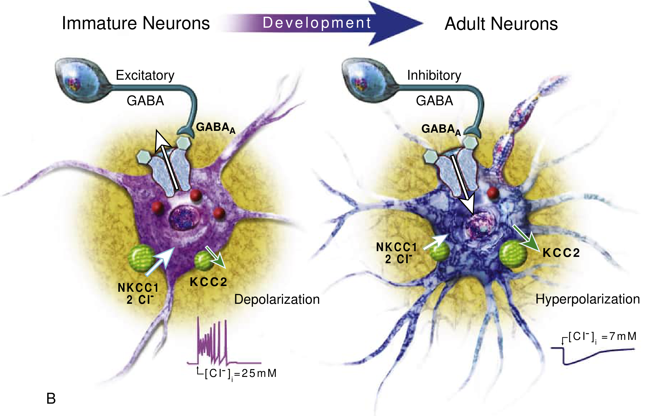
The neonatal brain has a pro-excitatory state due to immature ion transport:
| Feature | Immature Neuron | Mature Neuron |
|---|---|---|
| GABA-A receptor effect | Depolarization (excitatory!) | Hyperpolarization (inhibitory) |
| Cl- transporter dominant | NKCC1 (Cl- influx - high intracellular Cl-) | KCC2 (Cl- efflux) |
| Net effect | Cl- flows OUT on GABA-A activation | Cl- flows IN |
Why phenobarbital works poorly: It is a GABA-agonist, but in neonates GABA is actually EXCITATORY - hence the reduced response to phenobarbital.
3. ETIOLOGY BY DAY OF PRESENTATION
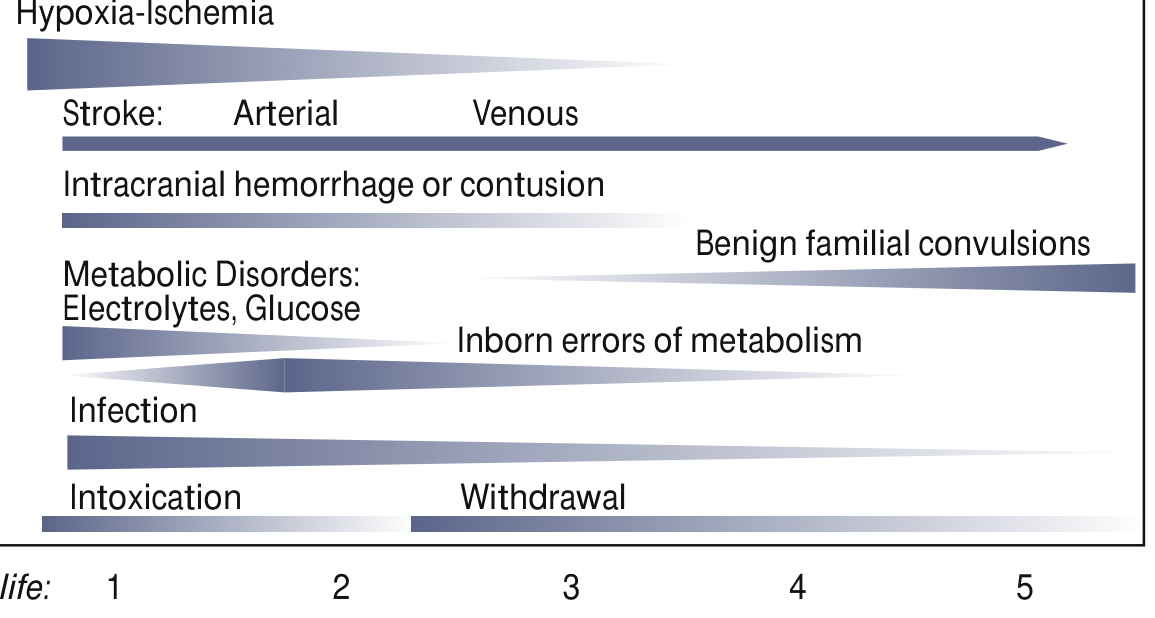
This is a HIGH-YIELD quiz topic!
| Day | Common Causes |
|---|---|
| Day 1 | Hypoxic-Ischemic Encephalopathy (HIE) - MOST COMMON OVERALL, Intracranial hemorrhage, Hypoglycemia, Hypo/hypernatremia, Intoxication |
| Day 1-3 | Metabolic (hypoglycemia, hypocalcemia, hypomagnesemia), Infection (meningitis), Stroke (arterial > venous) |
| Day 3-7 | Infection, Inborn errors of metabolism (IEM), Drug withdrawal |
| After Day 7 | Benign familial neonatal convulsions, Pyridoxine deficiency, IEM |
Mnemonic for Causes: "HIDE MAST"
- H - Hypoxic-Ischemic Encephalopathy (most common, ~50%)
- I - Intracranial hemorrhage (IVH, SDH, SAH)
- D - Drug withdrawal / intoxication
- E - Electrolyte abnormalities (hypocalcemia, hypomagnesemia, hyponatremia)
- M - Metabolic (hypoglycemia, IEM, pyridoxine deficiency)
- A - Anomalies of brain (cortical dysplasia, lissencephaly)
- S - Sepsis / Meningitis (bacterial, viral - HSV)
- T - Trauma (birth injury)
4. CLINICAL CLASSIFICATION (Volpe's Classification)
| Type | Features | Notes |
|---|---|---|
| Subtle (most common) | Eye deviation, blinking, fixed stare; repetitive mouth/tongue movements; apnea; bicycling/rowing limb movements | Most common in preterms; may NOT have EEG correlate |
| Clonic (focal/multifocal) | Rhythmic movements of muscle groups; slow clonic jerks (1-3/sec) | Suggests focal pathology (e.g., focal infarct) |
| Tonic (focal/generalized) | Sustained flexion or extension | Generalized tonic = bad prognosis; may be brainstem release phenomenon |
| Myoclonic (focal/multifocal/generalized) | Single or multiple rapid jerks | Generalized myoclonic = worst prognosis (Ohtahara syndrome) |
Key fact: Generalized tonic-clonic seizures as seen in older children are NOT seen in neonates due to immature synaptic connections.
5. JITTERINESS vs SEIZURES (High-yield comparison!)
| Feature | Jitteriness | Seizure |
|---|---|---|
| Stimulus-sensitive | YES | NO |
| Suppressible by holding/flexing | YES | NO |
| Eye deviation | NO | YES (common) |
| Autonomic changes | NO | YES |
| EEG abnormal | NO | YES |
| Dominant movement | Tremor (equal amplitude) | Clonic (fast + slow phase) |
| Consciousness | Alert | May be altered |
6. DIAGNOSIS
EEG (Gold Standard)
- Continuous conventional EEG is the gold standard (recommended by American Clinical Neurophysiology Society)
- Many neonatal seizures are subclinical - only 1/3 of EEG seizures have clinical signs
- Only 1/4 of clinically suspected seizures have EEG confirmation
- aEEG (amplitude-integrated EEG): Simplified bedside monitoring; less sensitive but useful when conventional EEG unavailable
Workup
- Bedside: Blood glucose, electrolytes (Na, Ca, Mg), blood gas
- Blood: CBC, culture, LFTs, metabolic screen (ammonia, lactate, amino acids)
- CSF: LP for meningitis/encephalitis (HSV PCR)
- Imaging: Cranial ultrasound (IVH), MRI brain (preferred - shows HIE, stroke, malformations)
- EEG: Continuous monitoring
7. MANAGEMENT
Step 1: Correct Underlying Cause First!
- Hypoglycemia: 10% dextrose 2 mL/kg IV bolus
- Hypocalcemia: 10% calcium gluconate 2 mL/kg IV slowly
- Hypomagnesemia: MgSO4 0.2 mL/kg of 50% solution IM
- Pyridoxine deficiency: Pyridoxine 100 mg IV (diagnostic and therapeutic)
Step 2: Antiepileptic Drugs
| Step | Drug | Dose |
|---|---|---|
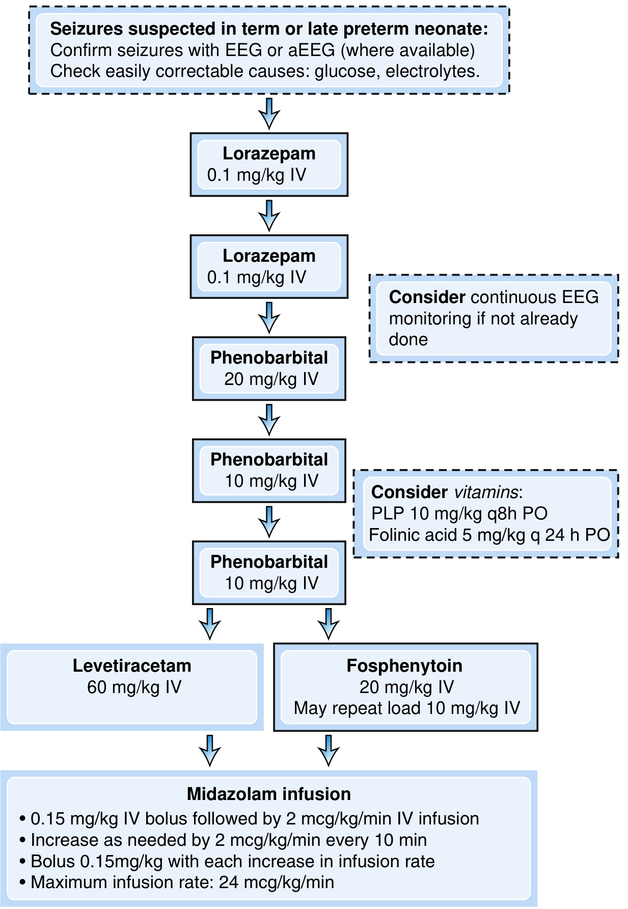
| First line | Phenobarbital | 20 mg/kg IV (can repeat 10 mg/kg x2, max 40-50 mg/kg) |
| Second line | Phenytoin / Fosphenytoin | 20 mg/kg IV (fosphenytoin preferred - fewer cardiac SE) |
| Alternative 2nd | Levetiracetam | 40-60 mg/kg IV |
| Third line | Midazolam infusion | 0.15 mg/kg bolus then 2 mcg/kg/min; max 24 mcg/kg/min |
| Metabolic seizures | Lorazepam | 0.1 mg/kg IV (benzodiazepine for acute control) |
Per OP Ghai / standard Indian pediatric practice:
- Phenobarbital remains the first-line drug in India
- Pyridoxine 100 mg IV should be given empirically if seizures are refractory (pyridoxine-dependent epilepsy)
8. NEONATAL EPILEPSY SYNDROMES
| Syndrome | Features |
|---|---|
| Benign Familial Neonatal Seizures | Autosomal dominant (KCNQ2/KCNQ3 gene); onset day 2-3; resolves by 6 weeks; good prognosis |
| Benign Neonatal Seizures (5th day fits) | Day 4-6 of life; clonic; no family history; resolves spontaneously; good prognosis |
| Ohtahara Syndrome | Suppression-burst EEG; onset in first weeks; tonic spasms; very poor prognosis |
| Early Myoclonic Encephalopathy | Erratic myoclonus + suppression-burst; metabolic causes; poor prognosis |
| Pyridoxine-Dependent Epilepsy | Refractory seizures; responds to pyridoxine 100 mg IV; mutation in ALDH7A1 gene |
9. PROGNOSIS
- Prognosis is primarily determined by the underlying etiology
- Of survivors with neonatal seizures: 25-70% develop subsequent epilepsy
- HIE - moderate mortality; high risk of neurodevelopmental disability
- Metabolic causes (hypocalcemia, hypoglycemia corrected promptly) - good prognosis
- Structural causes / Ohtahara / EME - poor prognosis
- Long-term risks: cerebral palsy, cognitive impairment, epilepsy
10. QUICK REVISION BULLETS (for last-minute quiz prep)
- Most common cause = HIE (~50%)
- Most common seizure type = Subtle (especially in preterms)
- Clonic seizures - most specific for focal pathology
- EEG is the gold standard - many seizures are subclinical
- Phenobarbital works poorly because GABA is excitatory in neonates (NKCC1 > KCC2)
- "5th day fits" = benign neonatal convulsions on day 4-6
- First line drug in India = Phenobarbital 20 mg/kg IV
- Refractory seizures - always trial Pyridoxine 100 mg IV
- Jitteriness is stimulus-sensitive + suppressible - seizures are NOT
- HSV encephalitis must be considered and acyclovir started empirically in CNS infection
Sources: Bradley & Daroff's Neurology in Clinical Practice (Neonatal Neurology, Ch. 110); Harrison's Principles of Internal Medicine 22e; OP Ghai Essential Pediatrics principles applied throughout

IMG_6770.jpg
Answer them i have class on them today
analyse_media
Your SGD covers two PEs today:
- PE 20.15 - Neonatal Seizures (already done above!)
- PE 20.20 - Common Surgical Conditions in Newborn: TEF, Oesophageal Atresia, Anal Atresia, Cleft Lip & Palate, CDH, Cause Abdomen (Prune Belly)
Let me now pull all the surgical conditions from the textbook library at once.
Finding Sources
Finding Sources
Finding Sources
Finding Sources
Reading File
Reading File
Reading File
Reading File
Reading File
Reading File
Finding Sources
Reading File
Reading File
Finding Sources
Loading Image
Reading File
Reading File
All content gathered. Now compiling the full SGD notes:
SGD 13 - PE 20.20: Surgical Conditions in Newborn
Date: 01-07-2026 | Complete Class Notes
1. TRACHEO-ESOPHAGEAL FISTULA (TEF) & ESOPHAGEAL ATRESIA (EA)
Definition
- EA = Congenital interruption of esophageal continuity -> proximal esophageal obstruction
- TEF = Pathological communication between esophagus and trachea
- Incidence: 1 in 1500-3000 live births; mild male predilection
Embryology
- 4th gestational week: Incomplete division of esophagotracheal diverticulum of foregut
- Normally the foregut divides into trachea (anterior) and esophagus (posterior)
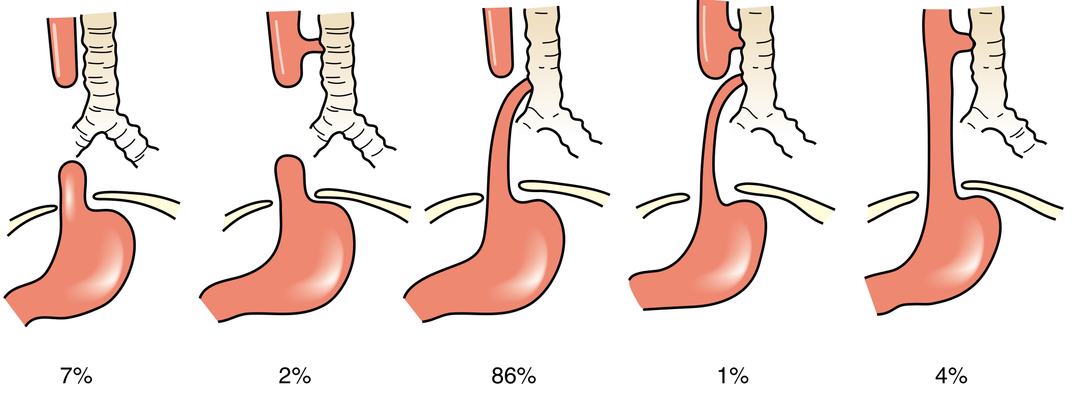
Gross Types (Vogt / Waterston Classification)
| Type | Description | Incidence |
|---|---|---|
| A | Pure EA - no fistula; both blind pouches | 7% |
| B | EA + proximal TEF | 2% |
| C (MOST COMMON) | Proximal EA + distal TEF | 86% |
| D | EA + both proximal and distal TEF | 1% |
| E (H-type) | TEF without EA (H-fistula) | 4% |
Memory trick: "C is the Common one = Coded 86%"
Associated Anomalies - VACTERL (in 60-70%)
- V - Vertebral
- A - Anorectal malformations
- C - Cardiac (most common associated anomaly - ~35%)
- T - Tracheal (tracheomalacia)
- E - Esophageal (the condition itself)
- R - Renal
- L - Limb (radial)
Clinical Features
- Maternal polyhydramnios (fetus can't swallow amniotic fluid - esp. isolated EA 86%)
- Excessive salivation / frothing at mouth from birth
- Choking, coughing, cyanosis on first feed
- Respiratory distress - aspiration pneumonia
- Scaphoid abdomen - in pure EA (no gas in abdomen)
- Gaseous distension of abdomen - in EA + distal TEF (air enters through fistula)
- H-type fistula: recurrent pneumonia, chronic cough - may present late
Key Diagnostic Sign
- Failure to pass nasogastric/orogastric tube (coils back at thoracic inlet)
- CXR showing the tube curled up in proximal blind pouch
- Air in abdomen on X-ray = confirms distal TEF
- No air in abdomen = pure EA (isolated)
- CONTRAINDICATED: Oral contrast study (aspiration risk!)
Management
Immediate (Preoperative):
- Replogle tube (double-lumen sump catheter) in upper pouch for continuous suction
- Position: Upright prone position (prevents aspiration)
- IV antibiotics (broad-spectrum)
- IV fluids - NPO
- Echocardiography + renal ultrasound (VACTERL workup)
- Avoid intubation if possible (positive pressure -> air into GI tract via TEF)
Surgical (Definitive):
- Primary repair: Right thoracotomy with extrapleural dissection, division of TEF, primary end-to-end esophageal anastomosis
- Minimally invasive: Thoracoscopic approach now preferred
- Long-gap EA: Staged repair; gastrostomy at birth, anastomosis at 4-6 months
- H-type fistula: Cervical approach for fistula ligation
2. CONGENITAL DIAPHRAGMATIC HERNIA (CDH)
Definition & Incidence
- Developmental diaphragmatic defect with herniation of abdominal contents into thoracic cavity
- Incidence: 1 in 2000-5000 births
- Disrupts lung and pulmonary vascular development
Types
| Type | Location | Frequency |
|---|---|---|
| Bochdalek (posterolateral) | Left side 85%, Right 13%, Bilateral 2% | 70-75% |
| Morgagni (anterior/retrosternal) | Right side mostly | 23-28% |
| Central | Rare | 2-7% |
Bochdalek = most common = Back/Posterior/Left
Pathophysiology
- Diaphragm develops from septum transversum + pleuroperitoneal folds - complete by 9 weeks
- Incomplete fusion -> defect -> bowel/organs herniate into thorax
- Pulmonary hypoplasia (both lungs affected, ipsilateral more severe)
- Pulmonary hypertension - thick arteriolar smooth muscle -> R-L shunting
Clinical Features
- Prenatal: Polyhydramnios, mediastinal shift on antenatal ultrasound, gastric bubble in chest
- At birth: Respiratory distress - grunting, dyspnea, retractions, cyanosis
- Classic triad on examination:
- Respiratory distress from birth
- Scaphoid (sunken) abdomen (bowel is in chest)
- Bowel sounds in the chest + Absent breath sounds on affected side
- Heart displaced to opposite side (R-side for L-CDH)
- Barrel-shaped chest
Investigations
- CXR: Bowel loops/gaseous shadows in chest + mediastinal shift (DIAGNOSTIC)
- Pulse oximetry: Pre-ductal vs post-ductal SpO2 difference (R-to-L shunting)
- Echo: Assess pulmonary hypertension and cardiac anomalies
- LHR (Lung-Head Ratio): Fetal sonographic prognostic marker
Management
Initial Stabilization (NOT emergency surgery!):
- Intubate and ventilate - "GENTLE ventilation" (permissive hypercapnia, avoid barotrauma)
- NGT for GI decompression
- Nitric oxide (iNO) - for pulmonary hypertension
- ECMO - if refractory respiratory failure
- Avoid mask ventilation (inflates bowel in chest)
- Agents: sildenafil, prostacyclin (PGI2), milrinone for pulmonary HTN
Surgical (once stabilized):
- Timing: Delayed repair (after stabilization) - NOT immediate emergency
- Approach: Subcostal/laparotomy; thoracoscopic repair also done
- Bochdalek: Primary repair if small; patch repair if large defect
- Survival: 65-90% in high-volume centers
Fetal intervention:
- FETO (Fetal Endoluminal Tracheal Occlusion): Balloon placed at 27-29 weeks -> lung growth by retention of lung fluid
3. ANAL ATRESIA / ANORECTAL MALFORMATIONS (ARM)
Definition & Incidence
- Failure of urorectal septum to divide the cloaca normally
- Incidence: 1 in 5000 live births; male predominance (58%)
Embryology
- 6th week: Urorectal septum descends to divide cloaca into:
- Anterior: urogenital sinus
- Posterior: anorectal canal
- Failure -> fistula between bowel and urinary tract (males) or introitus (females)
- Failure of anal membrane resorption -> anal stenosis or membrane
Classification (Wingspread / Peña)
LOW defects (below levator ani = GOOD prognosis):
- Perineal (cutaneous) fistula - most common low defect
- Anal stenosis
- Anal membrane
HIGH defects (above levator ani = WORSE prognosis):
| Males | Females |
|---|---|
| Rectourethral fistula (bulbar/prostatic) | Rectovestibular fistula |
| Recto-bladder neck fistula | Cloaca (rectum + vagina + urethra = single channel) |
| No fistula | No fistula |
Cloaca = single perineal opening in females (rectum + vagina + urethra fused)
Clinical Features
- Absent/abnormal anus on perineal inspection at birth
- No meconium passage in 24-48 hours
- Abdominal distension
- Meconium visible in urine (rectourinary fistula - males)
- Meconium at introitus/vestibule (females)
- Perineal fistula - meconium on perineum
Associated Anomalies - VACTERL (25-60% GU anomalies)
- Higher the defect = more associated anomalies
- Recto-bladder fistula / cloaca = 90% GU anomalies
- Low (perineal) fistula = only 10% GU anomalies
- Also: sacral anomalies (Currarino triad), spinal defects, cardiac, vertebral
Investigations
- Invertogram (Wangensteen-Rice): Lateral X-ray with baby inverted - gas bubble to skin distance (now rarely used)
- Distal colostogram (after colostomy) - gold standard to define fistula anatomy
- Renal ultrasound + VCUG
- Spinal ultrasound/MRI (sacral/spinal anomalies)
- Echocardiography
Management
LOW defects:
- Primary single-stage repair without colostomy
- Cutback anoplasty or limited PSARP (Posterior Sagittal Anorectoplasty)
- Anal stenosis: Serial dilatations
HIGH defects - THREE-STAGE approach:
- Stage 1 (Newborn): Divided loop sigmoid colostomy with mucous fistula
- Stage 2 (3-6 months): PSARP - Posterior Sagittal Anorectoplasty (Peña procedure)
- Electrical stimulation to identify sphincter center
- Midline incision from coccyx to perineum
- Fistula division + rectal mobilization + muscle reconstruction
- Stage 3 (Several weeks after Stage 2): Colostomy closure
- Followed by anal dilations for months
Key: PSARP (Peña procedure) = gold standard for high ARM
4. CLEFT LIP AND PALATE
Embryology
- Cleft lip: Failure of maxillary prominence to fuse with merged medial nasal prominences (5th-7th week)
- Cleft palate: Failure of lateral palatine processes (palatal shelves) to fuse (7th-12th week)
- Incisive fossa = landmark dividing anterior (primary) and posterior (secondary) palate
Classification
| Type | Description |
|---|---|
| Anterior/Primary palate defect | Cleft lip ± cleft of alveolus up to incisive fossa |
| Posterior/Secondary palate defect | Cleft of soft + hard palate behind incisive fossa |
| Complete | Through lip, alveolus, hard + soft palate |
| Incomplete | Partial |
| Unilateral vs Bilateral |
Epidemiology
- Cleft lip ± palate: 1 in 1000 births; 60-80% are MALE
- Cleft palate alone: 1 in 2000 births; more common in females
- Most common craniofacial anomaly
Clinical Features
- Visible cleft of lip +/- palate at birth
- Feeding difficulties - inability to create suction (especially cleft palate)
- Regurgitation through nose during feeding
- Speech problems (hypernasal speech) - if palate involved
- Risk of otitis media (abnormal eustachian tube function)
- Recurrent respiratory infections
- Dental anomalies
Associated conditions
- Pierre Robin sequence: cleft palate + micrognathia + glossoptosis -> airway obstruction
- Can be part of syndromes: Treacher Collins, Van der Woude, Stickler syndrome
Management - Team Approach (Rule of 10s)
| Timing | Procedure |
|---|---|
| 10 weeks / 10 lbs / Hb 10 g/dL | Cleft lip repair (Millard rotation-advancement technique) |
| 18 months (before speech development) | Cleft palate repair (Palatoplasty / Von Langenbeck) |
| 5-6 years | Alveolar bone grafting |
| Adolescence | Rhinoplasty / jaw correction |
"Rule of 10" for cleft lip repair: 10 weeks old, 10 lbs weight, Hb 10 g/dL
Feeding:
- Special wide-base nipples / Haberman feeder
- Nasogastric tube if severe feeding difficulty
- Mother's milk is preferred
5. PRUNE BELLY SYNDROME (Eagle-Barrett Syndrome / Triad Syndrome)
Definition
Characterized by a TRIAD of:
- Deficiency/absence of anterior abdominal wall muscles (wrinkled, prune-like abdomen)
- Bilateral cryptorchidism (undescended testes - intra-abdominal)
- Urinary tract anomalies - bilateral hydroureteronephrosis, megacystis, vesicoureteral reflux
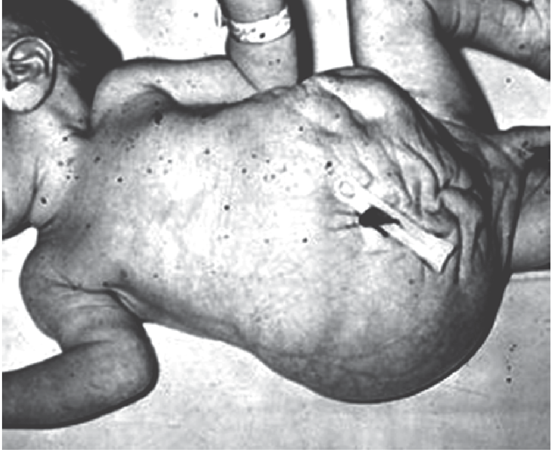
Epidemiology
- Almost exclusively in males (>95%)
- Females: urinary + abdominal wall defects but no cryptorchidism
- Rare: 1 in 35,000-50,000 births
Pathophysiology
- Likely due to fetal bladder outlet obstruction -> massive bladder distension -> compresses abdominal wall musculature -> prevents normal development
- Ureters: long, tortuous, dilated (mostly non-obstructive; due to decreased smooth muscle + increased collagen)
- ~80% have vesicoureteral reflux (VUR)
Clinical Features
- Wrinkled, lax, prune-like abdomen (abdominal wall is thin/deficient)
- Bilateral undescended testes (intra-abdominal)
- Dilated urinary tract (hydroureteronephrosis, megacystis)
- Respiratory difficulties (abdominal wall weakness affects breathing)
- Constipation (poor abdominal musculature)
- Orthopaedic: clubfoot, hip dislocation (oligohydramnios effect)
- Cardiac anomalies in some
Management
- No universal surgery for ureters (rarely obstructive)
- Bilateral orchidopexy at 6-12 months (along with abdominal wall repair)
- Abdominoplasty (Monfort procedure) - transverse lower abdominal incision, excision of redundant skin
- VUR management: prophylactic antibiotics; ureteral reimplantation if severe
- Infertility is common despite orchidopexy (abnormal spermatogenesis, retrograde ejaculation)
- Renal failure risk: abnormal kidneys + persistent pyelonephritis
QUICK COMPARISON TABLE - All Conditions
| Feature | TEF/EA | CDH | Anal Atresia | Cleft Lip/Palate | Prune Belly |
|---|---|---|---|---|---|
| Incidence | 1:1500-3000 | 1:2000-5000 | 1:5000 | 1:1000 | 1:35,000 |
| Key clinical sign | Can't pass NG tube | Scaphoid abdomen + bowel sounds in chest | Absent/abnormal anus | Visible facial cleft | Prune-like abdomen |
| Maternal history | Polyhydramnios | Polyhydramnios | - | - | Oligohydramnios |
| Associated syndrome | VACTERL | - | VACTERL | Pierre Robin | - |
| First surgical step | Replogle suction | Stabilize first | Colostomy (high) | Feeding support | Orchidopexy |
| Definitive surgery | Right thoracotomy + anastomosis | Subcostal repair | PSARP (Peña) | Millard repair at 10 wks | Abdominoplasty |
ALSO RECALL FROM PE 20.15 - NEONATAL SEIZURES (previously covered):
- Most common cause: HIE (~50%)
- Most common type: Subtle seizures
- First-line drug: Phenobarbital 20 mg/kg IV
- Why phenobarbital works poorly: GABA is excitatory in neonates (NKCC1 > KCC2)
- Jitteriness vs Seizure: Jitteriness is stimulus-sensitive + suppressible
- EEG = Gold Standard
- Refractory seizures: Trial Pyridoxine 100 mg IV
Sources: Sabiston Textbook of Surgery; Schwartz's Principles of Surgery; Bailey & Love's Short Practice of Surgery; The Developing Human - Clinically Oriented Embryology; Bradley & Daroff's Neurology in Clinical Practice
---Your complete SGD 13 notes are ready! Here's a quick orientation to what's covered:
What Your SGD Covers Today (PE 20.15 + PE 20.20):
PE 20.15 - Neonatal Seizures (covered in the previous message above)
PE 20.20 - 5 Surgical Conditions in Newborn:
| Condition | Must-Know High-Yield Points |
|---|---|
| TEF / EA | Type C = 86% (proximal EA + distal TEF); VACTERL; NG tube fails; CXR diagnosis; Replogle tube first; Right thoracotomy |
| CDH | Bochdalek = 70-75%, Left 85%; Scaphoid abdomen + bowel sounds in chest; Stabilize FIRST (not emergency surgery); iNO + gentle ventilation |
| Anal Atresia | 1:5000; PSARP (Peña) = gold standard; Low = primary repair; High = 3-stage (colostomy → PSARP → closure) |
| Cleft Lip & Palate | 1:1000; Males 60-80%; Rule of 10 for lip repair; Palatoplasty at 18 months; Pierre Robin sequence |
| Prune Belly | Triad = absent abdominal muscles + bilateral cryptorchidism + urinary anomalies; Almost all males; Orchidopexy + abdominoplasty |
Good luck in your SGD today! All the best for your class with Dr Satya Kumari, Dr Ramanamalli, and Dr Vijayalakshmi.
This is a shared conversation. Sign in to Orris to start your own chat.