
Complete claw hand
complete claw hand ulnar median nerve palsy deformity
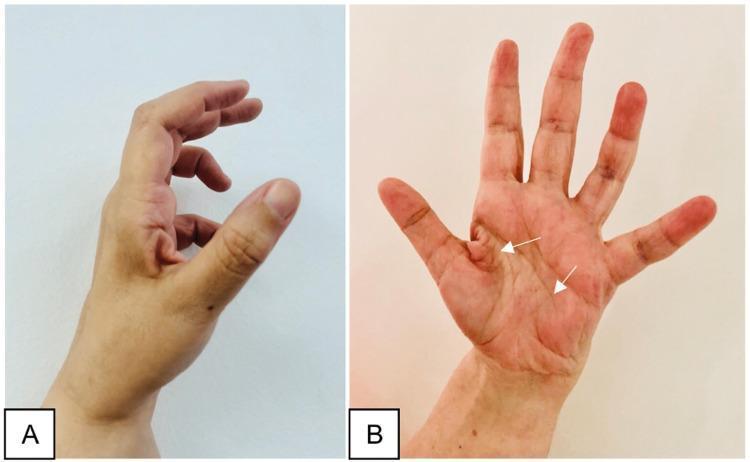
Two-panel clinical photograph demonstrating the late sequelae of a peripheral nerve injury in the hand, specifically illustrating features of ulnar nerve palsy. Image A (lateral view) shows a classic 'claw hand' deformity, characterized by hyperextension at the metacarpophalangeal (MCP) joints and flexion at the interphalangeal (IP) joints, most prominent in the ring and little fingers. The thumb appears adducted and flexed. Image B (anterior/palmar view) displays generalized muscle atrophy of the hand, with significant hollowing of the hypothenar eminence and interosseous spaces (indicated by white arrows). Notably, the thenar eminence remains relatively spared, suggesting the median nerve's innervation to the thenar muscles is intact. This combination of clawing and selective intrinsic muscle wasting is an important educational marker for localizing nerve lesions to the distal ulnar nerve (e.g., Guyon's canal or post-surgical complication). The images illustrate the clinical presentation of chronic motor deficit and the characteristic postural changes resulting from unbalanced muscle pull in the hand.
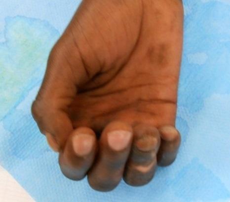
This clinical photograph displays a right hand exhibiting a classic ulnar claw deformity (griffe cubitale). The hallmark postural abnormality is evident in the 4th (ring) and 5th (little) fingers, which show hyperextension at the metacarpophalangeal (MCP) joints and marked flexion at the proximal interphalangeal (PIP) and distal interphalangeal (DIP) joints. This presentation is characteristic of ulnar nerve palsy, in this case, a sequela of leprosy. The palm shows hyperpigmented skin patches and induration of the integument, which are common dermatological manifestations of Hansen's disease. The thumb, index, and middle fingers appear relatively spared from the clawing posture, typical of a distal ulnar nerve lesion where the lumbricals of the first two fingers (innervated by the median nerve) remain functional. The image serves as an educational example of the neurological and musculoskeletal impact of chronic leprosy on hand function and morphology.
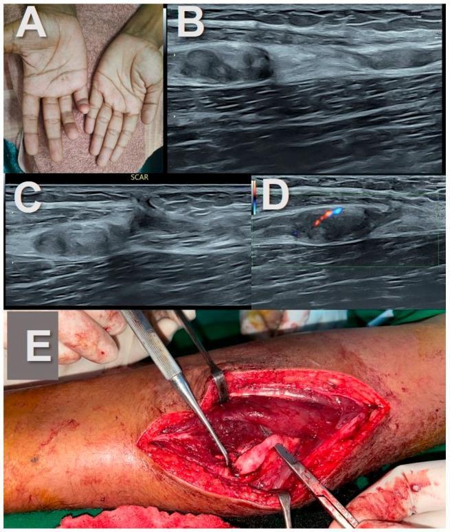
This composite educational image illustrates the clinical, sonographic, and surgical findings of ulnar nerve stump neuromas following traumatic transection. Panel A (Clinical Photograph): Demonstrates a 'claw hand' deformity involving the right 4th and 5th digits, a classic sign of distal ulnar nerve palsy. Panels B and C (Diagnostic Ultrasound): High-resolution longitudinal B-mode ultrasound images showing complete nerve transection. The proximal and distal nerve ends exhibit bulbous, hypoechoic, mass-like enlargements consistent with stump neuromas. A gap between the nerve ends is filled with hyperechoic adipose tissue in the intermuscular plane. Panel D (Power Doppler): Shows mild internal vascularity within the hypoechoic neuroma, represented by color flow signals. Panel E (Intra-operative Photograph): A surgical view of the mid-forearm displaying the bulbous, pale neural tissue during excision or repair. This sequence highlights the diagnostic pathway from physical examination to radiologic confirmation and surgical management of traumatic peripheral nerve injuries. Key concepts include post-traumatic neuroma formation, sonographic assessment of nerve continuity, and the 'split fat sign' in intermuscular neural lesions.
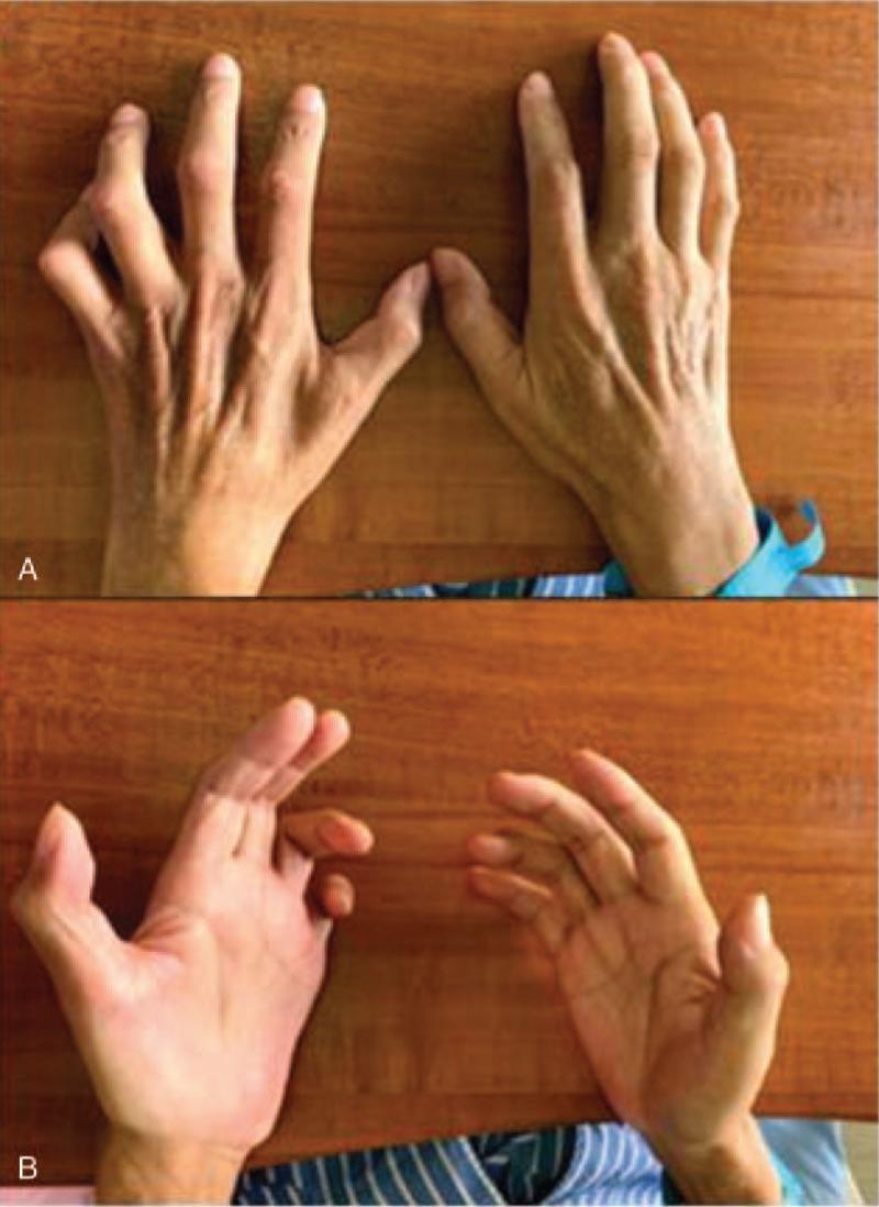
Clinical photograph of bilateral hands from a dorsal (A) and palmar (B) perspective, demonstrating characteristic features of ulnar nerve palsy. The primary visual finding is a 'claw hand' or 'benediction hand' deformity, most prominent on the left side, characterized by hyperextension at the metacarpophalangeal (MCP) joints and flexion at the proximal and distal interphalangeal (PIP and DIP) joints of the fourth and fifth digits. There is marked atrophy of the intrinsic hand muscles, specifically visible as hollowing of the interosseous spaces on the dorsal aspect and flattening of the hypothenar and thenar eminences on the palmar aspect. The skin appears thin with prominent tendons and vasculature, indicating loss of muscle bulk. This presentation is consistent with severe, chronic ulnar neuropathy, illustrating the loss of lumbrical and interossei muscle function. Educational concepts include the 'ulnar paradox' where distal lesions may appear more severe and the clinical manifestation of McGowan classification stages of nerve injury.
Complete Claw Hand
Definition
- Hyperextension at the metacarpophalangeal (MCP) joints
- Flexion at the proximal and distal interphalangeal (PIP and DIP) joints
Why "Complete" vs. "Partial/Ulnar" Claw?
| Feature | Partial (Ulnar) Claw | Complete Claw |
|---|---|---|
| Fingers affected | Ring + little only | All four fingers |
| Nerve(s) involved | Ulnar nerve alone | Ulnar + median nerve |
| Lumbricals affected | 3rd and 4th (ulnar-innervated) | All four lumbricals |
| Cause example | Isolated ulnar nerve injury | Combined nerve palsy, leprosy |
Anatomy and Mechanism
- Flex the MCP joints
- Extend the IP joints
- Lateral two lumbricals (1st and 2nd, for index and middle fingers) - innervated by the median nerve
- Medial two lumbricals (3rd and 4th, for ring and little fingers) - innervated by the ulnar nerve
- Ulnar nerve palsy alone → only ring and little fingers claw (partial/ulnar claw), because the median-innervated 1st and 2nd lumbricals partially compensate for the index and middle fingers
- Combined ulnar + median nerve palsy → all four lumbricals are lost → complete claw hand in all fingers
The Ulnar Paradox
- In a proximal ulnar nerve lesion (above the elbow, e.g., medial epicondyle fracture), the flexor digitorum profundus (FDP) to ring and little fingers is also paralyzed. Since there is no strong FDP to flex the IPs, the clawing of ring and little fingers is less pronounced than in a distal lesion.
- In a distal ulnar nerve lesion (e.g., at the wrist/Guyon's canal), FDP is intact, so it actively flexes the IPs and the claw is more marked.
Causes of Complete Claw Hand
- Combined median + ulnar nerve palsy - trauma, lacerations, fractures
- Leprosy - the classic cause; Mycobacterium leprae has a predilection for peripheral nerves, affecting both ulnar and median nerves, producing bilateral complete claw hand
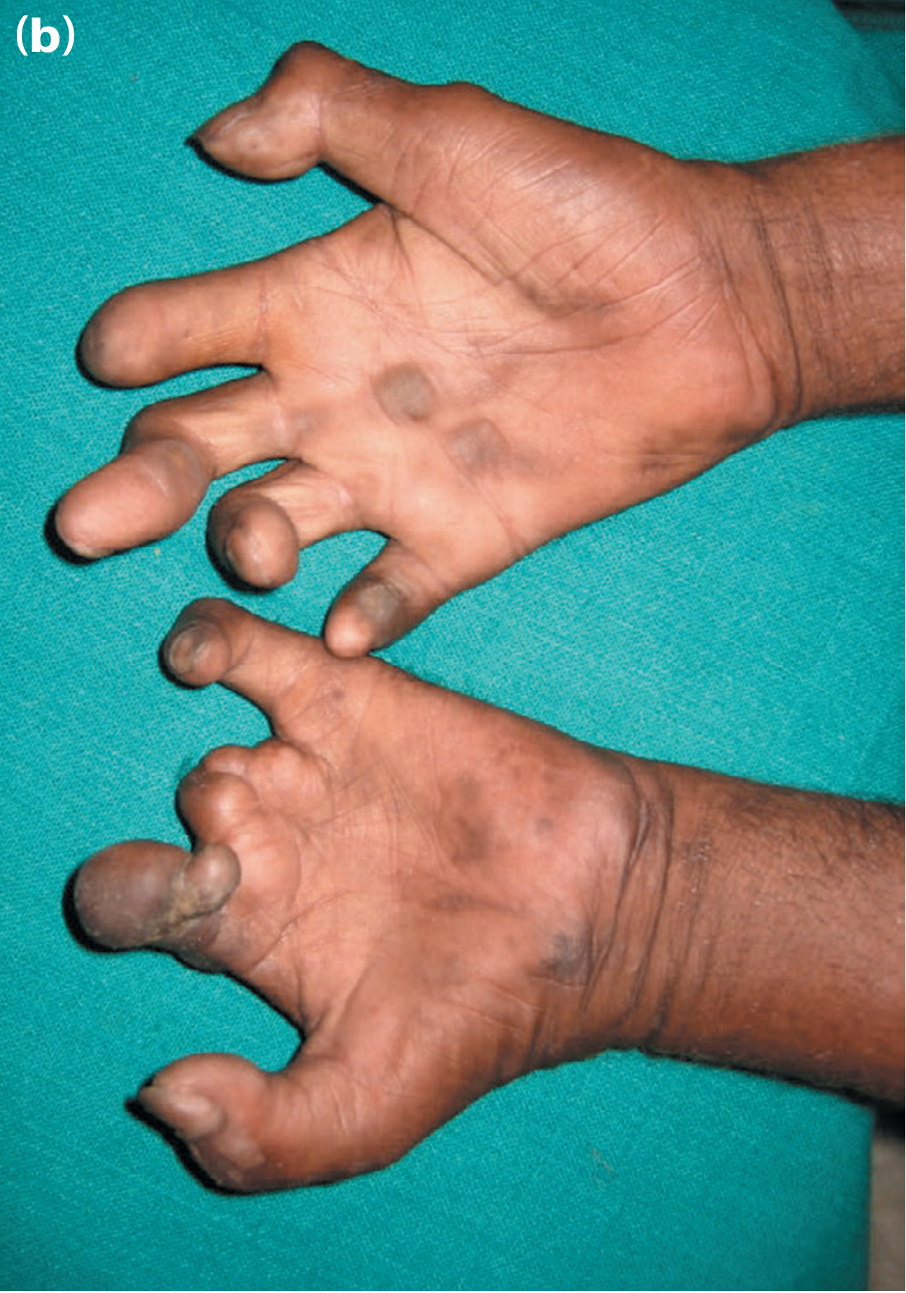
- Ischemic monomelic neuropathy - after arteriovenous fistula creation, can affect radial, median, and ulnar nerves simultaneously, producing claw hand deformity
- Volkmann's ischemic contracture (in severe cases with combined nerve involvement)
Additional Features
- Muscle wasting: Loss of interossei leads to hollowing of interosseous spaces in the metacarpus (dorsal guttering)
- Hypothenar and thenar atrophy in combined nerve palsy
- Thumb deformity: Loss of adductor pollicis (ulnar) + thenar muscles (median) causes the thumb to be adducted and lose the ability to oppose
- Sensory loss: Distribution depends on which nerves are affected - in complete palsy, the entire palmar surface and multiple digits are affected
Management
- Splints to prevent fixed contracture (dynamic MCP flexion splints)
- Physiotherapy
- Aim to restore lumbrical function - replacing the lost intrinsic muscle action
- Techniques pioneered by Paul Brand (especially for leprosy-related claw hand)
- Common transfers: Flexor digitorum superficialis (FDS) slips routed via the lumbrical canal to act as a lumbrical substitute
Summary Table
| Feature | Detail |
|---|---|
| Deformity | MCP hyperextension + IP flexion, all 4 fingers |
| Nerves involved | Ulnar (medial 2 lumbricals) + Median (lateral 2 lumbricals) |
| Classic cause | Leprosy (bilateral), combined nerve injury |
| Key muscle lost | All four lumbricals + all interossei |
| Sensory loss | Extensive palmar and digital surface |
| Treatment | Tendon transfers to restore intrinsic function |
Intrinsic muscleof hand
intrinsic muscles of hand anatomy diagram thenar hypothenar lumbricals interossei
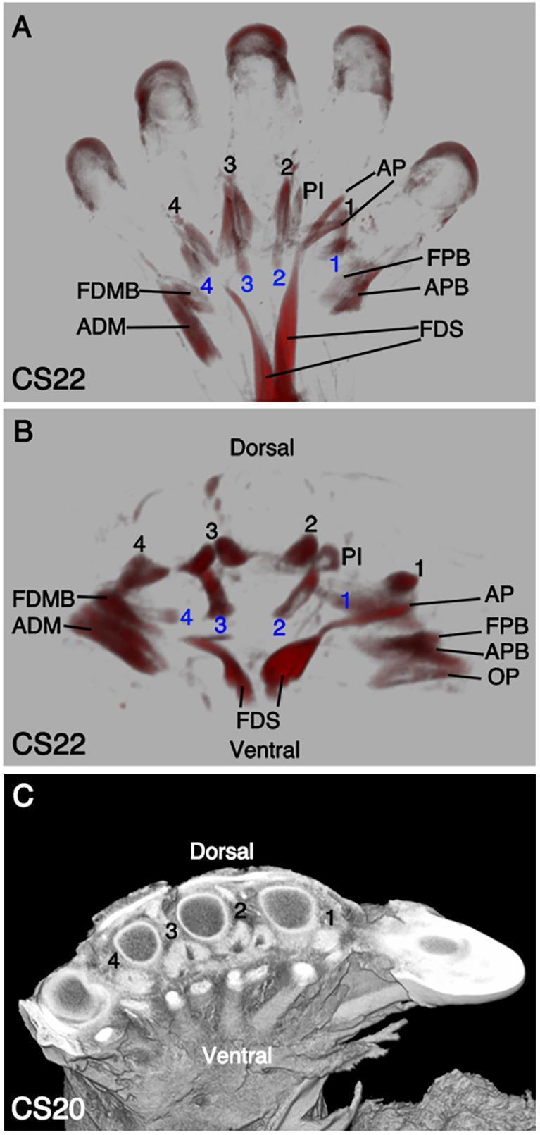
This composite educational graphic illustrates the embryonic development of human autopod (hand) musculature using Optical Projection Tomography (OPT) and high-resolution episcopic microscopy. Panel A displays a ventral view at Carnegie Stage 22 (CS22), highlighting the differentiation of intrinsic hand muscles. The thenar eminence (radial side) includes the flexor pollicis brevis (FPB), adductor pollicis (AP), and abductor pollicis brevis (APB), while the hypothenar eminence (ulnar side) shows the abductor digiti minimi (ADM) and flexor digiti minimi brevis (FDMB). The flexor digitorum superficialis (FDS) is visible with its proximal muscle belly and distal extensions. Panel B provides a distal-transverse view at CS22, showing the spatial arrangement of the four dorsal interossei (labeled 1-4 in black), palmar interossei (PI), and four lumbricals (labeled 1-4 in blue) relative to the ventral and dorsal surfaces. Panel C presents a CS20 cross-section demonstrating the anatomical relationship between developing metacarpal bones and surrounding soft tissue. This material serves as a reference for embryonic musculoskeletal development and the proximal translocation of extrinsic flexors.
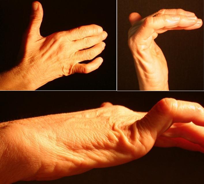
This composite of clinical photographs demonstrates a 'claw hand' (main en griffe) deformity, a characteristic sign of ulnar nerve neuropathy. The images show a right hand from dorsal, palmar, and lateral perspectives. Key visual features include hyperextension at the metacarpophalangeal (MCP) joints and flexion at the proximal and distal interphalangeal (IP) joints, most pronounced in the fourth and fifth digits. There is noticeable atrophy of the hypothenar eminence and hollowing of the interosseous spaces on the dorsal surface, indicating wasting of the intrinsic hand muscles (interossei and ulnar-innervated lumbricals). The thenar eminence appears relatively preserved, though subtle flattening of the adductor pollicis region may be present. This presentation is consistent with distal ulnar nerve compression, such as within Guyon’s canal, leading to motor deficit and muscle denervation. These clinical signs are essential for diagnosing peripheral nerve entrapment syndromes and distinguishing them from proximal lesions or central nervous system pathologies.
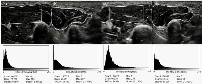
This diagnostic image displays four musculoskeletal ultrasound (US) frames and their corresponding grayscale intensity histograms, comparing the intrinsic hand muscles of a healthy control to a patient with moderate Carpal Tunnel Syndrome (CTS). Panels (a) and (c) depict the thenar muscle using a rectangular region of interest (ROI), while (b) and (d) show the hypothenar muscle with freehand traced ROIs. The visual echogenicity in the CTS patient's thenar muscle (c) appears heterogeneously increased compared to the healthy control (a). Quantitative analysis is provided through histograms below each US frame, showing pixel distribution across an intensity scale (0–255). Key metrics include Count, Mean, Standard Deviation (StdDev), and Mode. Notably, the thenar muscle in CTS (c) shows a significant rightward shift in its histogram, with a mean intensity of 40.839 compared to 20.262 in the control (a), indicating increased echo intensity (hyperechogenicity) which may correlate with denervation or fibrosis. The hypothenar histograms remain relatively similar between groups, serving as a comparative baseline. This clinical imaging demonstrates the use of quantitative grayscale analysis in evaluating muscle changes secondary to peripheral nerve compression.
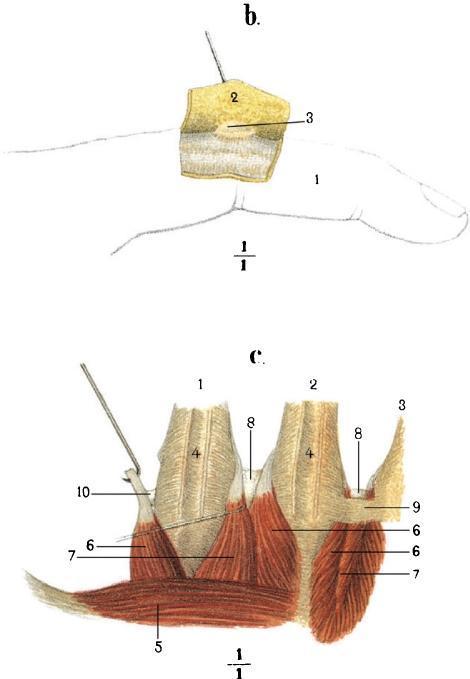
This historical anatomical illustration, likely from a 19th-century monograph, consists of two labeled diagrams (b and c) detailing the musculoskeletal and bursal anatomy of the hand. Diagram 'b' displays a lateral view of an index finger (1), with a flap of subcutaneous tissue (2) reflected upward by a surgical pin to reveal a subcutaneous mucous bursa (3) on the dorsal aspect. Diagram 'c' provides a deep dissection of the hand's dorsal metacarpal region. It illustrates the metacarpal bones II-IV (1-3) along with the associated dorsal interossei muscles (4, 6, 7) and the adductor pollicis (5). Key anatomical focuses include the intermetacarpophalangeal bursae (8) situated between the metacarpal heads, and specific tendinous or ligamentous attachments (9, 10) demonstrated via retraction. These illustrations serve as educational materials for understanding the spatial relationships between bones, intrinsic hand muscles, and the synovial bursae that facilitate movement in the digits and metacarpophalangeal joints.
Intrinsic Muscles of the Hand
- Thenar muscles (4)
- Hypothenar muscles (4)
- Lumbricals (4)
- Interossei (7 = 4 dorsal + 3 palmar)
1. THENAR MUSCLES
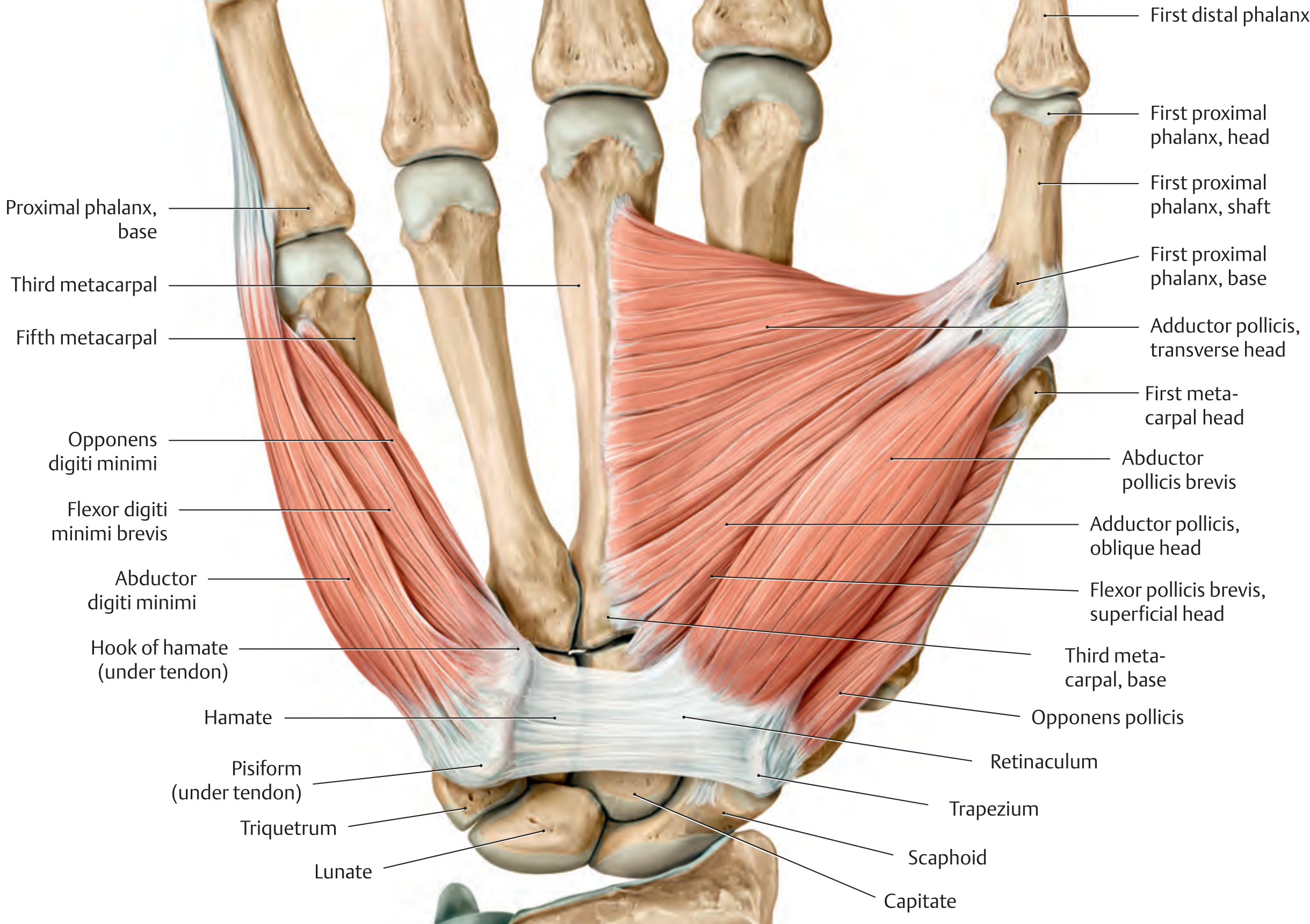
| Muscle | Origin | Insertion | Action | Nerve |
|---|---|---|---|---|
| Abductor pollicis brevis | Scaphoid, trapezium, flexor retinaculum | Base of proximal phalanx of thumb (via radial sesamoid) | Abduction of thumb | Median (C8, T1) |
| Flexor pollicis brevis | Superficial head: flexor retinaculum; Deep head: capitate, trapezium | Base of proximal phalanx of thumb (via radial sesamoid) | Flexion + opposition at CMC; flexion at MCP | Superficial: Median; Deep: Ulnar (C8, T1) |
| Opponens pollicis | Trapezium | Radial border of 1st metacarpal | Opposition at CMC joint | Median (C8, T1) |
| Adductor pollicis | Transverse head: palmar 3rd metacarpal; Oblique head: capitate, bases of 2nd & 3rd metacarpals | Base of proximal phalanx of thumb (via ulnar sesamoid) | Opposition at CMC; flexion at MCP | Ulnar (C8, T1) |
Memory tip - thenar muscles ("LOAF"): Lumbricals 1+2, Opponens pollicis, Abductor pollicis brevis, Flexor pollicis brevis = Median nerve. Adductor pollicis = Ulnar nerve.
2. HYPOTHENAR MUSCLES
| Muscle | Origin | Insertion | Action |
|---|---|---|---|
| Abductor digiti minimi | Pisiform | Ulnar base of proximal phalanx of 5th digit + dorsal digital expansion | MCP: flexion + abduction; PIP/DIP: extension |
| Flexor digiti minimi brevis | Hook of hamate, flexor retinaculum | Base of proximal phalanx of 5th digit | MCP: flexion |
| Opponens digiti minimi | Hook of hamate, flexor retinaculum | Ulnar border of 5th metacarpal | Draws 5th metacarpal palmarly (opposition) |
| Palmaris brevis | Ulnar border of palmar aponeurosis | Skin of hypothenar eminence | Tightens palmar aponeurosis (protective) |
3. LUMBRICALS (4 muscles)
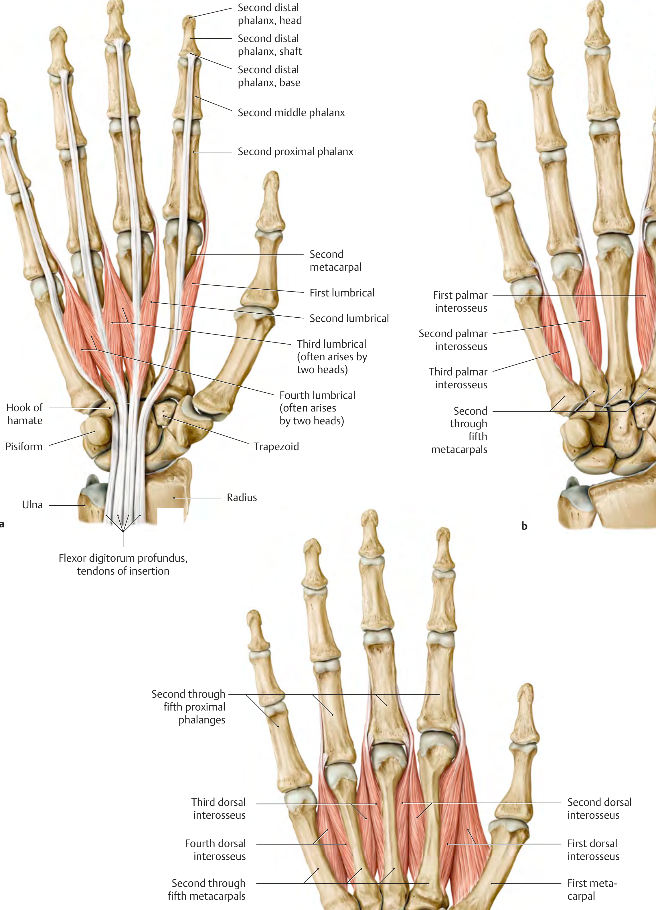
| Feature | Detail |
|---|---|
| Origin | Radial sides of the FDP tendons (1st & 2nd = unipennate; 3rd & 4th = bipennate) |
| Insertion | Dorsal digital expansion (extensor hood) of 2nd-5th digits |
| Action | Flex MCP joints (2nd-5th); Extend PIP and DIP joints |
| Nerve - 1st & 2nd | Median nerve (C8, T1) - for index and middle fingers |
| Nerve - 3rd & 4th | Ulnar nerve (C8, T1) - for ring and little fingers |
4. INTEROSSEI (7 muscles)
A. Dorsal Interossei (4) - "DAB"
| # | Origin | Insertion | Abducts |
|---|---|---|---|
| 1st | Adjacent sides of 1st and 2nd metacarpals | Radial side of 2nd proximal phalanx (index) | Index finger radially |
| 2nd | Adjacent sides of 2nd and 3rd metacarpals | Radial side of 3rd proximal phalanx (middle) | Middle finger radially |
| 3rd | Adjacent sides of 3rd and 4th metacarpals | Ulnar side of 3rd proximal phalanx (middle) | Middle finger ulnarly |
| 4th | Adjacent sides of 4th and 5th metacarpals | Ulnar side of 4th proximal phalanx (ring) | Ring finger ulnarly |
B. Palmar Interossei (3) - "PAD"
| # | Origin | Acts on |
|---|---|---|
| 1st | Ulnar side of 2nd metacarpal | Adducts index finger |
| 2nd | Radial side of 4th metacarpal | Adducts ring finger |
| 3rd | Radial side of 5th metacarpal | Adducts little finger |
Innervation Summary
| Nerve | Muscles |
|---|---|
| Median nerve (C8, T1) | Abductor pollicis brevis, Opponens pollicis, Flexor pollicis brevis (superficial head), Lumbricals 1 & 2 |
| Ulnar nerve (C8, T1) | Adductor pollicis, Flexor pollicis brevis (deep head), ALL hypothenar muscles, Lumbricals 3 & 4, ALL interossei (dorsal + palmar) |
Mnemonic - Ulnar nerve supplies: "All the little ones" - All interossei (7), Lumbricals 3 & 4, Hypothenar muscles, Adductor pollicis = LOAF muscles are median; everything else is ulnar.
Key Actions - The "Intrinsic Plus" Position
- MCP flexion (all joints)
- IP extension (PIP and DIP)
Compartments of the Hand
- Thenar compartment (3 thenar muscles)
- Adductor compartment (adductor pollicis alone)
- Hypothenar compartment (3 hypothenar muscles)
- Four dorsal interosseous compartments
- Three palmar interosseous compartments (sometimes grouped differently)
Horner syndrome
Horner syndrome ptosis miosis anhidrosis clinical photo
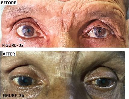
Two-panel clinical photograph displaying the 'Before' and 'After' ophthalmic findings in a patient with left-sided Horner's syndrome. The 'Before' image (Figure 3a) demonstrates classic clinical signs including left upper eyelid ptosis (drooping) and left-sided miosis (constricted pupil) compared to the right eye. The 'After' image (Figure 3b) illustrates the clinical response following three cycles of chemotherapy for an underlying superior sulcus tumor (Pancoast tumor). There is visible improvement in the left ocular presentation, characterized by the elevation of the left upper eyelid, reduction in ptosis severity, and a comparative increase in left pupil diameter (resolution of miosis). The image highlights the ocular manifestations of sympathetic nerve pathway disruption and its subsequent recovery during systemic treatment of the primary thoracic malignancy. Educational focus includes the recognition of Horner's syndrome triad (ptosis, miosis, and anhidrosis) and its clinical association with apical lung lesions.
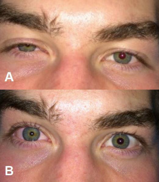
Two-panel clinical photograph (labeled A and B) demonstrating unilateral Horner's syndrome of the right eye following a nerve block procedure. Image A shows classic signs including significant ptosis (drooping of the upper eyelid), miosis (constriction of the pupil), and conjunctival injection (redness) of the right eye compared to the normal left eye. Image B captures the same patient with a higher upper eyelid position and larger pupil diameter in the right eye, illustrating the transition or comparison of clinical findings. The image serves as a teaching tool for recognizing the sympathetic nerve fiber disruption triad, typically presenting as ptosis, miosis, and anhidrosis. This specific case highlights iatrogenic Horner's syndrome as a potential complication of anesthesia/regional blocks involving the cervical plexus or stellate ganglion.
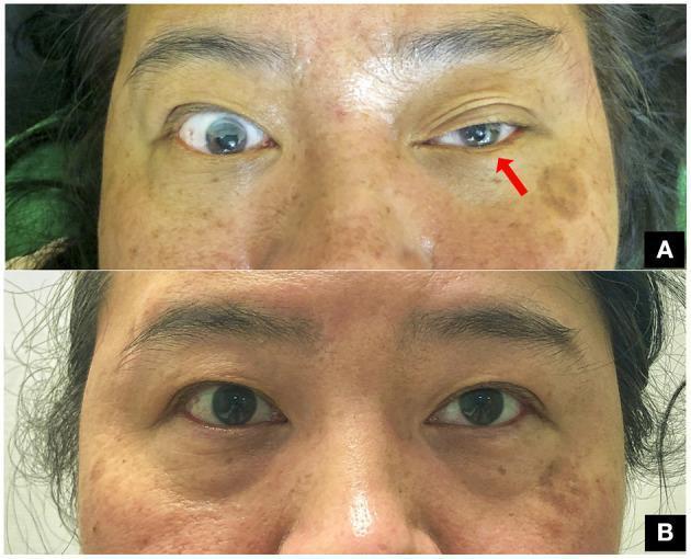
This clinical comparison photograph shows a 47-year-old woman presenting with classic oculofacial features of Horner’s syndrome following a clavicle fracture. Image A demonstrates the acute presentation of the oculosympathetic defect in the left eye, characterized by significant blepharoptosis (drooping of the upper eyelid) and ipsilateral miosis (constriction of the pupil) compared to the normal right eye. A red arrow points to the affected left eye to emphasize these findings. Image B shows the same patient at a three-month follow-up after surgical fixation of the fracture. It illustrates complete resolution of the syndrome, with symmetric eyelid positions and equal pupillary diameters (isocoria) in both eyes. This visual progression highlights the classic clinical triad (ptosis and miosis, with anhidrosis typically associated) and the potential for recovery following the management of the underlying traumatic compression of the preganglionic sympathetic pathway. The image is a valuable educational resource for neurology, ophthalmology, and trauma surgery.
Horner Syndrome
Definition
Classic Features (Triad)
| Sign | Mechanism | Muscle/Structure Affected |
|---|---|---|
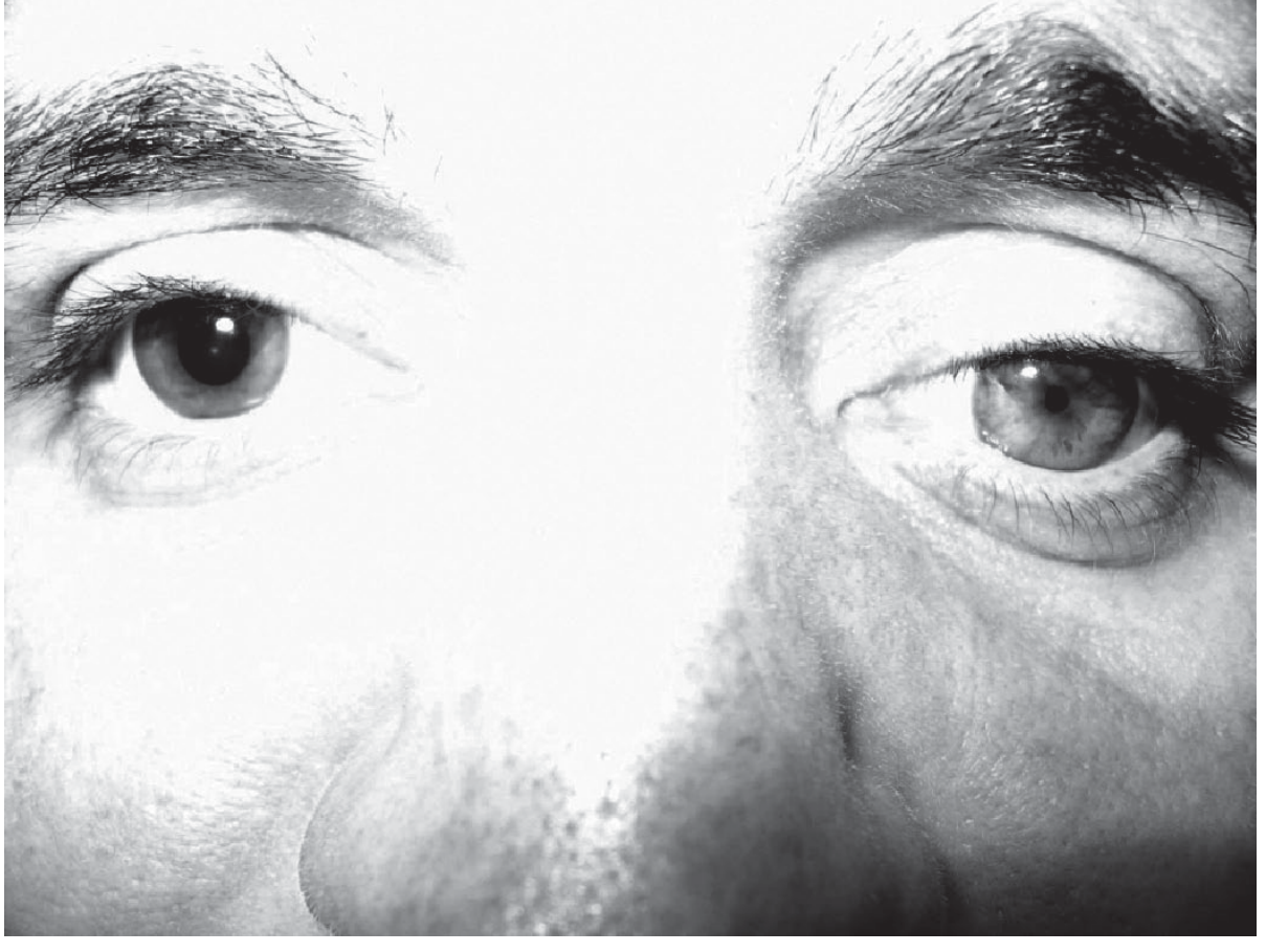
| Ptosis (partial) | Loss of Müller's smooth muscle innervation | Superior tarsal muscle (Müller's muscle) - elevates upper lid ~2mm |
| Miosis | Loss of pupillary dilator innervation | Dilator pupillae muscle |
| Anhidrosis | Loss of sympathetic innervation to sweat glands | Ipsilateral face (pattern depends on lesion level) |
- Apparent enophthalmos - actually an illusion created by narrowed palpebral fissure (the eyeball does not truly retract)
- Lower lid elevation (reverse ptosis / upside-down ptosis) - loss of inferior tarsal muscle
- Heterochromia iridis - depigmented (lighter) iris on affected side; classic in congenital cases
- Dilation lag - the miotic pupil dilates more slowly in the dark than the normal pupil
- Conjunctival hyperemia (transient, in acute phase)
The Three-Neuron Arc
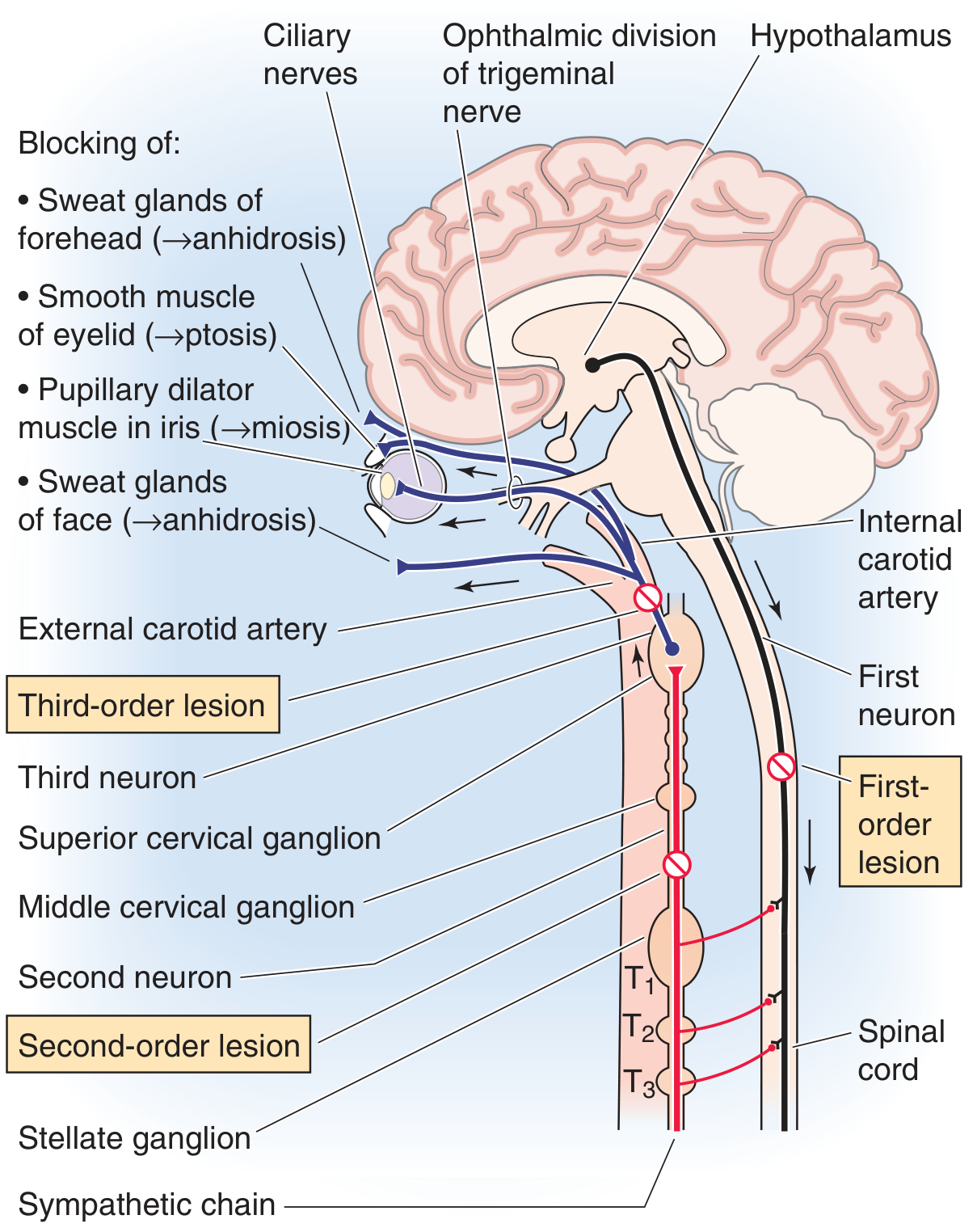
Neuron 1 - First Order (Central)
- Origin: Posterolateral hypothalamus
- Course: Descends ipsilaterally through brainstem tegmentum → lateral column of spinal cord → synapses at the ciliospinal center of Budge (C8-T2, mainly T1-T2)
- Lesion causes: Hypothalamic infarct, Wallenberg (lateral medullary) syndrome, spinal cord trauma, demyelination
Neuron 2 - Second Order (Preganglionic)
- Origin: Intermediolateral cell column (C8-T2)
- Course: Exits via ventral root → arches over the apex of the lung → ascends in the cervical sympathetic chain → synapses at the superior cervical ganglion
- Note: Sweating fibers for the face travel with the external carotid artery at this point
- Lesion causes: Pancoast (apical lung) tumor, cervical rib, thyroid surgery, neck trauma, aortic aneurysm
Neuron 3 - Third Order (Postganglionic)
- Origin: Superior cervical ganglion
- Course: Travels with the internal carotid artery → cavernous sinus → briefly with CN VI (abducens) → joins ophthalmic division of CN V → nasociliary branch → ciliary ganglion (passes through without synapsing) → reaches the eye via long ciliary nerves
- Lesion causes: Carotid artery dissection, cavernous sinus pathology (thrombosis, aneurysm, tumor), cluster headache, otitis media
Localization by Level
| Level | Anhidrosis Pattern | Key Associated Signs | Common Causes |
|---|---|---|---|
| 1st order (central) | Entire ipsilateral face and body | Contralateral hemiplegia, cerebellar signs, other brainstem signs | Wallenberg syndrome, spinal cord trauma, MS |
| 2nd order (preganglionic) | Entire ipsilateral face | Arm/chest pain (if Pancoast), brachial plexus signs | Pancoast tumor, cervical rib, post-thoracic surgery |
| 3rd order (postganglionic) | None or medial forehead/nose only (sweating fibers diverge with external carotid) | Ipsilateral headache (carotid dissection), CN VI palsy (cavernous sinus) | Carotid dissection, cavernous sinus lesion, cluster headache |
Key rule on anhidrosis: Lesions at or proximal to the common carotid bifurcation (1st and 2nd order) produce facial anhidrosis. Lesions distal to the bifurcation (3rd order) spare the face or only affect a small area, because sweat fiber supply travels with the external carotid, which branches off before the lesion. - Adams and Victor's Neurology
Pharmacological Testing
Step 1 - Confirm Horner: Apraclonidine (preferred) or Cocaine
- A weak direct alpha-1 agonist
- In normal eyes: no significant effect
- In Horner syndrome: due to denervation hypersensitivity, the miotic pupil dilates and the anisocoria reverses
- Works at any level of lesion (1st, 2nd, or 3rd order)
- Blocks norepinephrine reuptake → normal pupil dilates
- In Horner syndrome: pupil fails to dilate (regardless of lesion level) because norepinephrine is depleted at the synapse
- Anisocoria ≥0.8 mm = diagnostic
Step 2 - Localize: Hydroxyamphetamine 1% (Paredrine)
- Releases stored norepinephrine from intact nerve terminals
- Preganglionic (1st or 2nd order) Horner: 3rd-order neuron is intact → norepinephrine stored → pupil dilates
- Postganglionic (3rd order) Horner: 3rd-order neuron is damaged → no norepinephrine → pupil does not dilate
- Cannot differentiate 1st from 2nd order (no pharmacological test can)
Important Clinical Associations
| Cause | Clue |
|---|---|
| Pancoast tumor | 2nd-order; wasting of small muscles of hand (T1), arm pain, + chest X-ray finding at lung apex |
| Carotid artery dissection | 3rd-order; acute ipsilateral neck/face pain, pulsatile tinnitus, TIA risk - medical emergency |
| Wallenberg syndrome | 1st-order; vertigo, ipsilateral facial numbness, contralateral body pain/temp loss, dysphagia, ataxia |
| Cluster headache | 3rd-order; periodic severe unilateral headache, lacrimation, rhinorrhoea |
| Iatrogenic | After stellate ganglion block, internal jugular line, thyroid surgery |
| Congenital | Birth trauma to cervical sympathetics; heterochromia iridis; depigmented iris on affected side |
Key Differentials - Anisocoria
| Condition | Larger Pupil | Reactivity | Key Clue |
|---|---|---|---|
| Horner syndrome | Normal (contralateral) | Both react to light | Anisocoria worse in dark, dilation lag |
| CN III palsy | Abnormal (dilated) | Affected side does NOT react | Ptosis + ophthalmoplegia |
| Adie tonic pupil | Affected (dilated) | Poor to light, tonic to accommodation | Young women, absent ankle reflexes |
| Argyll Robertson | Bilateral small | No light reaction, accommodates | Neurosyphilis |
| Benign anisocoria | Variable | Both normal | Same asymmetry in all lighting |
Critical point: Horner anisocoria is more obvious in the dark (affected pupil fails to dilate), while CN III palsy anisocoria is more obvious in bright light (affected pupil fails to constrict). - Neuroanatomy through Clinical Cases, 3rd Ed.
Subacromial bursae
subacromial bursa anatomy shoulder rotator cuff diagram
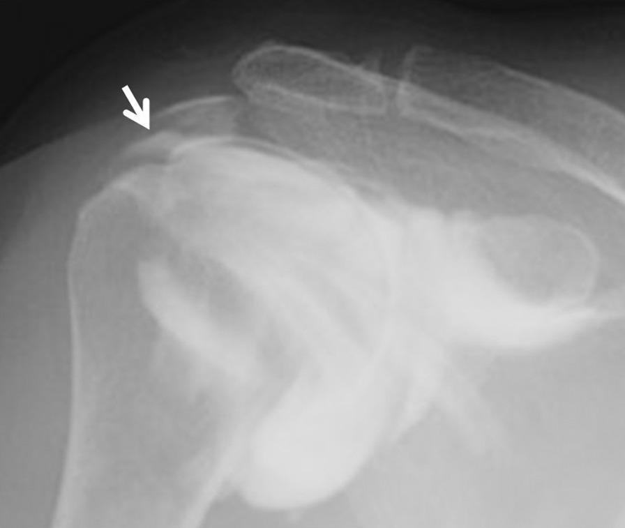
This diagnostic image is a subacromial bursography of the right shoulder, utilizing x-ray radiography with a contrast medium. The image depicts the shoulder joint anatomy, including the humeral head and the subacromial space. A key clinical finding is a localized focal pooling of radiopaque contrast medium within the tendon area of the rotator cuff, specifically indicated by a white arrow. This distribution of contrast indicates a bursal-side partial-thickness rotator cuff tear, likely involving the infraspinatus tendon. The contrast medium clearly delineates the soft tissue defect where the fluid has extravasated from the subacromial bursa into the tendon substance. This imaging modality is used here to identify pathology that may not be clearly visible on standard MRI, particularly for patients presenting with refractory shoulder impingement symptoms. The educational focus is on the diagnostic utility of bursography in identifying longitudinal bursal-side tears through characteristic contrast pooling.
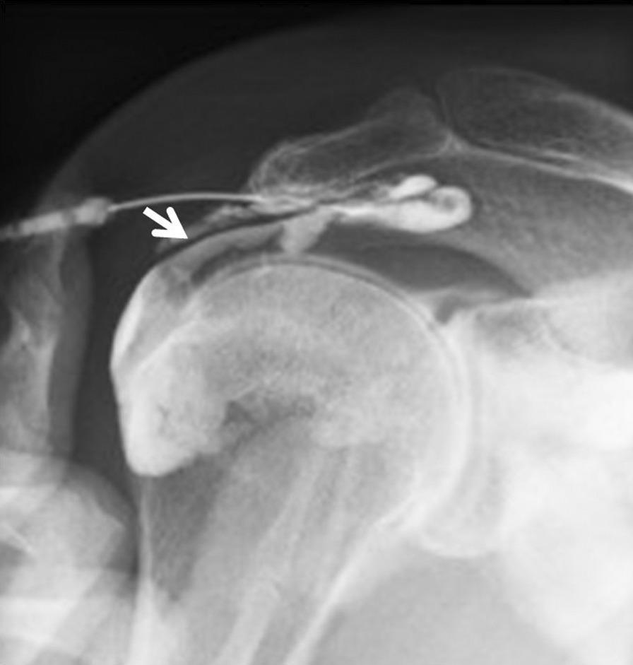
This diagnostic image is a subacromial bursography (fluoroscopic radiograph) of a human shoulder. The imaging modality highlights the subacromial space using radiopaque contrast medium. The bony anatomy includes the humeral head, greater tubercle, and the overlying acromion process. A needle is visible, through which contrast was administered. The key finding is a localized, abnormal accumulation or 'pooling' of contrast medium extending inferiorly from the subacromial bursa into the rotator cuff tendon area (indicated by a white arrow). This visual pattern is diagnostic of a bursal-side partial-thickness rotator cuff tear, specifically involving the infraspinatus or supraspinatus tendons, where the contrast fills the defect in the tendon fibers. The image demonstrates a critical diagnostic step for patients with refractory shoulder impingement syndrome where standard MRI findings might appear normal. It serves as an educational example of how dynamic contrast-enhanced imaging can reveal occult tendon pathology not seen on static radiography.
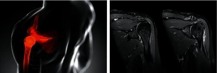
This composite educational graphic focuses on rotator cuff pathology and shoulder anatomy, featuring both an anatomical diagram and clinical diagnostic imaging. On the left, a schematic anatomical diagram of a human shoulder is shown in a coronal-style view; the humerus, scapula, and gleno-humeral joint are highlighted in red against a translucent body outline to indicate the anatomical region of interest, specifically the rotator cuff and subacromial space. On the right, two side-by-side diagnostic images are presented, consisting of Magnetic Resonance Imaging (MRI) scans in the coronal plane. These scans provide high-contrast visualization of the musculoskeletal structures, including the humeral head, acromion process, and the tendons of the rotator cuff (particularly the supraspinatus). The MRI images illustrate clinical findings such as tendon integrity, signal intensity changes suggestive of inflammation or fluid, and joint space morphology. This comparison serves to bridge basic anatomical concepts with real-world radiological diagnosis for orthopedic and sports medicine education.
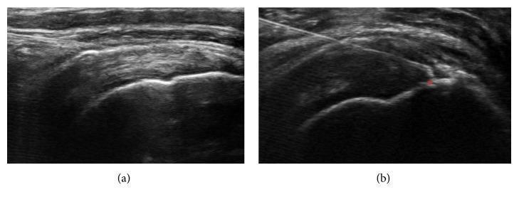
Two side-by-side musculoskeletal ultrasound images (long-axis view) demonstrating an ultrasound-guided injection for rotator cuff pathology. Image (a) shows the baseline anatomy of the shoulder, including the hyperechoic humeral head, the overlying supraspinatus tendon, and the subacromial-subdeltoid bursa. Image (b) illustrates a dynamic clinical procedure where a thin, hyperechoic, linear needle is seen entering from a lateral approach. The needle tip is precisely positioned at a focal hypoechoic defect within the supraspinatus tendon, which is marked with an asterisk to signify a partial-thickness tear. This diagnostic image highlights the use of real-time ultrasound for precise needle placement during regenerative medicine or corticosteroid injections in orthopedic clinical practice. Key features include the visualization of tendon architecture, cortical bone contours, and the characteristic reverberation artifact of the metal needle.
Create a comparison chart: subacromial bursitis vs. rotator cuff tear