
Cryptochorism
cryptorchidism undescended testis management
cryptorchidism undescended testis anatomy diagram
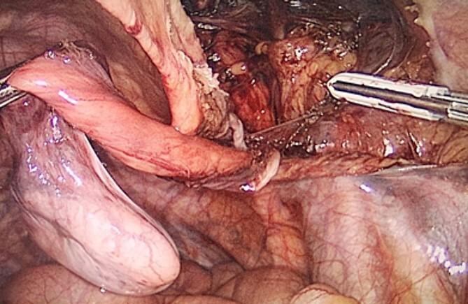
This laparoscopic photograph provides an intra-abdominal view of a surgical procedure for an undescended testis (cryptorchidism) associated with an inguinal hernia. The image prominently features a dissected, atrophic testicle, which appears as a pale, bulbous, and elongated structure being manipulated by surgical graspers. Attached to and surrounding the testis is a thin, glistening, and translucent membrane representing the hernia sac. Key anatomical structures visible include the spermatic cord and associated testicular vasculature, characterized by a network of fine reddish vessels. The operative field shows the inguinal canal region where the testis has been retracted into the peritoneal cavity for orchidectomy. The background depicts the surrounding peritoneal tissue and pelvic floor anatomy. This image illustrates a critical step in the laparoscopic management of adult cryptorchidism and inguinal hernioplasty (TAPP procedure), demonstrating the visual relationship between the ectopic gonad, its vascular supply, and the peritoneal defect.
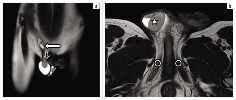
This diagnostic image consists of two T2-weighted magnetic resonance imaging (MRI) scans from a 9-month-old male child, illustrating cryptorchidism (undescended testis). Panel (a) is a coronal section showing a bright, oval T2-hyperintense structure representing the undescended right testis, localized at the level of the root of the penis and near the pubic tubercle (indicated by a black arrow). The inguinal canal is visible superiorly, marked with a white arrow. Panel (b) is an axial section further detailing the anatomical relationships; the undescended testis (star) is situated superficially. The crura of the penis are identified bilaterally (circles), located medial and inferior to the ectopic testicular position. These images demonstrate the high sensitivity of MRI in localizing non-palpable or ectopic testes by providing clear contrast between the hyperintense testicular tissue and surrounding pelvic anatomy, which is essential for preoperative surgical planning and preventing complications like atrophy or malignancy.
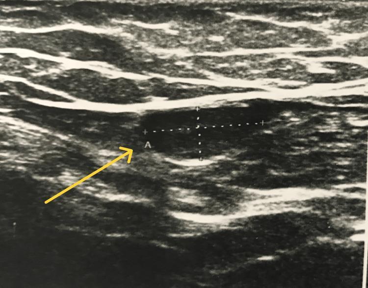
This diagnostic ultrasound image demonstrates an undescended testis (cryptorchidism) located within the inguinal canal. The testis is identified by a yellow arrow pointing to a well-defined, oval, hypoechoic structure relative to the surrounding soft tissue layers. Measurement calipers are positioned over the testicular parenchyma to assess its dimensions. The surrounding anatomical environment shows stratified layers of variable echogenicity, representing subcutaneous fat, fascia, and inguinal musculature. The testicular tissue itself appears relatively homogeneous in texture, a common sonographic finding for an ectopic or undescended testis. This imaging is characteristic of pediatric or adolescent urological evaluations where a testis is non-palpable in the scrotum. The educational focus of this image is the sonographic identification of undescended testes and the differentiation of testicular parenchyma from adjacent inguinal lymph nodes or surrounding soft tissues.
Cryptorchidism (Undescended Testis)
Note: The term "Cryptochorism" in your query is a misspelling of Cryptorchidism (from Greek: kryptos = hidden, orchis = testis).
Definition
Embryology and Normal Descent
- The testis begins as a thickening on the urogenital ridge at weeks 5-6 of embryonic life.
- During the 7th and 8th months, the testis descends along the inguinal canal into the upper scrotum, dragging the processus vaginalis with it.
- At birth, approximately 95% of infants have normally positioned testes.
- Schwartz's Principles of Surgery, 11th ed.
Incidence
| Population | Incidence |
|---|---|
| Preterm infants | ~30% |
| Term newborns | 1-3% |
| Adults (if untreated) | ~1% |
- Schwartz's Principles of Surgery, 11th ed.
Classification
| Type | Description |
|---|---|
| True undescended testis | Arrested along the normal path of descent (retroperitoneal, inguinal canal, at external ring) |
| Ectopic testis | Has passed through the external ring but lies in an abnormal location (over rectus abdominis, external oblique, medial thigh, or perineum behind the scrotum) |
| Retractile testis | Fully descended but temporarily pulled up by an overactive cremaster muscle - NOT truly undescended |
| Absent/vanishing testis | Failed normal development or intrauterine vascular accident |
- Schwartz's Principles of Surgery, 11th ed.
Clinical Features
- Examine in a warm room, patient supine and as relaxed as possible
- Try to milk the testis downward from the inguinal canal into the scrotum
- Cross-legged seated position can diminish the cremasteric reflex and help locate the testis
| Feature | Undescended | Retractile |
|---|---|---|
| Scrotum | Hypoplastic, poorly rugated | Normal |
| Testis in bath/sleep | Absent | Present |
| Able to milk to scrotum | No (or doesn't stay) | Yes, stays when relaxed |
| Cremasteric reflex | Present but pathologic | Overactive (normal variant) |
- More than 70% of cryptorchid testes are palpable on examination.
- The remaining ~30% (non-palpable) require further workup to confirm absence vs. intra-abdominal location.
- Bailey and Love's Short Practice of Surgery, 28th ed.; Smith and Tanagho's General Urology, 19th ed.
Complications
- Infertility - Scrotal temperature is 1-2°C lower than body temperature; elevated temperature impairs spermatogenesis. Histological changes visible from age 1. Bilateral cryptorchidism significantly worsens outcomes.
- Malignancy - Increased risk of testicular cancer (especially seminoma). The increased risk is thought to be an inherent abnormality predisposing to both incomplete descent and malignancy, not merely the abnormal environment. Orchidopexy reduces risk but does not eliminate it - however, scrotal placement facilitates earlier tumor detection.
- Torsion - Risk of testicular torsion, especially when testis lies at the pubic tubercle.
- Associated inguinal hernia - Due to patent processus vaginalis.
- Psychological impact of empty scrotum in a developing male.
- Schwartz's Principles of Surgery, 11th ed.; Bailey and Love, 28th ed.
Investigations
- Ultrasound: Poor sensitivity for localizing undescended testes; should not be routinely used. Useful exception: obese child where identifying an inguinal testis simplifies surgical approach. Also helpful in cases of ambiguous genitalia to detect Mullerian structures.
- MRI: Not the first-line investigation but superior for characterizing complex anomalies when ultrasound is insufficient.
- Laparoscopy: Gold standard for locating non-palpable, intra-abdominal testes.
- Campbell-Walsh-Wein Urology, 3-vol. set
Treatment
Timing
- Spontaneous descent is unlikely after 3 months of age.
- Histological changes (reduced spermatogonia) begin after age 2.
- Current recommendation: Orchidopexy by 1 year of age to optimize fertility potential and reduce malignancy risk.
- Schwartz's Principles of Surgery, 11th ed.
Surgical - Orchidopexy
- Surgical repositioning of the testis into the scrotum, secured in a dartos pouch.
- Standard orchidopexy - for palpable/inguinal testes.
- Fowler-Stephens orchidopexy - for intra-abdominal (high) testes; involves division of the testicular vessels, relying on the vasal blood supply. May be done in one or two stages.
- A recent meta-analysis (PMID 39030078) compared the Shehata technique vs. Fowler-Stephens orchidopexy for intra-abdominal testes (2024).
Hormonal (adjunct, limited role)
- hCG (human chorionic gonadotropin) may occasionally be effective, particularly in bilateral undescended testes (suggesting hormonal insufficiency).
- Combination of micropenis + bilateral cryptorchidism warrants hormonal evaluation and testosterone replacement.
- If no descent after 1 month of endocrine therapy, proceed to surgery.
- Schwartz's Principles of Surgery, 11th ed.
Post-pubertal management
- For undescended testes discovered after puberty, the management approach is more complex due to elevated malignancy risk. A 2024 systematic review (PMID 39399296) specifically addresses this.
Follow-up
- Retractile testes: yearly physical exam recommended (2-50% risk of becoming an acquired undescended testis).
- Even after orchidopexy, fertility remains ~2x lower than normal and malignancy risk persists - scrotal position enables earlier detection.
Summary Table
| Feature | Detail |
|---|---|
| Definition | Failure of testis to descend into scrotum |
| Incidence | 1-3% at term; 30% preterm |
| Most common location | Inguinal canal |
| Key complications | Infertility, malignancy, torsion, hernia |
| Recommended surgery age | By 12 months |
| Procedure | Orchidopexy (standard or Fowler-Stephens) |
| Imaging of choice | Laparoscopy for non-palpable (not USS) |