
Give me the notes from guyton textbook of physiology for movements of the small intestine
Reading File
Searching sources...
Finding Sources
Finding Sources
Found it. Let me read the full section now.
Finding Sources
Reading File
Good content so far. Let me continue reading the rest of this section.
Reading File
The section on movements of the small intestine ends around line 160 and then transitions to defecation. Let me fetch the image from the segmentation contractions figure, and also read the lines between 90-160 more carefully to make sure I captured the full small intestine section.
Reading File
Loading Image
I now have all the content needed. Let me also check lines 107-160 more carefully to capture the ileocecal valve section.
Reading File
I now have all the content. Here are the complete notes from Guyton and Hall on movements of the small intestine:
Movements of the Small Intestine
Guyton and Hall Textbook of Medical Physiology, Chapter 64 (p. 793-794)
Movements of the small intestine can be divided into mixing contractions and propulsive contractions. This separation is somewhat artificial because nearly all movements cause at least some degree of both mixing and propulsion.
1. Mixing Contractions (Segmentation Contractions)
- When a portion of the small intestine becomes distended with chyme, stretching of the intestinal wall elicits localized concentric contractions spaced at intervals along the intestine, lasting a fraction of a minute.
- These contractions cause "segmentation" of the small intestine, dividing it into spaced segments resembling a chain of sausages.
- As one set of segmentation contractions relaxes, a new set begins at new points between the previous contractions, "chopping" the chyme 2 to 3 times per minute and promoting progressive mixing with intestinal secretions.
Frequency:
- Maximum frequency in the duodenum and proximal jejunum: ~12 per minute (set by the basic electrical rhythm / slow waves of the intestinal wall)
- In the terminal ileum: 8 to 9 contractions per minute
Role of the enteric nervous system:
- Segmentation contractions become exceedingly weak when excitatory activity of the enteric nervous system is blocked by atropine
- Although slow waves initiate contractions, effective contraction requires background excitation mainly from the myenteric (Auerbach's) nerve plexus
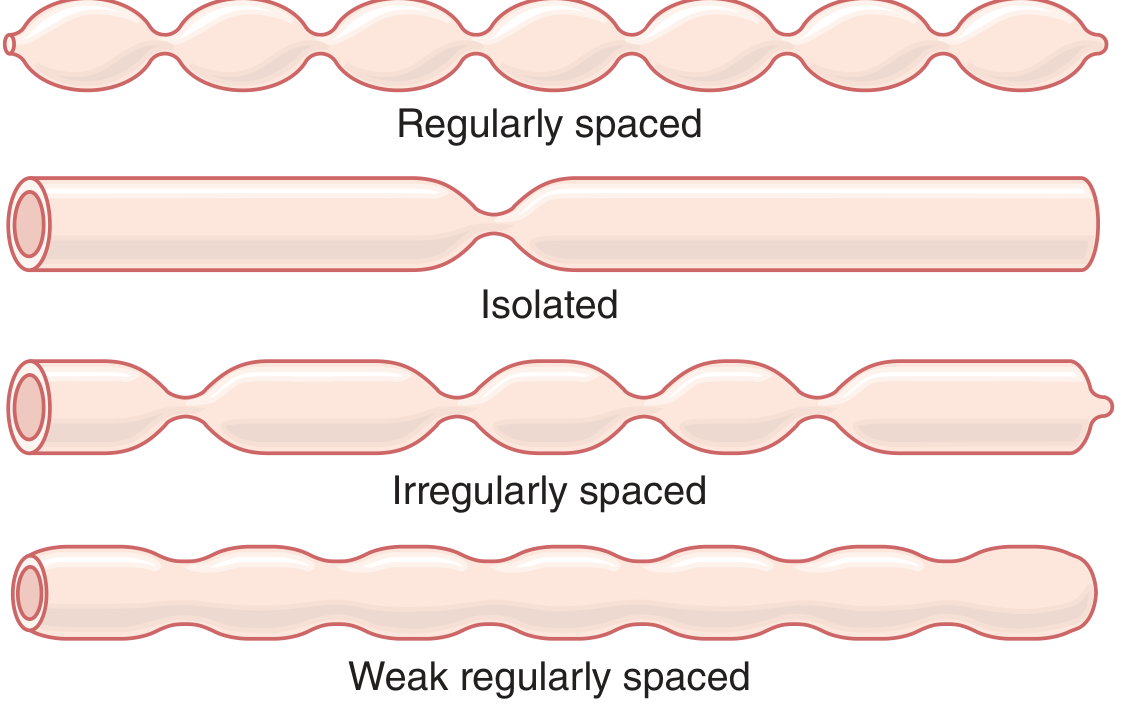
Figure 64.3 Segmentation movements of the small intestine
2. Propulsive Movements
A. Peristalsis
- Chyme is propelled by peristaltic waves that can occur anywhere in the small intestine and move toward the anus at a velocity of 0.5 to 2.0 cm/sec (faster proximally, slower terminally)
- Waves are normally weak and die out after traveling only 3 to 5 cm (rarely exceed 10 cm)
- Net forward movement of chyme averages only 1 cm/min
- Transit from pylorus to ileocecal valve takes 3 to 5 hours
- The function of peristalsis is both to propel chyme toward the ileocecal valve and to spread chyme along the intestinal mucosa for absorption
B. Control of Peristalsis - Nervous and Hormonal Signals
Stimulatory factors:
- Chyme entry into the duodenum causes stretching of the duodenal wall, increasing peristaltic activity
- Gastroenteric reflex - distention of the stomach, conducted through the myenteric plexus down along the small intestinal wall
- Hormones: Gastrin, CCK, insulin, motilin, and serotonin all enhance intestinal motility
Inhibitory factors:
- Hormones: Secretin, glucagon, and GLP-1 inhibit motility
(The physiological importance of individual hormonal factors in controlling small intestine motility remains uncertain)
C. Propulsive Effect of Segmentation Movements
- Segmentation movements, although lasting only seconds, often travel ~1 cm in the anal direction
- The distinction between segmentation and peristaltic movements is not always sharp - segmentation also contributes to propulsion
D. Peristaltic Rush
- Intense irritation of the intestinal mucosa (e.g., in severe infectious diarrhea) can cause powerful and rapid peristalsis - the "peristaltic rush"
- Initiated partly by autonomic nervous system/brainstem reflexes and partly by enhancement of myenteric plexus reflexes
- Powerful contractions travel long distances within minutes, sweeping intestinal contents into the colon
- Function: relieves the small intestine of irritative chyme and excessive distention
3. Movements Caused by the Muscularis Mucosae and Muscle Fibers of the Villi
- The muscularis mucosae can cause short mucosal folds that increase surface area exposed to chyme, thereby increasing absorption
- Individual muscle fibers extend into the intestinal villi, causing them to contract intermittently
- Villous contractions (shortening, elongating, shortening again) "milk" the villi, promoting lymph flow from the central lacteals into the lymphatic system
- These contractions are initiated mainly by local nervous reflexes in the submucosal (Meissner's) nerve plexus in response to chyme
4. The Ileocecal Valve - Preventing Backflow
- The ileocecal valve protrudes into the cecum lumen and is forcefully closed when cecal pressure rises - it resists reverse pressure of at least 50 to 60 cm of water
- The wall of the ileum for several centimeters upstream has a thickened circular muscle called the ileocecal sphincter, which remains mildly constricted, slowing emptying into the cecum
- After a meal, the gastroileal reflex intensifies ileal peristalsis and promotes emptying into the cecum
- Prolonged chyme retention in the ileum facilitates absorption; normally only 1500 to 2000 mL of chyme empty into the cecum each day
Feedback control by the cecum:
- When the cecum is distended: contraction of the ileocecal sphincter intensifies and ileal peristalsis is inhibited, delaying further emptying
- Cecal irritation (e.g., inflamed appendix) causes intense sphincter spasm and partial ileal paralysis, blocking emptying
- These reflexes are mediated via the myenteric plexus and extrinsic autonomic nerves (especially prevertebral sympathetic ganglia)
Source: Guyton and Hall Textbook of Medical Physiology, Chapter 64, pp. 793-794
This is a shared conversation. Sign in to Orris to start your own chat.