
Create a image of tetralogy of fallot pathophysiology of flow chart
Tetralogy of Fallot pathophysiology flowchart diagram showing the four defects: 1) Ventricular Septal Defect (VSD), 2) Pulmonary Stenosis (Right ventricular outflow tract obstruction), 3) Overriding Aorta, 4) Right Ventricular Hypertrophy. Show the pathophysiology flow: Pulmonary stenosis causes increased resistance to right ventricular outflow → Right ventricular pressure rises → Right-to-left shunt through VSD → Deoxygenated blood enters aorta via overriding aorta → Cyanosis (hypoxemia). Also show: Right ventricular hypertrophy develops as compensatory response. Include arrows showing blood flow direction, color-coded blue for deoxygenated blood and red for oxygenated blood. Clean medical education style with labeled boxes and connecting arrows in a vertical flowchart layout.
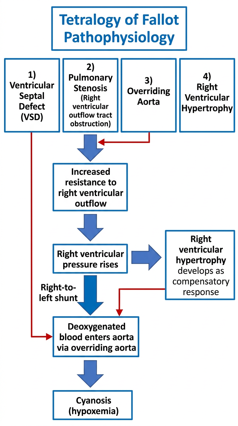
Key elements shown in the flowchart:
- Pulmonary stenosis (RVOTO - Right Ventricular Outflow Tract Obstruction)
- Right Ventricular Hypertrophy (compensatory)
- Overriding Aorta (straddles the VSD)
- Ventricular Septal Defect (VSD)
- Pulmonary stenosis → increased resistance to RV outflow
- RV pressure rises to match or exceed LV pressure
- Right-to-left shunt through the VSD
- Deoxygenated blood passes into the overriding aorta
- Systemic cyanosis (hypoxemia) results
- RV hypertrophy develops as a compensatory response to chronic pressure overload
Create a image of tetralogy of fallot pathophysiology of flow chart with diagram
A detailed medical education poster combining two sections side by side: LEFT SIDE - Anatomical Heart Diagram of Tetralogy of Fallot: Cross-section of the heart showing all four defects clearly labeled with arrows: (1) Large Ventricular Septal Defect (VSD) - hole between left and right ventricles, (2) Pulmonary Stenosis - narrowed pulmonary valve and right ventricular outflow tract, (3) Overriding Aorta - aorta positioned over the VSD straddling both ventricles, (4) Right Ventricular Hypertrophy - thickened RV wall. Show deoxygenated blood in blue flowing from RV through VSD into the aorta, and oxygenated blood in red. Label all heart chambers: RA, LA, RV, LV. Label pulmonary artery, aorta, pulmonary valve, tricuspid valve, mitral valve. RIGHT SIDE - Pathophysiology Flowchart with connected boxes and arrows: Box 1 (top): "4 Defects of Tetralogy of Fallot" ↓ Box 2: "Pulmonary Stenosis → ↑ RV Outflow Resistance" ↓ Box 3: "↑ Right Ventricular Pressure (≥ LV pressure)" ↓ Box 4: "Right-to-Left Shunt through VSD" ↓ Box 5: "Deoxygenated blood → Overriding Aorta → Systemic Circulation" ↓ Box 6 (red): "CYANOSIS (Hypoxemia)" Side arrow: "Compensatory RVH" pointing to Box 4 Bottom: "Tet Spells: ↑ infundibular spasm → ↑ R→L shunt → acute severe cyanosis" Clean white background, professional medical illustration style, color-coded with blue for deoxygenated blood and red for oxygenated, bold labels, suitable for medical students.
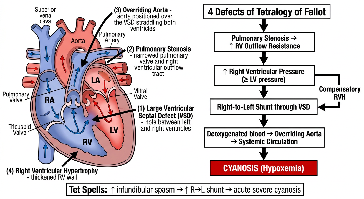
Summary of what's shown:
- Heart cross-section with all 4 defects labeled
- Blue = deoxygenated blood | Red = oxygenated blood
- Shows the right-to-left shunt pathway through the VSD into the overriding aorta
| Step | Event |
|---|---|
| 1 | Pulmonary stenosis → ↑ RV outflow resistance |
| 2 | RV pressure rises to match/exceed LV pressure |
| 3 | Right-to-left shunt through VSD |
| 4 | Deoxygenated blood enters overriding aorta |
| 5 | Cyanosis - "blue baby" presentation |
| Compensatory | Right Ventricular Hypertrophy (RVH) |
Pyloric stenosis give full details about this for child health nursing subject
pyloric stenosis infant hypertrophic
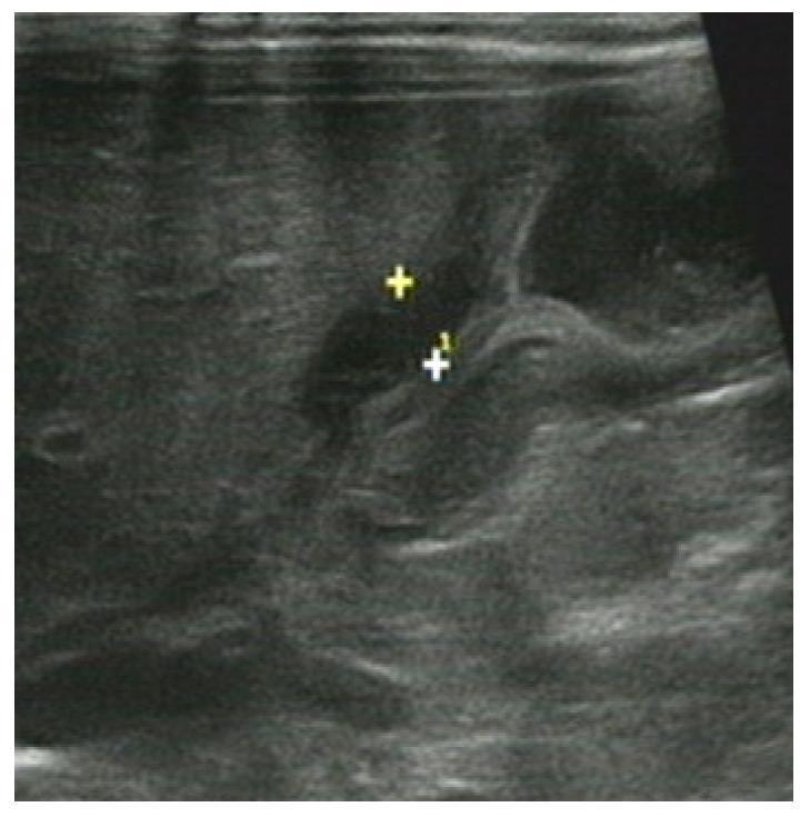
This abdominal ultrasound image depicts a case of Hypertrophic Pyloric Stenosis (HPS) in a 22-day-old male infant. The diagnostic image shows a longitudinal view of the pyloric canal, characterized by marked thickening of the pyloric muscularis layer. Electronic calipers, labeled '+' and '+1', are positioned to measure the muscle wall thickness (single-wall thickness), demonstrating a hypoechoic and homogenous muscular rim. The central pyloric mucosa appears compressed and echogenic, creating the typical 'olive' or 'doughnut' sign in cross-section (not shown) or an elongated canal in this longitudinal plane. Key clinical indices for diagnosis include a muscle thickness exceeding 3-4 mm and an overall pyloric canal length greater than 15-17 mm. This visual evidence is a classic hallmark of neonatal gastric outlet obstruction requiring surgical intervention.
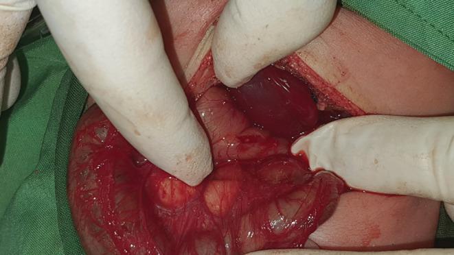
This clinical photograph captures an intraoperative view of a pediatric abdominal exploration for a Ramstedt pyloromyotomy. The surgical field, accessed via a supraumbilical incision, demonstrates the classic appearance of hypertrophic pyloric stenosis. The pylorus is visible as a thickened, olive-shaped, reddish-tan mass being manually stabilized by gloved fingers. The serosa appears glistening and hyperemic with fine superficial vasculature. Adjacent anatomical structures include the liver, which presents a dark reddish-brown color, and sections of the small intestine. In this specific case, the surgical approach is through a left-leaning incision due to situs inversus totalis, where the pylorus and liver are positioned in mirror-image to normal anatomy. The incision site shows pale skin edges with underlying subcutaneous tissue and minimal blood, surrounded by green surgical drapes. This visual highlights the surgical presentation of infant hypertrophic pyloric stenosis and its anatomical management in the context of rare congenital variations like situs inversus.
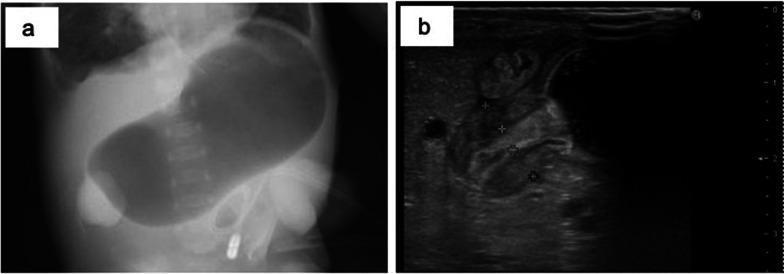
This composite diagnostic image illustrates classic findings of infantile hypertrophic pyloric stenosis (HPS) using two modalities. Image (a) is a supine abdominal X-ray (radiography) of an infant demonstrating a severely distended, air-filled stomach (the 'single bubble' appearance), occupying the majority of the upper abdomen and crossing the midline. This indicates gastric outlet obstruction. Image (b) is a transverse abdominal ultrasonography scan of the same patient. It shows a significantly thickened and elongated pyloric muscle, which appears as a hypoechoic (dark) ring surrounding a narrow central lumen. Caliper markers on the ultrasound indicate a muscle wall thickness of 4-5 mm, exceeding the standard diagnostic threshold (typically >3 mm). The combination of these images serves as a primary diagnostic tool for HPS, demonstrating both the secondary effect (gastric dilation) and the primary pathology (pyloric muscular hypertrophy).
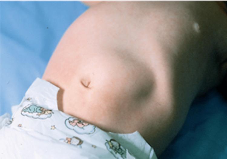
This clinical photograph displays the abdominal region of an infant in a supine position, highlighting a distinct physical examination finding. A visible, localized bulge is evident in the right upper quadrant (RUQ), situated superior and lateral to the centrally located umbilicus. This protrusion represents the classically described 'olive-like' mass associated with Infantile Hypertrophic Pyloric Stenosis (IHPS). The abdominal contour exhibits asymmetry due to this mass and a generalized epigastric fullness, which may indicate gastric distension proximal to the pyloric obstruction. The skin appears intact with fair pigmentation and no signs of acute inflammation or surgical scarring. A small, incidental white macule is visible on the upper right thoracic-abdominal junction. This image serves as a key pedagogical example for pediatric physical diagnosis, illustrating the visible signs of a thickened pyloric muscle and subsequent mechanical gastric outlet obstruction.
Pyloric Stenosis - Complete Guide for Child Health Nursing
1. Definition
- The Developing Human - Clinically Oriented Embryology, p.612
- Tintinalli's Emergency Medicine, p.2626
2. Anatomy
- The pyloric muscularis propria hypertrophies (mainly circular muscle)
- This narrows the pyloric canal
- Mucosal and submucosal edema worsens obstruction
- The stomach becomes markedly distended proximal to the obstruction
3. Epidemiology / Incidence
| Parameter | Detail |
|---|---|
| Incidence | 1 in 300-900 live births (~0.2% general population) |
| Male : Female ratio | 4:1 (males predominantly affected) |
| Birth order | More common in firstborn children |
| Siblings | ~6% risk if sibling affected |
| Twins | High concordance in monozygotic twins |
| Age of onset | 2-10 weeks (classic: 3-6 weeks) |
- Current Surgical Therapy 14e
- Robbins & Cotran Pathologic Basis of Disease
4. Etiology & Risk Factors
- Genetic factors - High monozygotic twin concordance; genome-wide association studies have identified genes related to GI development
- Erythromycin / Azithromycin exposure in the first 2 weeks of life (oral or via breast milk) - strongly associated
- Turner syndrome and Trisomy 18 - increased risk
- Young maternal age and prematurity - associated risk factors
- Family history - maternal family history in particular increases risk
5. Pathophysiology
Hypertrophy of pyloric circular muscle
           ↓
Narrowing of pyloric canal (stenosis)
           ↓
Gastric outlet obstruction
           ↓
Food/milk cannot pass into duodenum
           ↓
Stomach becomes distended with food + secretions
           ↓
Forceful PROJECTILE NON-BILIOUS VOMITING
           ↓
Loss of HCl (gastric acid) in vomitus
           ↓
Hypochloremic, Hypokalemic METABOLIC ALKALOSIS
           ↓
Progressive DEHYDRATION + MALNUTRITION
           ↓
Failure to thrive, weight loss
6. Clinical Features (Signs & Symptoms)
Cardinal Features:
| Feature | Description |
|---|---|
| Projectile vomiting | Non-bilious, forceful, occurs at end of or shortly after feeding; increases in frequency and force daily |
| Hunger after vomiting | Infant appears hungry and wants to re-feed immediately (distinguishes from infection) |
| Visible gastric peristalsis | Left-to-right peristaltic waves visible across upper abdomen just before vomiting |
| Palpable "olive" mass | Firm, ovoid 1-2 cm mass in right upper quadrant (epigastric); nearly pathognomonic - found in up to 90% of cases |
| Dehydration | Sunken fontanelle, dry mucous membranes, decreased urine output, poor skin turgor |
| Weight loss / failure to thrive | Due to caloric loss from repeated vomiting |
| Jaundice | Unconjugated hyperbilirubinemia (indirect) may occur in some cases |
| Constipation | Little stool due to reduced oral intake reaching the bowel |
Vomiting Characteristics (important for nursing assessment):
- Non-bilious (bile-free) - obstruction is above the ampulla of Vater
- Projectile - may shoot several feet
- Non-bloody
- Occurs after feeding, not at birth
- Infant remains alert and hungry after vomiting
7. Diagnostic Investigations
A. Ultrasound (Gold Standard)
- Pyloric muscle wall thickness ≥ 4 mm
- Pyloric channel length ≥ 16 mm (some sources: ≥ 14 mm)
- Inability of fluid to pass through pylorus during exam
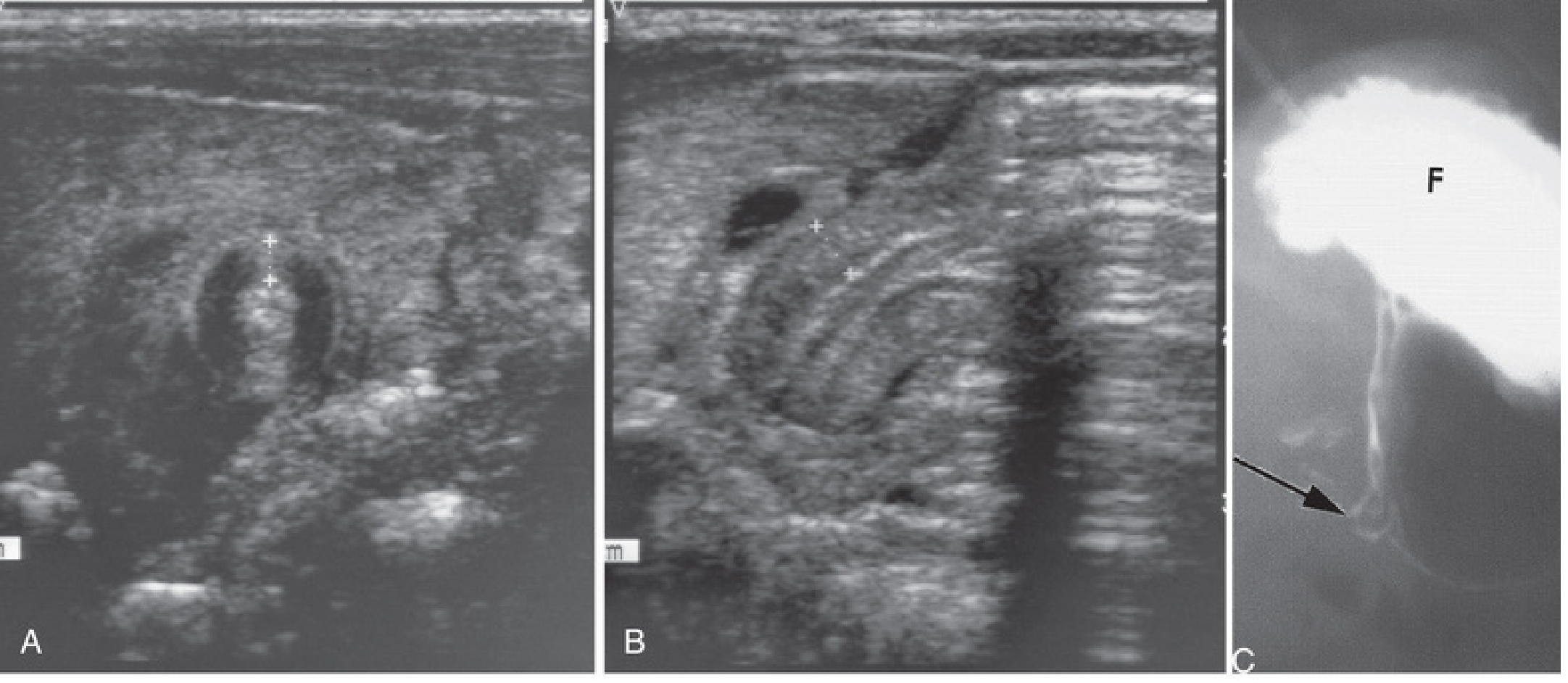
B. Upper GI Series (Barium/Contrast Study)
- Used when ultrasound findings are equivocal
- Shows "string sign" - narrow elongated pyloric channel
- "Shoulder sign" - impression on antrum
- Contrast stays in distended stomach
C. Laboratory Investigations (Metabolic Panel)
| Lab Finding | Result | Reason |
|---|---|---|
| Serum Chloride | ↓ (Hypochloremia) | Lost in HCl vomitus |
| Serum Potassium | ↓ (Hypokalemia) | Lost in vomitus + kidneys exchange K+ for H+ |
| Serum Bicarbonate | ↑ (> 30 mEq/L) | Metabolic alkalosis |
| Blood pH | ↑ | Metabolic alkalosis |
| Serum Sodium | ↓ (often) | Dehydration |
| BUN/Creatinine | ↑ | Dehydration |
| Urine | Paradoxical aciduria | Kidneys conserve H+ over K+ to maintain Na+ |
D. Clinical Test Feed
- Observe infant feeding: gastric peristalsis from left to right visible
- Palpate relaxed, feeding baby for pyloric "olive" in right upper quadrant
8. Clinical Images
9. Medical Management (Pre-operative)
Step 1: Fluid & Electrolyte Correction
- Insert nasogastric tube (8-10Fr) to decompress the stomach - feeds discontinued
- IV fluids: 0.9% Normal Saline with 0.15% KCl in 5% dextrose at 6-7.5 mL/kg/hour
- Two boluses of 20 mL/kg Normal Saline for dehydration
- Maintenance rate: 1.5× normal maintenance
- Check electrolytes every 6-12 hours until normal
Step 2: Monitoring Endpoints Before Surgery
- Chloride ≥ 90-100 mEq/L
- Bicarbonate ≤ 30 mEq/L (bicarbonate > 30 mEq/L = risk for post-op respiratory depression and apnea)
- Wet diapers returning (urine output)
- Correction takes 24-48 hours typically
Warning: Do NOT take infant to anaesthesia with bicarbonate > 30 mEq/L - diminished respiratory drive poses risk of post-operative apnea and respiratory arrest.
- Current Surgical Therapy 14e
10. Surgical Management - Ramstedt's Pyloromyotomy
Procedure:
- Approach: Laparoscopic (preferred) OR open (right upper quadrant/periumbilical/supraumbilical incision)
- Laparoscopic technique: 3 ports - one at umbilicus (with port) + two 3-mm stab incisions without ports
- Pylorus stabilized with grasper
- A longitudinal serosal incision is made through the anterior wall of the pyloric canal
- Muscular layer split from stomach to duodenal side (where muscle softens)
- Intact submucosa preserved - mucosa should bulge through the myotomy
- Leak test: 30-60 mL air injected through orogastric tube to confirm:
- Air passes through pylorus
- No air escapes through mucosa (rules out perforation)
Post-operative Feeding:
- IV fluids continued until feeds re-established (within 24 hours)
- Ad lib feeding with 60-mL limit - most practical approach
- Most infants discharged within 36 hours of tolerating 2 consecutive feeds
- Early post-op vomiting is normal - gastroesophageal reflux (GOR) is more likely than incomplete myotomy if it persists
11. Complications
Surgical Complications:
| Complication | Description | Management |
|---|---|---|
| Incomplete myotomy | Myotomy not extended far enough onto stomach side; presents as ongoing projectile vomiting | Return to OR for revision |
| Mucosal perforation | Accidental entry into mucosa | Close perforation + omental buttress OR close, rotate pylorus 180°, redo myotomy; if unrecognized → sepsis |
Medical Complications (if untreated):
- Severe dehydration and shock
- Electrolyte imbalances (metabolic alkalosis)
- Failure to thrive and malnutrition
- Aspiration pneumonia (from repeated vomiting)
12. Nursing Management (Child Health Nursing Focus)
A. Pre-operative Nursing Care
- Monitor weight daily (assess weight loss and nutritional status)
- Assess for signs of dehydration: sunken fontanelle, dry mucous membranes, skin turgor, urine output
- Observe and document vomiting: frequency, force, amount, character (non-bilious? projectile?)
- Monitor vital signs including temperature (detect infection)
- Observe for visible gastric peristalsis waves
- NPO (Nil Per Os) - Discontinue oral feeds; maintain NG tube for gastric decompression
- IV access - Maintain patent IV line for fluid and electrolyte replacement
- Fluid monitoring - Strict intake and output chart; monitor urine output (wet diapers)
- Electrolyte monitoring - Assist with blood draws every 6-12 hours; monitor lab results
- Positioning - Keep head elevated to reduce aspiration risk
- Comfort and non-nutritive sucking - Offer pacifier to satisfy sucking needs while NPO
- Parental education - Explain condition, investigations, surgical procedure, and recovery to parents
- Emotional support - Reassure anxious parents; encourage bonding
B. Post-operative Nursing Care
- Monitor vital signs every 1-2 hours initially
- Assess surgical wound for signs of infection (redness, swelling, discharge)
- Monitor for return of bowel activity
- Observe feeds for tolerance (frequency of vomiting post-op)
- Assess pain using appropriate infant pain scale (e.g., CRIES scale, NIPS)
- Pain management - Administer prescribed analgesics; non-pharmacological comfort measures
- Wound care - Inspect and care for incision site; keep dry and clean
- Gradual re-feeding - Introduce feeds as per protocol (small, frequent feeds initially)
- Position - Keep semi-upright after feeds to reduce reflux
- Monitor for complications - Watch for ongoing vomiting (incomplete myotomy), fever, abdominal distension (perforation)
- Parental education - Teach parents about post-operative care, feeding technique, wound care, and warning signs
C. Key Nursing Diagnoses
- Deficient fluid volume r/t persistent vomiting and inability to maintain oral intake
- Imbalanced nutrition: less than body requirements r/t vomiting and NPO status
- Risk for aspiration r/t projectile vomiting
- Acute pain r/t surgical procedure
- Anxiety (parental) r/t child's illness and hospitalization
- Risk for infection r/t surgical wound
13. Differential Diagnosis
| Condition | Key Distinguishing Feature |
|---|---|
| Gastroesophageal Reflux (GOR) | Vomiting starts at birth, waxes and wanes, not truly projectile |
| Pylorospasm | Intermittent, ultrasound shows no fixed thickening |
| Gastroenteritis | Diarrhea present; infant unwell/febrile; bilious/non-projectile |
| Meningitis/UTI | Unwell infant, fever, altered sensorium |
| Duodenal atresia | Bilious vomiting, "double bubble" sign on X-ray |
| Rumination disorder | Onset after 3 months, associated with autism spectrum disorder |
14. Prognosis
- Outcomes after pyloromyotomy are excellent
- Surgery is curative in virtually all cases
- Mortality is extremely rare with proper pre-operative resuscitation
- Long-term GI function is normal
15. Quick Summary Table
| Feature | Details |
|---|---|
| Age | 2-10 weeks (peak: 3-6 weeks) |
| Sex | Male > Female (4:1) |
| Vomiting | Projectile, non-bilious, post-prandial |
| Sign | Palpable pyloric "olive" (RUQ mass) |
| Metabolic abnormality | Hypochloremic, hypokalemic metabolic alkalosis |
| Diagnosis | Ultrasound (muscle ≥4mm, canal ≥16mm) |
| Pre-op treatment | IV fluid & electrolyte correction |
| Surgery | Ramstedt's pyloromyotomy |
| Prognosis | Excellent - curative |
Draw a nursing care plan for baby kumari based on three priorities problem
What condition does Baby Kumari have? (Based on our previous discussion, shall I base this on Pyloric Stenosis?)
🏥 NURSING CARE PLAN
PRIORITY PROBLEM 1
Nursing Diagnosis: Deficient Fluid Volume
| Assessment (Subjective & Objective Data) | Nursing Goal / Expected Outcome | Nursing Interventions | Rationale | Evaluation |
|---|---|---|---|---|
| S: Mother reports Baby Kumari has been vomiting forcefully after every feed for the past 2 weeks | Baby Kumari will show signs of adequate hydration within 24-48 hours as evidenced by moist mucous membranes, return of skin turgor, and urine output ≥1-2 mL/kg/hour | 1. Monitor and record strict Intake & Output - Count wet diapers every 4-6 hours; document all IV fluids administered | Accurate I&O tracking identifies fluid balance and guides replacement therapy | Goal Met / Partially Met / Not Met (evaluate after 24 hours) |
| O: Sunken anterior fontanelle | 2. Assess hydration status every 2-4 hours - fontanelle, mucous membranes, skin turgor, capillary refill, eyes | Frequent reassessment detects improvement or deterioration in hydration promptly | ||
| O: Dry mucous membranes | 3. Administer IV fluids as prescribed - 0.9% Normal Saline with 0.15% KCl in 5% Dextrose at 6-7.5 mL/kg/hour; give 20 mL/kg NS bolus for dehydration as ordered | Corrects fluid and electrolyte losses (Cl⁻, K⁺) caused by gastric acid loss in vomitus | ||
| O: Skin turgor reduced (poor skin tenting) | 4. Insert and maintain NG tube (8-10 Fr) - Keep on free drainage; measure and document aspirate | Decompresses the stomach, prevents further vomiting, and allows measurement of ongoing gastric losses | ||
| O: Urine output < 1 mL/kg/hour | 5. Monitor electrolytes (Chloride, Potassium, Bicarbonate, Sodium) every 6-12 hours as ordered | Identifies and tracks correction of hypochloremic, hypokalemic metabolic alkalosis; surgery cannot proceed until Cl⁻ ≥ 90 mEq/L and HCO₃⁻ ≤ 30 mEq/L | ||
| O: Serum Cl⁻ low, K⁺ low, HCO₃⁻ elevated | 6. Maintain IV access - Check cannula site for patency, phlebitis, infiltration every 4 hours | Ensures uninterrupted delivery of IV fluids and medications | ||
| O: Weight loss noted from birth weight | 7. Weigh Baby Kumari daily at same time on same scale | Daily weight is the most accurate indicator of fluid balance in infants | ||
| 8. Keep Baby Kumari NPO (Nil Per Os) - Nothing by mouth; explain to mother | Prevents further vomiting and gastric distension; allows the stomach to rest |
PRIORITY PROBLEM 2
Nursing Diagnosis: Imbalanced Nutrition: Less Than Body Requirements
| Assessment (Subjective & Objective Data) | Nursing Goal / Expected Outcome | Nursing Interventions | Rationale | Evaluation |
|---|---|---|---|---|
| S: Mother reports Baby Kumari vomits everything she feeds and then cries with hunger | Baby Kumari will maintain/improve nutritional status as evidenced by stable weight and readiness for post-operative feeding within 48 hours | 1. Maintain NPO status and NG tube - Ensure all oral feeds are withheld; provide mouth care with moist swabs | NPO prevents further caloric losses through vomiting; oral care maintains mucosal comfort | Goal Met / Partially Met / Not Met |
| O: Birth weight not regained; weight loss noted | 2. Administer IV dextrose-containing fluids as prescribed (D5/NS + electrolytes) | Provides caloric support (glucose) and prevents hypoglycaemia during NPO period | ||
| O: Infant appears hungry, irritable; re-feeds eagerly after each vomit | 3. Monitor blood glucose 4-6 hourly | Infants have limited glycogen stores; prolonged NPO risks hypoglycaemia | ||
| O: No weight gain documented over past week | 4. Offer a pacifier / non-nutritive sucking | Satisfies the infant's strong sucking reflex and provides comfort without introducing fluids into the stomach | ||
| O: Visible left-to-right gastric peristaltic waves across upper abdomen | 5. Record and document vomiting episodes - Time, volume, frequency, character (non-bilious, projectile) | Accurate records communicate severity to the surgical team and track any improvement | ||
| O: Pale, irritable, reduced subcutaneous fat noted | 6. Educate and support mother - Explain why feeds are withheld, reassure that IV nutrition is supporting Baby Kumari, encourage skin-to-skin contact | Reduces maternal anxiety and guilt; promotes bonding and infant comfort | ||
| 7. Coordinate with dietitian/surgical team regarding post-operative feeding plan | Ensures smooth transition to oral feeding after pyloromyotomy | |||
| 8. Elevate head of cot by 30° | Semi-upright positioning reduces the discomfort of gastric distension |
PRIORITY PROBLEM 3
Nursing Diagnosis: Risk for Aspiration
| Assessment (Subjective & Objective Data) | Nursing Goal / Expected Outcome | Nursing Interventions | Rationale | Evaluation |
|---|---|---|---|---|
| S: Mother reports vomit "shoots across the room" after feeds | Baby Kumari will not experience aspiration as evidenced by clear lung sounds, absence of respiratory distress, and O₂ saturation ≥ 95% throughout pre-operative period | 1. Keep Baby Kumari NPO and maintain NG tube on free drainage or intermittent aspiration | Empty stomach dramatically reduces aspiration risk; NG drainage prevents gastric pooling | Goal Met / Partially Met / Not Met |
| O: Forceful, projectile, non-bilious vomiting after every feed | 2. Position Baby Kumari at 30-45° head elevation (anti-Trendelenburg); prone or right lateral position if awake and monitored | Gravity assists in keeping gastric contents away from airway; right lateral positioning promotes gastric emptying | ||
| O: Grossly distended upper abdomen / epigastric region | 3. Aspirate NG tube regularly and before any procedures; document amount and colour of aspirate | Ensures adequate decompression; prevents sudden regurgitation during handling or procedures | ||
| O: Crying, restless infant who may inhale during vomiting | 4. Monitor respiratory status - Respiratory rate, O₂ saturation (SpO₂), breath sounds every 2-4 hours | Early detection of aspiration signs: tachypnoea, crackles, desaturation, coughing | ||
| O: High risk period during and after feeding before NG tube insertion | 5. Keep suction equipment at bedside at all times (appropriate-size suction catheter + Yankauer) | Enables immediate suctioning of oropharynx if vomiting occurs | ||
| 6. Do not leave Baby Kumari unattended after any feed or procedure | Constant supervision allows immediate response to sudden vomiting | |||
| 7. Educate parents - Do not attempt to feed Baby Kumari orally; demonstrate safe positioning; teach signs of respiratory distress (grunting, nasal flaring, chest retractions) | Empowers parents to maintain safety measures and recognise warning signs | |||
| 8. Ensure emergency equipment is available - Oxygen, bag-valve-mask, suction at bedside | Readiness for rapid response to aspiration event |
SUPPLEMENTARY NURSING CONSIDERATIONS
Pre-Operative Checklist for Baby Kumari:
| Item | Action |
|---|---|
| Electrolytes | Cl⁻ ≥ 90 mEq/L, K⁺ normal, HCO₃⁻ ≤ 30 mEq/L before proceeding to OR |
| Hydration | Wet diapers, moist mucous membranes restored |
| IV access | Patent, secure, labeled |
| NG tube | In situ, draining, position confirmed |
| Consent | Informed surgical consent obtained from parents/guardian |
| Weight | Documented pre-operative weight |
| Parental education | Surgery explained (Ramstedt's pyloromyotomy), expectations discussed |
| Blood group & cross-match | Done as per surgical protocol |
Key Safety Reminders:
⚠️ Do NOT take Baby Kumari to theatre if serum bicarbonate > 30 mEq/L - risk of post-operative respiratory depression and apnea.
⚠️ Pyloric Stenosis is NOT a surgical emergency - adequate pre-operative resuscitation takes priority over urgency of surgery.
⚠️ Vomitus is non-bilious - bilious vomiting in a neonate suggests a different, more urgent diagnosis (e.g., midgut volvulus) and must be escalated immediately.
Parental Education Points:
- Explain the diagnosis in simple terms - "the muscle at the exit of the stomach is too thick and blocks food from passing"
- Reassure that surgery (pyloromyotomy) has an excellent outcome and is curative
- Explain why Baby Kumari cannot be fed orally and why she appears hungry
- Teach safe positioning and aspiration precautions
- Address maternal guilt about not being able to breastfeed during NPO period
- Discuss expected hospital stay and post-operative feeding
Tracheoesophaeal fistula
tracheoesophageal fistula esophageal atresia types diagram
tracheoesophageal fistula newborn chest xray
Tracheoesophageal Fistula (TEF) - Complete Guide for Child Health Nursing
1. Definition
TEF is the most common birth defect of the lower respiratory tract.
- The Developing Human - Clinically Oriented Embryology, p.576
2. Embryology (How It Happens)
| Week | Normal Development | In TEF |
|---|---|---|
| Week 4 | Foregut begins to divide into ventral respiratory and dorsal esophageal segments | Division is incomplete |
| Week 4-5 | Tracheoesophageal folds fuse in midline to form the tracheoesophageal septum | Defective septum forms |
| Week 6-7 | Complete separation of trachea and esophagus | Abnormal connection persists |
- Incomplete fusion of lateral tracheoesophageal folds
- Imbalance in growth of cranial and caudal folds
- Deficiency in Sonic Hedgehog (Shh) gene signaling
- Disturbances in apoptosis or epithelial proliferation
3. Incidence & Epidemiology
| Parameter | Data |
|---|---|
| Incidence | 1 in 3,000-4,500 live births |
| Sex | Slight male predominance (M:F = 1.3:1) |
| Recurrence risk (sibling) | 0.5-2% |
| Recurrence (parent affected) | 3-5% |
| Associated anomalies | ~50% of infants have associated anomalies |
| Chromosomal | Trisomy 18, Trisomy 21 in 5-10% |
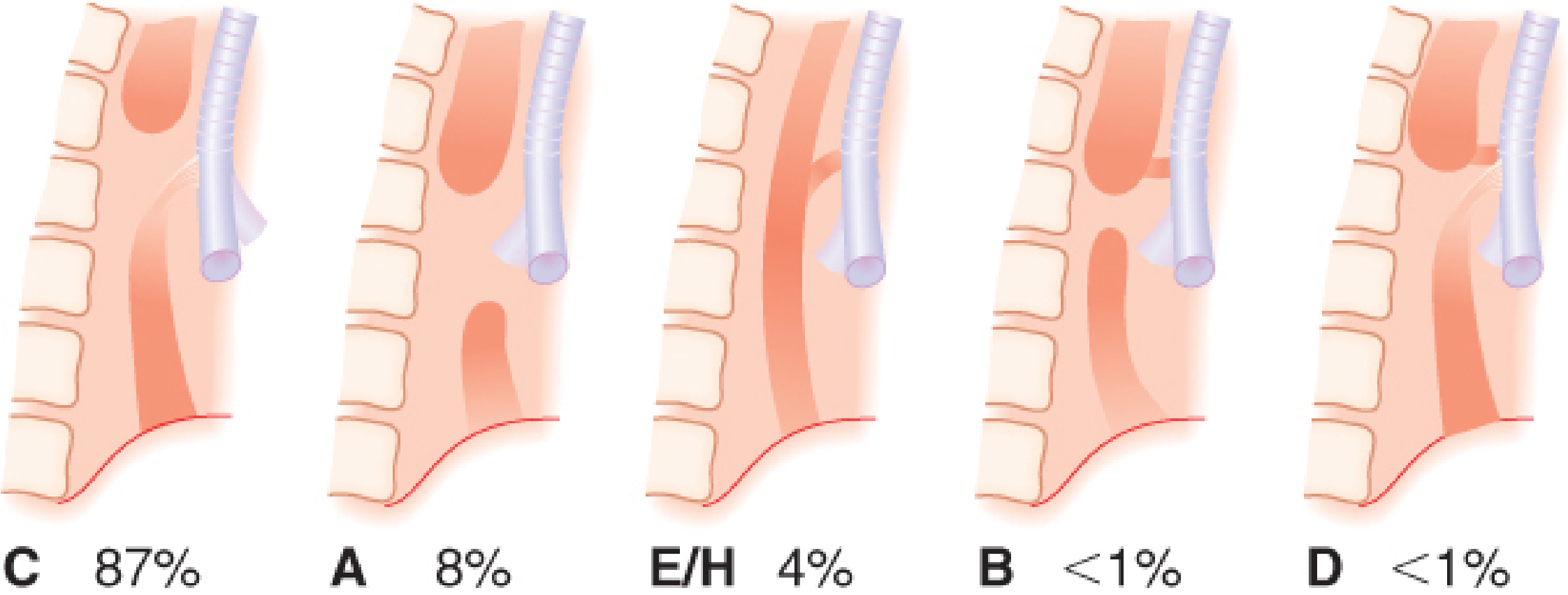
4. Classification - Gross Classification (Types A-F)
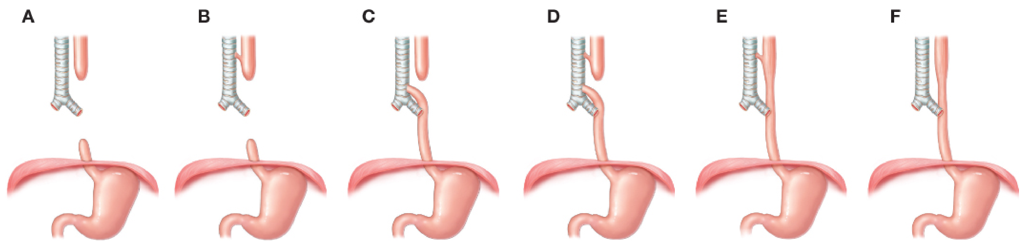
| Type | Description | Frequency |
|---|---|---|
| Type C (Gross C) | EA with DISTAL TEF - Upper esophagus ends blindly (proximal pouch), lower esophagus connects to trachea at carina | 87-88% - MOST COMMON |
| Type A (Gross A) | Pure EA (no fistula) - Both ends blind, no connection to trachea; gasless abdomen on X-ray | 7-8% |
| Type E / H-type | TEF without EA - H-shaped fistula; esophagus is patent, presents later with recurrent chest infections | 3-4% |
| Type B (Gross B) | EA with PROXIMAL TEF - Upper pouch connects to trachea | <1% |
| Type D (Gross D) | EA with BOTH proximal and distal TEF | <1% |
| Type F | Congenital esophageal stenosis without atresia or TEF | Rare |
5. Associated Anomalies - VACTERL Syndrome
| Letter | Anomaly | Frequency in TEF |
|---|---|---|
| V | Vertebral anomalies | 6-20% |
| A | Anorectal malformations / Anal atresia | 10-15% |
| C | Cardiac anomalies (most common associated defect) | 15-35% |
| TE | Tracheoesophageal fistula | - |
| R | Renal and urinary anomalies | 5-15% |
| L | Limb anomalies (radial aplasia, polydactyly) | 5-20% |
6. Pathophysiology
Failure of tracheoesophageal septum to form completely
                    ↓
Abnormal connection (fistula) between trachea and esophagus
                    ↓
┌─────────────────────────────────────────────────────────┐
│                  TWO MAJOR PROBLEMS                      │
└──────────────────────┬──────────────────────────────────┘
                       │
       ┌───────────────┴────────────────┐
       ↓                                ↓
ESOPHAGEAL ATRESIA              FISTULOUS CONNECTION
(Upper pouch blind)             (Trachea ↔ Esophagus)
       ↓                                ↓
Infant CANNOT swallow      Air enters stomach from trachea
       ↓                   (gastric distension)
Saliva pools in pouch       Gastric acid refluxes INTO lungs
       ↓                                ↓
ASPIRATION of secretions    CHEMICAL PNEUMONITIS
       ↓                                ↓
Coughing, choking,          Respiratory distress
cyanosis on feeding         SpO₂ drops
       ↓
Fetus cannot swallow amniotic fluid
→ POLYHYDRAMNIOS (prenatal)
7. Clinical Features
Prenatal Signs:
- Polyhydramnios (excess amniotic fluid - fetus cannot swallow)
- Absent or small stomach bubble on fetal ultrasound
Postnatal / Neonatal Signs (Classic "3 Cs"):
| Feature | Description |
|---|---|
| Coughing | On feeding or with secretions |
| Choking | Episodes when attempting to feed |
| Cyanosis | Episodic - especially during feeds |
| Excessive drooling / frothy secretions | Saliva pools in blind esophageal pouch |
| Regurgitation of feeds immediately | Food cannot reach stomach |
| Respiratory distress | Tachypnoea, nasal flaring, intercostal retractions |
| Abdominal distension | Air passes through fistula into stomach |
| Inability to pass NG/OG tube | Tube coils in upper esophageal pouch - PATHOGNOMONIC |
In H-type TEF (without atresia) - presents LATER:
- Recurrent chest infections / pneumonia
- Coughing after feeds
- Abdominal bloating (air enters stomach)
- May not be diagnosed until weeks/months after birth
8. Diagnosis
A. Bedside Test (Most Important First Step)
- Tube coils in the upper esophageal pouch and CANNOT be advanced to stomach
- X-ray shows tube curling back in the mediastinum - diagnostic
B. Chest + Abdominal X-ray
| Finding | Significance |
|---|---|
| OG tube coiled in superior mediastinum | Confirms EA |
| Gas in abdomen (bowel gas present) | Confirms DISTAL TEF (most common, Type C) |
| Gasless abdomen | Suggests isolated EA (Type A) or proximal TEF only |
| Pneumonia changes | Aspiration pneumonitis |
| Vertebral abnormalities | VACTERL workup |
C. Contrast Study
- Only if H-type TEF suspected (esophagus is patent but fistula present)
- Barium swallow may show fistula connection
- Bronchoscopy/esophagoscopy can directly visualize the fistula
D. Echocardiogram (Mandatory Pre-op)
- Identify congenital heart disease
- Identify right-sided vs. left-sided aortic arch (determines surgical approach)
- Identify vascular rings or aberrant vessels
E. Renal and Spinal Ultrasound
- Complete VACTERL workup
F. Complete Blood Count + Metabolic Panel
- Assess for infection (aspiration pneumonia)
- Baseline before surgery
9. Grading / Risk Classification (Waterston / Spitz Classification)
| Group | Criteria | Survival |
|---|---|---|
| Group I | Birth weight > 1500g, no major cardiac anomaly | ~97% |
| Group II | Birth weight < 1500g OR major cardiac anomaly | ~59% |
| Group III | Birth weight < 1500g AND major cardiac anomaly | ~22% |
10. Management
A. Pre-operative (Immediate) Management
- NPO - Nil by mouth immediately - stop all oral feeds
- Replogle tube (double-lumen sump suction catheter) inserted into upper esophageal pouch - continuous low-pressure suction to prevent aspiration of secretions
- Head-of-bed elevation at 30-45° - prevents gastric acid reflux through fistula into lungs
- IV access - IV fluids + electrolytes for hydration
- Supplemental oxygen - for respiratory support
- Antibiotics - broad-spectrum for aspiration pneumonia
- Echocardiogram + renal/spinal ultrasound - VACTERL workup
- Transfer to NICU in a tertiary pediatric surgical center
B. Surgical Management
- If infant in good condition: Primary repair within 24-48 hours
- If severe aspiration pneumonia: Gastrostomy first → improve condition → delayed repair
| Approach | Description |
|---|---|
| Open Right Thoracotomy (traditional) | Posterior lateral thoracotomy, right chest approach (if left aortic arch); fistula ligated + end-to-end esophageal anastomosis |
| VATS (Video-Assisted Thoracoscopic Surgery) | Minimally invasive, thoracoscopic repair; preferred when technically feasible; avoids musculoskeletal sequelae |
- Ligation and division of the fistula
- Primary end-to-end esophageal anastomosis (connecting the two esophageal ends)
- Trans-anastomotic feeding tube placed under direct visualization
- Azygos vein typically ligated to gain access
- Gastrostomy may be placed if anastomotic tension is high (long-gap EA)
C. Post-operative Management
- Mechanical ventilation: low pressure, low volume, high rate
- Minimize neck extension to protect anastomosis
- Trans-anastomotic tube feeds commenced within 24-48 hours
- Contrast esophagram at Day 5-7 to check anastomosis before oral feeds
- Oral feeds once anastomosis confirmed intact
11. Complications
Early Complications:
| Complication | Description |
|---|---|
| Anastomotic leak | Saliva/contrast leaks at repair site; managed conservatively (most resolve) |
| Aspiration pneumonia | Pre/post-operative |
| Respiratory failure | Prematurity + lung disease |
| Recurrent TEF | Fistula reforms |
Late Complications:
| Complication | Description |
|---|---|
| Anastomotic stricture | Narrowing at repair site; causes dysphagia; treated by dilation |
| Gastroesophageal Reflux (GERD) | Very common (40-70%); due to disrupted esophageal motility |
| Tracheomalacia | Floppy trachea causing expiratory stridor; "dying spell" (paroxysmal, severe) |
| Esophageal dysmotility | Long-term swallowing difficulties |
| Musculoskeletal (open approach) | Winged scapula, breast deformity, scoliosis |
12. Nursing Management
Pre-operative Nursing Care:
- Risk for Aspiration r/t pooling of secretions in blind esophageal pouch and gastric acid reflux through fistula
- Impaired Gas Exchange r/t aspiration pneumonitis and respiratory distress
- Risk for Deficient Fluid Volume r/t inability to take oral feeds
| Intervention | Rationale |
|---|---|
| Maintain Replogle tube on continuous low suction | Drains pooled secretions from upper pouch; prevents aspiration |
| Position infant at 30-45° head elevation; prone if appropriate | Gravity reduces reflux of gastric acid through fistula into lungs |
| Keep infant NPO; maintain IV fluids | Prevents any aspiration from oral feeds |
| Monitor respiratory status every 1-2 hours (SpO₂, RR, breath sounds) | Detects early respiratory deterioration |
| Have suction equipment immediately available at bedside | Rapid response to secretion accumulation or vomiting |
| Administer supplemental O₂ as prescribed | Maintains adequate oxygenation |
| Administer antibiotics as ordered | Treats/prevents aspiration pneumonia |
| Perform gentle oropharyngeal suctioning as needed | Clears secretions accumulating in mouth and pharynx |
| Warm isolette / thermoregulation | Preterm/neonatal thermoregulation is compromised |
| Parental education and psychological support | Reduce parental anxiety; explain condition, surgery, and prognosis |
Post-operative Nursing Care:
| Intervention | Rationale |
|---|---|
| Maintain neck in neutral/slight flexion position | Avoids tension on the anastomotic suture line |
| No oral/NG suctioning without surgeon's order | Risk of damaging fresh anastomosis |
| Commence trans-anastomotic tube feeds per protocol | Nutrition while anastomosis heals |
| Monitor chest tube drainage (if present) for blood, chyle, or saliva | Detects anastomotic leak or chylothorax early |
| Wound care and incision monitoring | Infection prevention |
| Monitor for respiratory distress, stridor (tracheomalacia) | Tracheomalacia causes characteristic barking cough/stridor |
| Introduce oral feeds only after esophagram confirms no leak | Patient safety |
| GERD precautions: upright positioning after feeds, small frequent feeds | Reduces reflux which is very common post-repair |
13. Prognosis
- With timely diagnosis and surgical repair, overall survival exceeds 90% in well-resourced settings
- Group I (Spitz) infants have near-normal survival (~97%)
- Prognosis is significantly worsened by major cardiac anomalies, extreme prematurity, and long-gap EA
- Long-term quality of life is generally good, though GERD, dysphagia, and tracheomalacia require ongoing management
14. Quick Summary
| Feature | Details |
|---|---|
| Incidence | 1 in 3,000-4,500 births |
| Most common type | Type C - EA + distal TEF (87%) |
| Key sign | NG tube cannot be passed to stomach |
| Prenatal clue | Polyhydramnios + absent stomach bubble on USS |
| Classic symptoms | Choking, coughing, cyanosis + excessive drooling on feeding |
| Associated syndrome | VACTERL |
| Immediate management | NPO + Replogle sump suction + head-up position |
| Surgery | Fistula ligation + end-to-end esophageal anastomosis |
| Approach | Right thoracotomy or VATS |
| Prognosis | >90% survival overall; excellent in term infants without cardiac anomalies |
Create a image of tracheoesophagus fistula types with diagram
Medical education diagram showing all 5 types of Tracheoesophageal Fistula (TEF) with Esophageal Atresia using the Gross Classification system. Clean white background, professional anatomical illustration style. Show 5 side-by-side labeled diagrams, each with trachea (grey/blue tube with cartilage rings) on the left and esophagus (pink/salmon tube) on the right, with stomach shown at the bottom of esophagus: TYPE C (87%) - Most Common: Upper esophagus ends in blind pouch (proximal pouch, no connection to stomach), lower esophagus connects to trachea just above carina via fistula. Stomach has gas. Large bold label "TYPE C - 87% MOST COMMON". Red arrow showing fistula connection. TYPE A (7-8%) - Pure Esophageal Atresia: Both upper and lower esophagus end blindly, NO connection to trachea at all. Wide gap between two ends. Stomach is gasless/empty. Label "TYPE A - 7-8% Pure EA, No Fistula" TYPE E / H-type (3-4%) - H-type Fistula: Esophagus is FULLY PATENT (no atresia), but there is a horizontal fistula connecting trachea to esophagus midway. Stomach has gas. Label "TYPE E/H - 3-4% H-type, No Atresia" TYPE B (<1%): Upper esophagus connects to trachea via proximal fistula AND lower esophagus ends blindly. Label "TYPE B - <1% Proximal TEF + Distal EA" TYPE D (<1%): BOTH proximal and distal fistulas present - upper esophagus connects to trachea AND lower esophagus connects to trachea. Label "TYPE D - <1% Double Fistula" Below each diagram: small text box with key clinical feature. Bottom of image: Title "GROSS CLASSIFICATION OF TRACHEOESOPHAGEAL FISTULA (TEF)" in bold. Color code: Trachea = light blue with white cartilage rings, Esophagus = pink/salmon, Stomach = peach, Fistula connections = red dashed lines, Blind ends = capped/closed tips shown clearly.
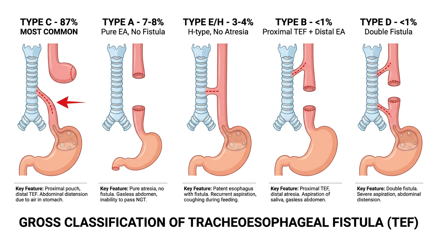
Quick Reference - All 5 Types:
| Type | Description | Frequency | Key Feature |
|---|---|---|---|
| Type C | EA + Distal TEF | 87% | Blind upper pouch + lower esophagus joins trachea near carina; bowel gas present on X-ray |
| Type A | Pure EA, no fistula | 7-8% | Both ends blind; gasless abdomen on X-ray; NG tube coils in upper mediastinum |
| Type E/H | H-type fistula, no atresia | 3-4% | Esophagus fully patent; presents later with recurrent chest infections + bloating |
| Type B | EA + Proximal TEF | <1% | Upper pouch connects to trachea; air may enter upper airway directly |
| Type D | EA + Double (proximal + distal) TEF | <1% | Both ends connected to trachea; most complex form |
Nursing Memory Aid - "3 Cs" of TEF presentation:
- Choking on feeds
- Coughing episodes
- Cyanosis (episodic, especially during feeding)
Signs and Symptoms
Signs and Symptoms of Tracheoesophageal Fistula (TEF)
Prenatal Signs (Before Birth)
| Sign | Significance |
|---|---|
| Polyhydramnios | Excess amniotic fluid because the fetus CANNOT swallow and absorb it (esophagus is blocked) |
| Absent or small stomach bubble on fetal USS | No fluid reaches the stomach due to esophageal atresia |
| Dilated proximal esophageal pouch | Visible on detailed fetal scan; high specificity for EA |
When polyhydramnios is present, an orogastric tube must be passed immediately after delivery to exclude TEF before the first feed.
- Barash Clinical Anesthesia 9e
At Birth / Immediately Postnatal Signs
1. Inability to Pass Nasogastric / Orogastric Tube
- The most important diagnostic sign
- The tube coils back in the upper esophageal pouch and cannot be advanced to the stomach
- On chest X-ray: tube seen curled at level of T2-T4 in upper mediastinum
- Pathognomonic of esophageal atresia
2. Chest X-Ray Finding
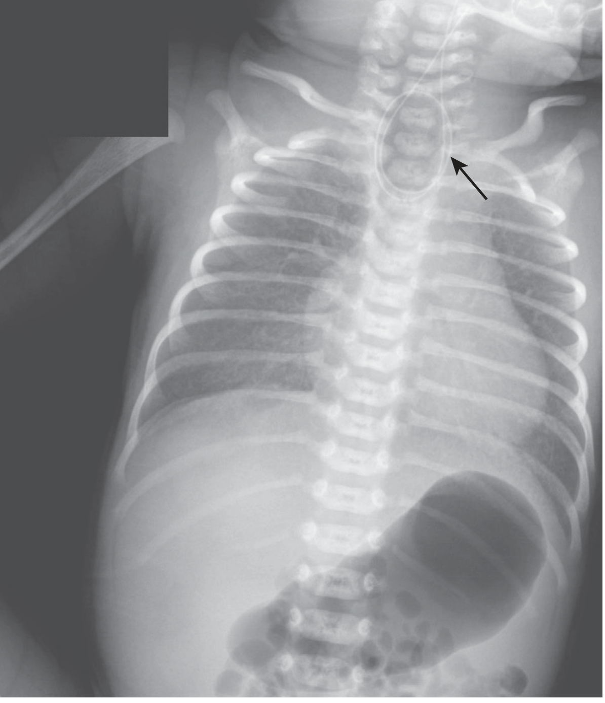
| X-ray Finding | Meaning |
|---|---|
| Tube coiled in upper mediastinum | Esophageal atresia confirmed |
| Gas-filled abdomen (bowel gas present) | Distal TEF present (Type C - most common) |
| Gasless abdomen | Pure EA (Type A) - no fistula to stomach |
Classic "3 Cs" of TEF - Cardinal Signs
     ┌──────────────────────────────────────┐
     │         THE 3 Cs OF TEF              │
     │                                      │
     │   1. COUGHING                        │
     │   2. CHOKING                         │
     │   3. CYANOSIS                        │
     │                                      │
     │   Triggered on first / every feed    │
     └──────────────────────────────────────┘
Complete Signs & Symptoms - System by System
A. Respiratory Signs (Most Prominent)
| Sign/Symptom | Mechanism | What You Observe |
|---|---|---|
| Coughing immediately on feeding | Fluid/milk passes through fistula into trachea | Sudden, forceful cough when milk is given |
| Choking on first feed | Aspiration of feed | Infant gags, splutters, cannot swallow |
| Cyanosis (blueness) | Aspiration → airway obstruction → ↓ O₂ | Lips, tongue, fingertips turn blue during feeds |
| Tachypnoea (fast breathing) | Aspiration pneumonitis, reduced lung volume | Respiratory rate > 60/min |
| Nasal flaring | Increased work of breathing | Nostrils widen with each breath |
| Intercostal / subcostal retractions | Accessory muscle use for breathing | Chest wall sucks in between ribs |
| Grunting respirations | Infant generating PEEP to maintain open airways | Audible grunt on expiration |
| Stridor / noisy breathing | Secretion pooling in airway or tracheomalacia | Harsh inspiratory noise |
| Apnoea | Severe aspiration | Breathing stops temporarily |
| SpO₂ drops during feeds | Aspiration | Pulse oximeter reading falls |
B. Gastrointestinal Signs
| Sign/Symptom | Mechanism | What You Observe |
|---|---|---|
| Excessive drooling / frothy secretions | Saliva pools in blind esophageal pouch and overflows | Bubbles/foam around mouth and nose - FIRST sign noticed by nurse |
| Immediate regurgitation of feeds | Food cannot pass to stomach; returns through blind pouch | Milk appears immediately after feed attempt |
| Abdominal distension | Air passes through fistula into stomach with each cry/breath | Abdomen becomes distended and tympanic |
| Inability to swallow | Upper esophagus ends in blind pouch | Infant appears to swallow but feed returns |
| Absent bowel gas (in pure EA, Type A) | No connection to GI tract | Gasless abdomen on X-ray |
C. General Signs
| Sign/Symptom | What You Observe |
|---|---|
| Inability to pass NG tube | Tube meets resistance at ~10-12 cm; cannot be advanced |
| Respiratory distress at rest | Present even between feeds due to accumulated secretions |
| Recurrent pneumonia | Particularly in H-type (Type E) TEF - delayed presentation |
| Poor feeding / failure to feed | Every attempt to feed results in coughing and cyanosis |
| Restlessness and irritability | Discomfort from secretion pooling and respiratory distress |
Signs by TEF Type - Comparison Table
| Type | Age at Presentation | Key Presenting Signs | X-ray Finding |
|---|---|---|---|
| Type C (EA + distal TEF) 87% | At birth / day 1 | Excessive drooling, coughing + cyanosis on first feed, abdominal distension | NG tube coiled in pouch + gas in abdomen |
| Type A (pure EA) 7-8% | At birth / day 1 | Excessive drooling, immediate regurgitation, NO abdominal distension | NG tube coiled in pouch + gasless abdomen |
| Type E / H-type 3-4% | Weeks to months later (can be diagnosed in adulthood) | Recurrent chest infections, coughing after feeds, bloating, bronchiectasis - NO obvious regurgitation | Normal esophagus on X-ray; diagnosed by contrast study or bronchoscopy |
| Type B (EA + proximal TEF) <1% | At birth | Recurrent pneumonia, air entering airway directly from upper pouch | Tube coiled + abnormal air entry |
| Type D (double fistula) <1% | At birth | Most severe; combination of Types B + C features | Variable |
- Sleisenger & Fordtran's GI and Liver Disease
How Symptoms Develop - Step-by-Step Mechanism
Baby attempts to feed (milk/fluid given)
              ↓
Fluid CANNOT pass down blind esophageal pouch
              ↓
Fluid overflows → enters trachea via fistula
              ↓
   ┌──────────────────────────────────┐
   │    ASPIRATION INTO LUNGS         │
   └───────────┬──────────────────────┘
               ↓
   ┌───────────┴──────────────────────┐
   ↓                                  ↓
COUGHING / CHOKING            CYANOSIS (↓ SpO₂)
(airway irritation)           (impaired gas exchange)
               ↓
   Gastric acid REFLUXES up through fistula
               ↓
   Chemical PNEUMONITIS (lung inflammation)
               ↓
   Respiratory distress worsens
               ↓
   ATELECTASIS → further ↓ in lung function
Very First Sign Noticed - Nursing Pearl
"Frothy white bubbles/foam around the mouth and nose of a newborn" is often the FIRST sign that prompts the bedside nurse to suspect TEF - before the first feed is even attempted.In many cases, it is the nursing staff who first identify the condition when they notice the accumulation of oral secretions during routine newborn care.
- Schwartz's Principles of Surgery 11e
Signs That Indicate Complications Have Set In
| Complication | Signs |
|---|---|
| Aspiration Pneumonitis | Fever, ↑ work of breathing, crackles on auscultation, haziness on chest X-ray |
| Severe respiratory failure | Persistent SpO₂ < 90%, grunting, severe retractions, apnoea |
| Dehydration | Sunken fontanelle, dry mucous membranes, ↓ urine output (Type A - cannot swallow at all) |
| Abdominal compartment syndrome | Tense, grossly distended abdomen making breathing impossible |
Differential Diagnosis (Conditions with Similar Signs)
| Condition | Differentiating Feature |
|---|---|
| Choanal atresia | Cyanosis relieved by crying (opens mouth); NG tube cannot pass through nose |
| Laryngomalacia | Stridor present from birth; worsens with feeds; no feeding failure |
| GOR (reflux) | Vomiting starts later, is not immediate; NG tube passes easily |
| Cleft palate | Visible on examination; nasal regurgitation of feeds |
| Laryngotracheoesophageal cleft | Similar symptoms + aphonia (cannot make sound/cry) |
Summary Table: Signs & Symptoms at a Glance
| Category | Signs & Symptoms |
|---|---|
| Prenatal | Polyhydramnios, absent stomach bubble on USS |
| First sign at birth | Frothy secretions/drooling from mouth |
| On first feed attempt | Coughing, choking, cyanosis (3 Cs) |
| Respiratory | Tachypnoea, nasal flaring, retractions, grunting, apnoea |
| GI | Regurgitation, abdominal distension (Type C), inability to feed |
| Diagnostic sign | NG tube cannot be passed to stomach |
| H-type (delayed) | Recurrent pneumonia, cough after feeds, bronchiectasis |
Medical and nursing management
Medical and Nursing Management of Tracheoesophageal Fistula (TEF)
SECTION A: MEDICAL MANAGEMENT
PHASE 1 - Immediate Stabilization (At Birth / On Diagnosis)
KEY PRINCIPLE: Surgical repair of TEF is NEVER an immediate emergency. Stabilization and workup come first.
- Schwartz's Principles of Surgery 11e
Step 1: Airway and Respiratory Stabilization
| Intervention | Dose / Detail | Rationale |
|---|---|---|
| Supplemental Oxygen | Via hood, nasal prongs or mask | Corrects hypoxia from aspiration |
| Positioning | Head-of-bed elevated ≥ 30° (upright or semi-prone) | Gravity prevents gastric acid reflux through fistula into lungs |
| NPO immediately | Nothing orally from time of diagnosis | Stops aspiration from feeds |
| Intubation + ventilation | Only if severe respiratory failure; use low pressure, low volume, high rate | High-pressure ventilation forces air through fistula → gastric distension → worsens lung expansion |
| Avoid mask positive pressure ventilation | If possible | Air enters stomach through fistula → acute gastric distension → diaphragm pushed up → respiratory collapse |
Step 2: Replogle Tube (Upper Pouch Suction) - Most Important Intervention
- Insert a double-lumen sump suction catheter (Replogle tube) into the upper esophageal pouch
- Connect to continuous low-pressure suction (~80 mmHg)
- Irrigate regularly with 1-2 mL normal saline to maintain patency
- Purpose: Prevents accumulation and aspiration of saliva pooling in the blind esophageal pouch
"A sump catheter is placed in the upper pouch on continuous suction. Ensuring the patency of the sump catheter minimizes coughing, bronchospasm, and desaturation episodes."
- Schwartz's Principles of Surgery 11e
Step 3: IV Access and Fluids
| Intervention | Detail |
|---|---|
| IV access | Avoid the right upper extremity - it may interfere with surgical positioning during repair |
| IV fluids | Warmed electrolyte solution (e.g., dextrose-saline) at maintenance rate |
| Central line | Considered for all patients - facilitates antibiotics and TPN if needed |
| Blood glucose monitoring | 4-6 hourly - neonates have poor glycogen reserves |
| Electrolytes | Monitor and correct sodium, potassium, chloride |
Step 4: Antibiotics
| Drug | Rationale |
|---|---|
| Broad-spectrum IV antibiotics (e.g., Ampicillin + Gentamicin) | Treat/prevent aspiration pneumonitis from pool of secretions and gastric acid reflux |
Step 5: Pre-operative Investigations (VACTERL Workup)
| Investigation | Purpose |
|---|---|
| Echocardiogram | Identify congenital heart disease (present in 15-35%); identify side of aortic arch (determines surgical approach - right or left chest) |
| Abdominal ultrasound | Detect renal anomalies |
| Spinal X-ray / USS | Identify vertebral anomalies |
| Chest + abdominal X-ray | Confirm tube position, check bowel gas, assess lungs |
| Blood group and cross-match | Pre-operative preparation |
| Full blood count + metabolic panel | Baseline values |
| Examination of anus | Confirm patent anus (exclude anal atresia) |
| Examination of limbs | Identify radial/limb anomalies |
PHASE 2 - Surgical Management
Timing of Surgery
| Patient Condition | Timing |
|---|---|
| Stable, well-oxygenating, no major cardiac anomaly | Definitive repair within 24-48 hours |
| Severe aspiration pneumonia present | Gastrostomy first → stabilize for days → then repair |
| Premature infant on ventilator | May need gastrostomy to decompress stomach first; delayed repair |
| Long-gap EA | Staged repair; esophageal lengthening procedures |
Surgical Options
| Step | Action |
|---|---|
| 1 | Position: left lateral decubitus (right side up) |
| 2 | Right extrapleural approach through 4th intercostal space |
| 3 | Identify and ligate and divide the fistula |
| 4 | Mobilize upper and lower esophageal ends |
| 5 | End-to-end esophagoesophagostomy (primary anastomosis) |
| 6 | Place trans-anastomotic feeding tube under direct vision |
| 7 | Secure anastomosis with interrupted sutures |
| 8 | Leave retropleural drain near anastomosis |
Note: Left thoracic approach used in the 5% of infants with a right-sided aortic arch.
- 3 ports in right chest
- CO₂ insufflation to deflate lung for visualization
- Same operative steps as open: fistula ligation + anastomosis
- Advantages: avoids musculoskeletal complications (winged scapula, scoliosis, breast deformity)
- Requires experienced pediatric surgical team
| Stage | Procedure |
|---|---|
| Stage 1 | Gastrostomy (stomach opened to skin for feeds + decompression) |
| Stage 2 | Esophageal lengthening (Foker technique: traction sutures to stretch esophageal ends over weeks) |
| Stage 3 | Delayed primary anastomosis once gap is bridged |
Post-operative Medical Management
| Intervention | Detail |
|---|---|
| Mechanical ventilation | Low pressure, low volume, high rate settings; avoid neck extension |
| Trans-anastomotic tube feeding | Commenced 24-48 hours post-op; bypasses anastomosis |
| IV antibiotics | Continued post-operatively |
| Contrast esophagram | Performed at Day 5-7 post-op to confirm anastomosis integrity before oral feeds |
| Oral feeding | Started only after esophagram confirms no leak |
| Proton pump inhibitor / antacid | Ranitidine or Omeprazole - prevents GERD (nearly universal after repair) |
| Retropleural drain | Left in situ; removed once no leak confirmed |
Medical Management of Complications
| Complication | Medical / Surgical Treatment |
|---|---|
| Anastomotic leak (10-20%) | Keep NBM, leave drain in situ, IV antibiotics, serial contrast studies; most heal without re-operation |
| Anastomotic stricture (up to 40%) | Balloon or bougie dilation under endoscopic/fluoroscopic guidance (may need repeated sessions) |
| Recurrent TEF (up to 10%) | Endoscopic ablation with fibrin glue/electrocautery OR surgical re-division with tissue interposition |
| GERD (nearly all patients) | Thickened feeds, positioning, PPI/H₂ blocker; surgical fundoplication if refractory |
| Tracheomalacia (~10%) | Mild - improve with time; Severe - aortopexy (surgical suspension) or tracheostomy; severe "dying spells" require intubation |
| Esophageal dysmotility | Thickened feeds, upright positioning, prokinetics |
SECTION B: NURSING MANAGEMENT
PRE-OPERATIVE NURSING CARE
Priority Nursing Diagnoses (Pre-op):
- Risk for Aspiration r/t pooled secretions in blind esophageal pouch and gastric acid reflux through fistula
- Impaired Gas Exchange r/t aspiration pneumonitis
- Risk for Deficient Fluid Volume r/t NPO status and inability to feed
- Anxiety (parental) r/t sudden serious diagnosis and surgical plan
- Hypothermia Risk r/t neonatal inability to regulate temperature
Pre-operative Nursing Interventions
| Nursing Action | Rationale |
|---|---|
| Position infant at 30-45° head elevation at all times | Gravity reduces reflux of gastric acid through fistula; reduces aspiration risk |
| Maintain Replogle tube patency - check position, irrigate with 1-2 mL NS, ensure continuous suction | Keeps blind pouch drained; single most important measure to prevent aspiration |
| Gentle oropharyngeal suctioning with small catheter as needed | Clears secretions pooling in mouth and pharynx |
| Suction equipment at bedside at ALL times | Immediate response to sudden secretion accumulation or vomiting |
| Monitor SpO₂ continuously with pulse oximeter | Detects desaturation early; TEF infants can deteriorate rapidly |
| Assess respiratory rate, rhythm, breath sounds every 1-2 hours | Identifies developing aspiration pneumonitis (crackles, wheeze, tachypnoea) |
| Administer supplemental oxygen as prescribed | Maintains SpO₂ ≥ 94-95% |
| Keep bag-valve-mask and oxygen at bedside | Emergency respiratory support |
| Nursing Action | Rationale |
|---|---|
| Maintain strict NPO - no oral feeds under any circumstances | Any oral feed causes immediate aspiration |
| Maintain and document IV fluid administration | Only source of hydration and glucose pre-operatively |
| Strict intake and output chart - count wet diapers (note: urine output ≥ 1-2 mL/kg/hr expected) | Monitors fluid balance; dehydration can develop rapidly |
| Monitor blood glucose 4-6 hourly | Neonates at risk of hypoglycaemia; dextrose in IV fluids essential |
| Weigh infant daily on same scale | Detects fluid loss or retention |
| Offer pacifier (non-nutritive sucking) | Satisfies infant's sucking reflex; provides comfort |
| Nursing Action | Rationale |
|---|---|
| Place infant in warm isolette or under radiant warmer | Neonates cannot thermoregulate; hypothermia worsens respiratory status |
| Monitor axillary temperature every 2-4 hours (normal: 36.5-37.5°C) | Detects hypo/hyperthermia |
| Minimize unnecessary exposure during procedures | Prevents heat loss |
| Nursing Action | Rationale |
|---|---|
| Administer IV antibiotics as prescribed (on time) | Treats aspiration pneumonitis; prevents sepsis |
| Maintain aseptic technique for all procedures | Immunocompromised neonate; high infection risk |
| Monitor for signs of infection: fever, ↑ WBC, lethargy, poor perfusion | Early identification of sepsis |
| Parameter | Frequency | Normal Values |
|---|---|---|
| Vital signs (HR, RR, BP, Temp) | Every 1-2 hours | HR: 120-160; RR: 30-60; Temp: 36.5-37.5°C |
| SpO₂ | Continuous | ≥ 94-95% |
| Blood glucose | Every 4-6 hours | 2.6-6.0 mmol/L |
| Replogle tube drainage | Every 4 hours | Document amount, colour, consistency |
| Urine output | Every 4-6 hours | ≥ 1-2 mL/kg/hour |
| Electrolytes | As ordered | Monitor Na, K, Cl, HCO₃ |
| Item | Action |
|---|---|
| Consent | Informed written consent obtained from parents/guardian |
| IV access | Patent, secure - avoid right arm |
| Blood group and cross-match | Sent to lab |
| Replogle tube | In situ, patent, on suction |
| NPO confirmed | No oral feeds |
| Pre-op weight documented | Baseline for surgery and drug dosing |
| Echocardiogram done | Confirmed cardiac status and aortic arch side |
| VACTERL workup completed | All investigations done |
| Parental education | Surgery explained, consent process completed |
| Identification bands | Correctly labelled on infant |
| Pre-operative blood results | Reviewed and reported to surgeon |
| Action | Detail |
|---|---|
| Explain TEF in simple terms | "The food pipe and airway are abnormally connected; surgery will separate them" |
| Reassure about surgical outcomes | >90% survival; most children lead normal lives |
| Explain why NPO and Replogle tube are needed | Parents often distressed seeing baby with tubes |
| Encourage parents to touch, talk to, and bond with baby | Skin-to-skin if possible and safe |
| Teach parents to recognize warning signs | Sudden worsening of breathing → call nurse immediately |
| Address guilt and anxiety | Many parents blame themselves; reassure it is not caused by anything they did |
POST-OPERATIVE NURSING CARE
Priority Nursing Diagnoses (Post-op):
- Acute Pain r/t surgical incision and chest drain
- Risk for Impaired Gas Exchange r/t post-operative atelectasis and secretions
- Risk for Infection r/t surgical wound, chest drain, and IV access sites
- Imbalanced Nutrition: Less Than Body Requirements r/t NPO status and surgical recovery
- Risk for Aspiration r/t anastomotic leak and GERD
Post-operative Nursing Interventions
| Nursing Action | Rationale |
|---|---|
| Keep neck in neutral or slightly flexed position at all times | Neck extension puts tension on fresh anastomotic suture line → anastomotic dehiscence risk |
| Place a neck roll under shoulders to maintain neutral position | Prevents inadvertent neck extension during sleep |
| Continue head elevation at 30° | Reduces GERD and aspiration risk |
| Document and communicate positioning requirements to ALL staff and parents | Prevents accidental neck hyperextension |
| Nursing Action | Rationale |
|---|---|
| Monitor SpO₂ continuously; observe for respiratory distress | Post-op atelectasis and secretions are common |
| Gentle chest physiotherapy as ordered | Loosens secretions and prevents atelectasis |
| DO NOT perform blind nasopharyngeal/nasogastric suctioning without surgeon's explicit order | Risk of traumatizing or perforating the fresh esophageal anastomosis |
| Monitor for stridor or barking cough | May indicate tracheomalacia |
| Assess breath sounds every 2-4 hours | Monitors for pneumonia or pneumothorax |
| Wean oxygen as tolerated per SpO₂ | Gradual removal of respiratory support |
| Nursing Action | Rationale |
|---|---|
| Inspect thoracotomy/VATS wound every 4-8 hours | Signs of infection: redness, warmth, swelling, discharge |
| Maintain retropleural chest drain - document amount and character of drainage | Saliva or milky fluid in drain = anastomotic leak (report immediately) |
| Keep drain below level of chest; avoid kinking | Ensures effective drainage |
| Perform aseptic dressing changes | Infection prevention |
| Do not remove drain until surgeon confirms anastomosis is intact on contrast study | Premature removal risks collection formation |
| Nursing Action | Rationale |
|---|---|
| Assess pain using infant pain scale (CRIES / NIPS / FLACC) every 2-4 hours | Neonates cannot verbalize pain |
| Administer prescribed analgesics on schedule (Paracetamol, Morphine/Fentanyl infusion) | Adequate pain control reduces stress response and improves recovery |
| Non-pharmacological comfort: skin-to-skin, pacifier, swaddling, gentle touch | Reduces opioid requirement; promotes bonding |
| Monitor for respiratory depression with opioid use | Neonates are sensitive to opioid respiratory side-effects |
| Nursing Action | Rationale |
|---|---|
| Administer trans-anastomotic tube feeds as prescribed (commenced 24-48 hours post-op) | Bypasses anastomosis; provides nutrition while it heals |
| Start at low rate; increase gradually per feeding protocol | Prevents gastric distension and aspiration |
| Document feed tolerance: abdominal distension, aspirate, vomiting | Identifies feeding intolerance early |
| Prepare for and assist with contrast esophagram at Day 5-7 | Confirms anastomosis is intact before oral feeds are introduced |
| Introduce oral feeds only after clear esophagram confirmed by surgeon | Patient safety - prevents feeding into a leaking anastomosis |
| Position upright after all feeds | Reduces GERD (present in nearly all post-repair patients) |
| Monitor weight daily | Tracks nutritional recovery |
| Parameter | Frequency |
|---|---|
| Vital signs | Every 1-2 hours initially, then every 4 hours when stable |
| SpO₂ | Continuous |
| Chest drain output | Every 4 hours - note amount, colour, character |
| Urine output | Every 4-6 hours (target ≥ 1-2 mL/kg/hr) |
| Blood glucose | Every 6-8 hours until feeding established |
| Wound inspection | Every 8 hours |
| Pain assessment | Every 2-4 hours |
| Feed tolerance | With each feed |
NURSING MANAGEMENT OF POST-OP COMPLICATIONS
| Complication | Nursing Signs to Detect | Nursing Response |
|---|---|---|
| Anastomotic leak | Saliva/milky fluid in chest drain; fever; respiratory deterioration; tachycardia | Immediately inform surgeon; keep NBM; ensure drain patent; prepare for investigation |
| Anastomotic stricture | Gagging/choking on feeds (weeks-months later); feed refusal; regurgitation | Document and report; prepare for contrast study or endoscopic dilation |
| Tracheomalacia | Barking/brassy cough; expiratory wheeze; "dying spell" (sudden severe cyanosis, stridor, unconsciousness when agitated) | Dying spell = emergency: intubate immediately; call surgeon; prepare for resuscitation; keep infant calm |
| GERD | Vomiting after feeds; respiratory symptoms; arching after feeds | Positioning; thicken feeds per order; administer PPI; document |
| Recurrent TEF | Return of coughing/choking on feeds after previously tolerating well | Report immediately; prepare for bronchoscopy/esophagram |
| Wound infection | Redness, warmth, swelling, discharge from incision | Culture wound; administer antibiotics; wound care |
| Pneumonia | Fever, ↑ respiratory rate, crackles, SpO₂ drop | Chest physiotherapy; antibiotics; O₂ support; report to team |
DISCHARGE PLANNING AND PARENTAL EDUCATION
| Topic | Teaching Points |
|---|---|
| Feeding | Small, frequent feeds; thicken feeds if advised; upright positioning for 30 min after feeds |
| Positioning | Avoid neck extension; upright after feeds |
| GERD medications | Explain purpose, dose, frequency of PPI/antacid |
| Warning signs | Return to hospital immediately for: choking on feeds, blueness, breathing difficulty, fever, refusal to feed |
| Follow-up | Regular outpatient review for contrast studies, dilation if stricture develops, developmental assessment |
| Feeding difficulties | Referral to speech therapist for feeding assessment and support |
| Emotional support | Connect family with TEF support groups; acknowledge the journey has been difficult |
Summary: Management at a Glance
DIAGNOSIS CONFIRMED
        ↓
IMMEDIATE STABILIZATION
• Head-up 30-45°
• Replogle tube on suction
• IV access (not right arm)
• NPO strictly
• Supplemental oxygen
• IV antibiotics
• IV fluids + glucose
        ↓
VACTERL WORKUP
• Echo (cardiac + aortic arch)
• Renal/spinal USS
• Vertebral X-ray
        ↓
SURGICAL REPAIR (24-48 hrs if stable)
• Open thoracotomy OR VATS
• Fistula ligation + esophageal anastomosis
        ↓
POST-OPERATIVE CARE
• Neutral neck position
• Trans-anastomotic feeds 24-48 hrs
• Contrast esophagram Day 5-7
• Oral feeds when anastomosis confirmed intact
• GERD management
• Monitor for tracheomalacia
        ↓
DISCHARGE + LONG-TERM FOLLOW-UP
• GERD, stricture, dysmotility surveillance
• Speech therapy
• Developmental assessment
Meningitis child health nursing essay
bacterial meningitis child brain inflammation CSF
meningitis rash purpura petechiae child neck stiffness
MENINGITIS IN CHILDREN
A Comprehensive Essay for Child Health Nursing
INTRODUCTION
1. DEFINITION
- Bacterial meningitis - the most serious form; requires urgent antibiotic therapy
- Viral (aseptic) meningitis - most common type; usually self-limiting
- Tuberculous meningitis - chronic form; caused by Mycobacterium tuberculosis
- Fungal meningitis - in immunocompromised children
"Meningitis is inflammation of the meninges, the membranes that cover the brain and spinal cord. They contain the CSF, which bathes the brain. To enter this space, pathogens must cross the blood-brain barrier."
- Tietz Textbook of Laboratory Medicine 7e
2. EPIDEMIOLOGY AND INCIDENCE
| Parameter | Data |
|---|---|
| Most vulnerable age group | Neonates, infants under 2 years, and adolescents |
| Peak incidence | Neonates (first month of life) and infants |
| Global burden | A major cause of childhood mortality worldwide |
| Mortality with treatment | Reduced to < 10% with antibiotics and supportive care |
| Long-term disability | Up to 20-30% of survivors have neurological sequelae |
3. ETIOLOGY - CAUSATIVE ORGANISMS BY AGE GROUP
| Age Group | Common Organisms |
|---|---|
| Neonates (0-30 days) | Group B Streptococcus (GBS), Escherichia coli, Listeria monocytogenes, Herpes Simplex Virus (HSV), Klebsiella, Enterobacter |
| Infants (1-3 months) | GBS, E. coli, S. pneumoniae, N. meningitidis, H. influenzae |
| Older infants & children (3 months - 12 years) | S. pneumoniae (pneumococcus), N. meningitidis (meningococcus), H. influenzae type b (now rare due to vaccine) |
| Adolescents | N. meningitidis, S. pneumoniae |
| Immunocompromised | Cryptococcus neoformans (fungal), Mycobacterium tuberculosis, Listeria |
"S. pneumoniae and N. meningitidis are the most common causes of community-acquired acute bacterial meningitis... colonizing the nasopharyngeal mucosa, which provides a route of entry."
- Bradley and Daroff's Neurology in Clinical Practice
Risk Factors for Meningitis in Children:
- Prematurity and low birth weight
- Immunodeficiency (congenital or acquired)
- Maternal colonization with GBS or HSV
- Delivery complications
- Anatomical defects: dermal sinus tracts, open neural tube defects, cochlear implants
- Sinusitis, otitis media (direct spread)
- Trauma or neurosurgical procedures
- Unvaccinated status
- Crowded living conditions
- Viral respiratory infections (increase nasopharyngeal colonization risk)
4. PATHOPHYSIOLOGY
Colonization of NASOPHARYNX by organism
(S. pneumoniae, N. meningitidis, etc.)
               ↓
Organism invades bloodstream → BACTEREMIA
               ↓
Organism crosses BLOOD-BRAIN BARRIER
(barrier breaks down due to infection/inflammation)
               ↓
Organism enters SUBARACHNOID SPACE
               ↓
CSF has LOW COMPLEMENT, LOW ANTIBODIES
→ Organisms multiply rapidly without immune control
               ↓
Bacterial toxins and cell wall components released
               ↓
INFLAMMATORY RESPONSE triggered
(cytokines, leukocyte infiltration, ↑ vascular permeability)
               ↓
    ┌──────────────────────────────────────┐
    ↓                                      ↓
CEREBRAL OEDEMA              RAISED INTRACRANIAL PRESSURE (ICP)
(brain swelling)              (herniation risk)
    ↓                                      ↓
MENINGEAL IRRITATION         IMPAIRED CEREBRAL PERFUSION
(neck stiffness, Kernig,      (altered consciousness, seizures)
Brudzinski signs)
               ↓
COMPLICATIONS: Hydrocephalus, SIADH, Septicaemia,
Waterhouse-Friderichsen syndrome, DIC, Hearing loss
"Meningitis starts with breakdown of the blood-brain barrier. Once organisms enter the subarachnoid space, they multiply quickly because the CSF has low levels of complement, antibodies, and other host defenses. Bacterial cell wall components and toxins produce an inflammatory response responsible for much of the damage."
- Tintinalli's Emergency Medicine
5. CLINICAL FEATURES - SIGNS AND SYMPTOMS
A. In Neonates (0-30 days) - Signs are NON-SPECIFIC:
| Sign / Symptom | Description |
|---|---|
| Fever OR hypothermia | Neonates may not mount a fever (temperature instability) |
| Lethargy / poor activity | Baby less responsive than usual |
| Poor feeding | Refuses feeds or feeding poorly |
| Bulging fontanelle | Most important sign in neonates - indicates raised ICP |
| Abnormal cry | High-pitched, inconsolable cry |
| Seizures | Focal or generalized convulsions |
| Mottling / poor perfusion | Signs of sepsis |
| Respiratory distress / grunting | Non-specific but concerning |
| Jaundice | May be present |
| Vomiting and diarrhea | Non-specific GI signs |
| Increased or decreased tone | Neurological involvement |
"Some neonates present with few symptoms early in the course of their illness, so maintain a high degree of suspicion for early meningitis when confronted with a potentially sick newborn."
- Tintinalli's Emergency Medicine
B. In Older Infants (>90 days) and Children (>3 years):
Classic Triad of Meningitis:
FEVER + NECK STIFFNESS + ALTERED CONSCIOUSNESS
| Sign / Symptom | Description / Mechanism |
|---|---|
| Fever | High grade; may be sudden onset |
| Severe headache | Due to meningeal irritation and raised ICP |
| Neck stiffness (nuchal rigidity) | Resistance to passive neck flexion; meningeal irritation |
| Photophobia | Sensitivity to light; meningeal irritation |
| Phonophobia | Sensitivity to sound |
| Vomiting | Projectile; from raised ICP; not preceded by nausea |
| Altered level of consciousness | Drowsiness, confusion, coma |
| Seizures | Generalized or focal; raised ICP + cortical irritation |
| Irritability | Especially in infants - increases when picked up/held |
| Bulging fontanelle | In infants; indicates raised ICP |
| Petechial / purpuric rash | EMERGENCY - suggests N. meningitidis (meningococcal) septicaemia |
C. Two Key Clinical Signs (Meningeal Irritation Signs):
| Sign | How to Test | Positive Result |
|---|---|---|
| Kernig's Sign | Patient supine; flex hip to 90°; attempt to extend the knee | Inability to extend knee fully without pain = POSITIVE |
| Brudzinski's Sign | Patient supine; passively flex the neck forward | Involuntary flexion of both hips and knees = POSITIVE |
"Nuchal rigidity and Kernig or Brudzinski signs may be absent in children with meningitis even up to the age of 2 years old. A bulging fontanelle, vomiting, irritability that increases when the infant is held, inconsolability, or a complex febrile seizure may be the only signs suggestive of meningitis."
- Tintinalli's Emergency Medicine
D. Waterhouse-Friderichsen Syndrome (Meningococcal Emergency):
| Feature | Description |
|---|---|
| Causative organism | Neisseria meningitidis |
| Key sign | Rapidly spreading non-blanching purpuric rash (purpura fulminans) |
| Associated features | Shock, adrenal haemorrhage, DIC, multi-organ failure |
| Prognosis | Very high mortality if not treated within hours |
6. DIAGNOSIS AND INVESTIGATIONS
A. Lumbar Puncture and CSF Analysis (Gold Standard)
| CSF Finding | Bacterial Meningitis | Viral Meningitis | Normal |
|---|---|---|---|
| Appearance | Turbid/cloudy/purulent | Clear or slightly turbid | Clear |
| Opening pressure | Elevated (>200 mmH₂O) | Normal or slightly elevated | 70-180 mmH₂O |
| WBC count | >1000/mm³ (predominantly neutrophils) | 10-500/mm³ (lymphocytes) | 0-5/mm³ |
| Protein | Markedly ↑ (>100 mg/dL) | Normal or slightly ↑ | 15-45 mg/dL |
| Glucose | ↓ (<40 mg/dL or <50% serum) | Normal | 50-80 mg/dL |
| Gram stain | Organisms visible (60-90%) | Negative | Negative |
| Culture | Positive for organism | Negative | Negative |
| Finding | Points |
|---|---|
| Positive CSF Gram stain | 2 |
| CSF protein > 80 mg/dL | 1 |
| Blood absolute neutrophil count ≥ 10,000/mm³ | 1 |
| Seizure at or before presentation | 1 |
| Score ≥ 2 = High risk bacterial meningitis |
B. Blood Investigations
| Test | Purpose |
|---|---|
| Blood cultures (x2, before antibiotics if possible) | Isolate causative organism |
| Full Blood Count | ↑ WBC (neutrophilia in bacterial); ↓ WBC (poor prognosis) |
| Blood glucose (bedside) | Compare with CSF glucose; rule out hypoglycaemia |
| CRP / Procalcitonin | Elevated in bacterial infection |
| Serum electrolytes | Detect hyponatraemia (SIADH) |
| Coagulation screen (PT, APTT) | Detect DIC (in meningococcal disease) |
| Renal and liver function | Baseline; monitor organ function |
| Blood urea, creatinine | Monitor renal function |
C. Other Investigations
| Investigation | Purpose |
|---|---|
| CT Brain (before LP if signs of raised ICP) | Exclude herniation risk before LP; detect abscess, hydrocephalus |
| Chest X-ray | Identify pneumonia as primary source |
| Urine culture | Identify urinary source in neonates |
| MRI Brain | Detailed assessment of complications (cerebritis, abscess, ventriculitis) |
| EEG | If seizures occur |
| Audiometry (later) | Assess for sensorineural hearing loss post-recovery |
Note: "Although establishing a diagnosis is important, patients who are unstable but suspected of having bacterial meningitis should receive antibiotics as quickly as possible. Defer lumbar puncture until the child is stabilized."
- Tintinalli's Emergency Medicine
7. MEDICAL MANAGEMENT
Principle: TIME IS BRAIN - Antibiotics must NOT be delayed
SUSPECT MENINGITIS
       ↓
Do NOT delay antibiotics for LP if child is unstable
       ↓
BLOOD CULTURES → ANTIBIOTICS → LP (when stable) → CT if needed
A. Antibiotic Therapy
| Age Group | Empiric Antibiotic Choice | Rationale |
|---|---|---|
| Neonates (0-30 days) | Ampicillin + Cefotaxime OR Ampicillin + Gentamicin | Covers GBS, E. coli, Listeria |
| Infants 1-3 months | Ampicillin + Cefotaxime + Vancomycin | Covers neonatal + older childhood organisms |
| Children > 3 months | Ceftriaxone or Cefotaxime + Vancomycin | Covers S. pneumoniae, N. meningitidis, H. influenzae |
| Suspected HSV | Add Acyclovir | HSV encephalitis/meningitis in neonates and infants |
| Tuberculosis suspected | Isoniazid + Rifampicin + Pyrazinamide + Ethambutol (4-drug) | Tuberculous meningitis protocol |
| Immunocompromised | Add Amphotericin B (if fungal suspected) | Cryptococcal meningitis |
"Antibiotics may have difficulty penetrating the blood-brain barrier. Therefore, doses used to treat meningitis are frequently higher than the doses used for other pediatric infections."
- Tintinalli's Emergency Medicine
- N. meningitidis: 5-7 days
- H. influenzae: 7-10 days
- S. pneumoniae: 10-14 days
- GBS / gram-negative bacilli (neonatal): 14-21 days
- Tuberculous meningitis: 9-12 months
B. Dexamethasone (Corticosteroids)
| Aspect | Detail |
|---|---|
| Dose | 0.15 mg/kg IV every 6 hours for 2-4 days |
| Timing | Must be given before or with the first dose of antibiotics to be effective |
| Indication | H. influenzae type b meningitis (reduces hearing loss); consider for S. pneumoniae (reduces mortality in high-income settings) |
| Mechanism | Reduces CNS inflammatory response; decreases labyrinthitis → less sensorineural hearing loss |
| Limitation | Not routinely recommended for all bacterial meningitis types by the American Academy of Pediatrics |
C. Supportive Medical Management
| Intervention | Detail | Rationale |
|---|---|---|
| IV fluids | Isotonic fluids (0.9% NS); careful fluid management | Avoid fluid overload (worsens cerebral oedema); maintain perfusion |
| Management of SIADH | Fluid restriction; monitor urine output and serum Na | SIADH (inappropriate ADH) → hyponatraemia → cerebral oedema |
| Anti-seizure medications | Phenobarbitone (neonates), Levetiracetam or Phenytoin (older children) as prescribed | Control seizures; prevent secondary brain injury |
| Management of raised ICP | Head elevation 30°; mannitol (0.25-1 g/kg IV) if herniation risk; hyperventilation if needed | Prevent cerebral herniation |
| Glucose management | Monitor blood glucose 4-6 hourly; dextrose if hypoglycaemic | Sepsis causes glucose depletion; brain is glucose-dependent |
| Management of shock | 20 mL/kg isotonic saline bolus; inotropes if required (Dopamine/Dobutamine) | Meningococcal septicaemia causes profound septic shock |
| Respiratory support | Oxygen; intubation + mechanical ventilation if GCS ≤ 8 or respiratory failure | Maintains cerebral oxygenation; protects airway |
| Chemoprophylaxis (contacts) | Rifampicin OR Ciprofloxacin for close contacts of N. meningitidis | Eradicates nasopharyngeal carriage; prevents secondary cases |
8. NURSING MANAGEMENT
Priority Nursing Diagnoses:
- Risk for Ineffective Cerebral Tissue Perfusion r/t cerebral oedema and raised ICP
- Hyperthermia r/t infectious process and inflammatory response
- Acute Pain r/t meningeal irritation (headache, photophobia, neck stiffness)
- Risk for Injury: Seizures r/t cerebral irritation
- Deficient Fluid Volume / Risk for Fluid Volume Excess r/t fever, vomiting, SIADH
- Disturbed Sensory Perception r/t photophobia, phonophobia, altered consciousness
- Anxiety (parental) r/t sudden life-threatening illness
A. Neurological Nursing Care
| Nursing Action | Rationale |
|---|---|
| Perform neuro observations every 1-2 hours using Glasgow Coma Scale (GCS) - or AVPU scale for young children | Detects early deterioration in level of consciousness; rising ICP causes progressive GCS decline |
| Monitor for signs of raised ICP: ↑ BP + ↓ HR + irregular respirations (Cushing's triad); bulging fontanelle; dilated, unresponsive pupils; posturing | Signs of impending brain herniation = medical emergency |
| Elevate head of bed 30° in midline position | Promotes venous drainage from brain; reduces ICP |
| Assess and document neurological observations: pupil size, reaction, equality; eye movements; limb movements; tone | Focal deficits indicate localized brain damage |
| Protect from falls / injury - padded cot sides, call bell within reach | Altered consciousness and seizures increase fall/injury risk |
| Seizure precautions: padded side rails; oxygen and suction at bedside; seizure protocol ready | Seizures occur in up to 30% of children with meningitis |
| During seizure: time it, protect airway, turn to recovery position, DO NOT restrain, call for help | Prevents aspiration and injury; documents seizure duration |
| Administer anti-seizure medications as prescribed; monitor for side effects | Controls seizures; prevents further brain injury |
B. Fever Management
| Nursing Action | Rationale |
|---|---|
| Monitor temperature every 2-4 hours (axillary in neonates, oral/rectal in older children) | High fever increases cerebral metabolic demand and worsens brain injury |
| Administer antipyretics as prescribed (Paracetamol 15 mg/kg every 4-6 hours) | Reduces fever; reduces cerebral oxygen consumption |
| Tepid sponging (30-32°C water) - do NOT use cold water or alcohol | Reduces body temperature without causing vasoconstriction or shivering |
| Remove excess clothing and bedding | Facilitates heat dissipation |
| Maintain adequate fluid intake (IV) | Fever increases insensible fluid loss |
| Monitor for febrile seizures | High fever is a common seizure trigger in children under 6 years |
C. Comfort and Environmental Care
| Nursing Action | Rationale |
|---|---|
| Darken the room / dim lights | Reduces photophobia pain |
| Reduce noise - close doors, speak softly, limit visitors | Reduces phonophobia discomfort |
| Position in comfortable position - side-lying with knees slightly flexed, neck in comfortable neutral position | Reduces tension on meninges; reduces headache and neck pain |
| Administer analgesics as prescribed (Paracetamol, Ibuprofen; Opioids for severe pain) | Meningeal irritation causes severe headache and neck pain |
| Handle infant gently; avoid unnecessary stimulation | Irritability increases when held or stimulated |
| Maintain quiet environment and cluster nursing care activities | Reduces frequency of disturbing the child; minimizes ICP spikes |
D. Fluid and Electrolyte Management
| Nursing Action | Rationale |
|---|---|
| Maintain strict intake and output chart; weigh daily | Monitors fluid balance; SIADH causes fluid retention |
| Monitor electrolytes - especially serum sodium | SIADH → hyponatraemia → worsening cerebral oedema; target Na 135-145 mEq/L |
| In confirmed SIADH: fluid restrict as ordered | Prevents dilutional hyponatraemia from worsening |
| Monitor urine output (target ≥ 1 mL/kg/hr) | Assesses hydration and renal function |
| Administer IV fluids as prescribed (isotonic); avoid hypotonic fluids | Hypotonic fluids worsen hyponatraemia and cerebral oedema |
| Monitor blood glucose 4-6 hourly; treat hypoglycaemia with dextrose | Brain requires constant glucose supply; sepsis depletes glucose |
E. Infection Control and Isolation
| Nursing Action | Rationale |
|---|---|
| Droplet precautions for all bacterial meningitis (especially N. meningitidis) for first 24 hours of antibiotics | N. meningitidis spreads via respiratory droplets; protect staff, family, other patients |
| Surgical mask for anyone within 1 metre of child | Droplet transmission prevention |
| Strict hand hygiene (before and after each contact) | Prevents transmission of causative organisms |
| Administer antibiotics at prescribed times exactly | Maintaining therapeutic blood/CSF drug levels is critical |
| Monitor antibiotic administration: ensure IV access is patent; check allergies | Ensures uninterrupted delivery of life-saving treatment |
| Chemoprophylaxis: notify public health; ensure household contacts receive prophylaxis (N. meningitidis) | Prevents secondary cases in close contacts |
| Lift droplet precautions after 24 hours of effective antibiotic therapy | Organism is typically no longer transmissible |
F. Monitoring and Vital Signs
| Parameter | Frequency | Alert Values |
|---|---|---|
| Vital signs (HR, BP, RR, Temp) | Every 1-2 hours (ICU) or every 4 hours (stable) | Cushing's triad: ↑BP + ↓HR + irregular RR |
| GCS / AVPU | Every 1-2 hours | GCS ≤ 8 = secure airway |
| Pupil assessment | Every 1-2 hours | Unequal, dilated, non-reactive pupils = herniation |
| SpO₂ | Continuous | < 94% = increase O₂; report |
| Blood glucose | Every 4-6 hours | < 2.6 mmol/L = hypoglycaemia; treat |
| Serum sodium | 6-12 hourly | < 130 mEq/L = severe hyponatraemia |
| Urine output | Hourly if critically ill | < 1 mL/kg/hr = inadequate |
| Rash assessment | Every 1-2 hours | New/spreading petechiae/purpura = emergency |
G. Lumbar Puncture (LP) Nursing Role
| Before LP | During LP | After LP |
|---|---|---|
| Explain procedure to parents | Assist child into correct position (lateral foetal position OR seated leaning forward) | Monitor puncture site for bleeding or CSF leak |
| Ensure consent obtained | Maintain position firmly and calmly; reassure child | Nurse flat for 1-2 hours (reduces post-LP headache) |
| Check for contraindications (raised ICP, coagulopathy) | Label and send CSF specimens immediately to lab (in correct order: tube 1-biochemistry, tube 2-microbiology, tube 3-cell count) | Monitor for headache, neurological changes |
| Gather equipment aseptically | Observe for distress or deterioration | Encourage fluids (if permitted) |
H. Parental Support and Education
| Topic | Nursing Action |
|---|---|
| Explain diagnosis | Use simple, clear language; "The covering of the brain is infected and inflamed" |
| Explain treatment plan | Antibiotics, monitoring, duration of stay |
| Encourage involvement | Parents can hold infant (if stable), talk to child, participate in care |
| Address fear and anxiety | Acknowledge severity; reassure about treatment; do not give false promises about outcomes |
| Chemoprophylaxis | Teach family members about need for rifampicin/ciprofloxacin prophylaxis |
| Warning signs of complications | Hearing difficulty, developmental delay, seizures - report at any point during recovery |
| Vaccination | Educate about meningitis vaccines (pneumococcal, meningococcal, Hib) for prevention |
| Discharge planning | Follow-up hearing test (audiology), developmental assessment, outpatient neurology |
9. COMPLICATIONS
| Complication | Description | Nursing Consideration |
|---|---|---|
| Sensorineural Hearing Loss | Most common; caused by labyrinthitis | Arrange post-discharge audiology testing |
| Hydrocephalus | Obstruction of CSF flow by inflammatory exudate | Monitor head circumference in infants; assess fontanelle; arrange imaging |
| Seizures | Cortical irritation; may become chronic epilepsy | Anti-epileptic therapy; seizure safety education |
| Cognitive impairment / Learning disability | Direct brain damage | Long-term developmental monitoring; school support |
| Cerebral abscess | Localised collection of pus | Monitor for focal neurological signs; prepare for surgical drainage |
| Subdural effusion / empyema | Fluid collection under dura | Serial imaging; surgical drainage if symptomatic |
| Septic shock / DIC | Meningococcal disease especially | Intensive monitoring; blood products; vasopressors |
| SIADH | Inappropriate ADH → hyponatraemia | Fluid restriction; electrolyte monitoring |
| Waterhouse-Friderichsen syndrome | Bilateral adrenal haemorrhage in meningococcal disease | Hydrocortisone replacement; ICU care |
| Death | < 10% with treatment; higher with delayed diagnosis | Early recognition saves lives |
"Survivors can experience sensorineural hearing loss, visual impairment, seizures, hydrocephalus, cognitive impairment, learning disabilities, and emotional problems. Factors predicting mortality include coma, seizures, shock, respiratory distress, neutropenia, and a high protein level in CSF."
- Tintinalli's Emergency Medicine
10. PREVENTION AND VACCINATION
| Vaccine | Protects Against | Schedule |
|---|---|---|
| PCV13 / PCV15 (Pneumococcal conjugate vaccine) | S. pneumoniae | 6 weeks, 10 weeks, 14 weeks, booster at 9 months |
| Hib vaccine | H. influenzae type b | Primary immunisation series |
| MenACWY vaccine | N. meningitidis A, C, W, Y | Adolescents; high-risk groups |
| MenB vaccine | N. meningitidis type B | Adolescents; high-risk groups |
| MMR vaccine | Viral causes (measles, mumps) | 9-15 months, 15-18 months |
"With the development of an extremely effective Haemophilus influenzae type b (Hib) vaccine... The S. pneumoniae vaccine has also decreased the incidence of meningitis in children by decreasing the incidence of otitis media."
- Bradley and Daroff's Neurology in Clinical Practice
11. PROGNOSIS
- Bacterial meningitis mortality has been reduced to < 10% with timely antibiotics and supportive care
- Viral meningitis is generally self-limiting with full recovery in 7-10 days
- Tuberculous meningitis carries high morbidity; prolonged treatment required
- Poor prognostic factors: Coma at presentation, seizures, shock, respiratory distress, low CSF WBC, low CSF glucose, high CSF protein, neutropenia, S. pneumoniae as causative agent
- Long-term follow-up is essential for developmental monitoring, audiological assessment, and educational support