
Teach me about eyelid pathologies like never before. With simplest explainatiojs and in enough details to make me a topper in my next ophthalmology theory exams. Use diagrams illustrations to make things remember
Reading File
Now let me search the medical library for comprehensive eyelid pathology content in parallel:
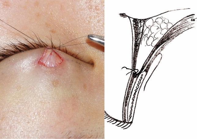
Kanski's Clinical Ophthalmology is the key textbook. Let me pull the main eyelid pathology sections from it in parallel:
Finding Sources
Finding Sources
Searching Images
ptosis entropion ectropion eyelid clinical photo
All in block1 of Kanski's. Let me read all the key sections in parallel:
Reading File
Reading File
Reading File
Excellent content flowing in. Let me read the ectropion, entropion sections and the tumour/chalazion sections in parallel:
Reading File
Reading File
Reading File
Now let me get the tumours/neoplasms and congenital sections, and search for key images:
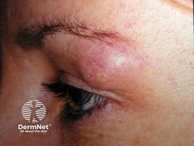
I now have excellent, rich content from Kanski's. Let me fetch the key textbook images and get the congenital/misc sections:
Loading Image
Loading Image
Loading Image
I now have a comprehensive set of content and images from Kanski's Clinical Ophthalmology. Let me now write the complete, exam-ready guide.
🎯 EYELID PATHOLOGIES - The Complete Exam Guide
Based on Kanski's Clinical Ophthalmology 10th Edition
🧱 PART 1: ANATOMY FIRST (The Foundation of Everything)
Before pathology makes sense, burn this anatomy into your memory.
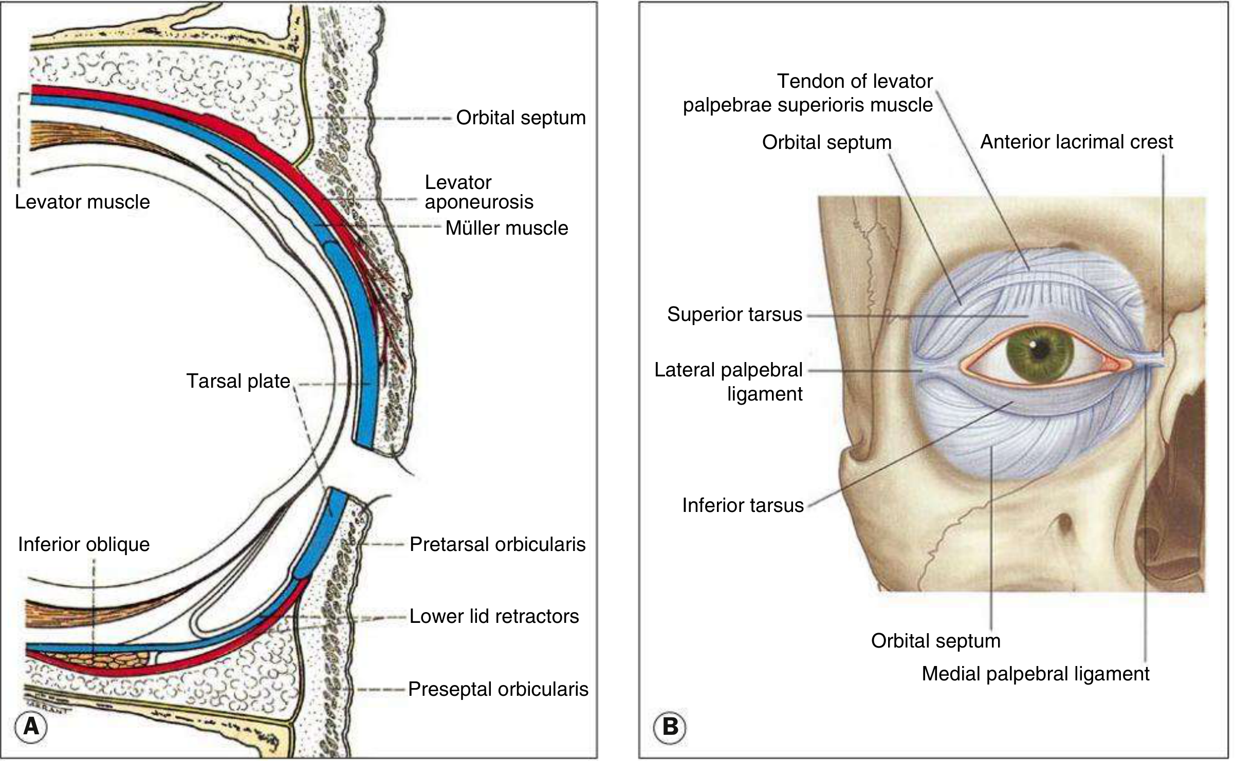
The Layers (Front to Back - "SMCTO")
SKIN (thinnest in body)
↓
MUSCLE (Orbicularis oculi - closes lid, CN VII)
↓
CONNECTIVE TISSUE (orbital septum + tarsal plate)
↓
TARSAL PLATE (upper = 10mm; lower = 5mm) - contains Meibomian glands
↓
CONJUNCTIVA (palpebral)
Openers vs Closers
| Action | Muscle | Nerve |
|---|---|---|
| Opens upper lid (main) | Levator palpebrae superioris | CN III (oculomotor) |
| Opens upper lid (assists) | Müller muscle (superior tarsal m.) | Sympathetic |
| Closes lid | Orbicularis oculi | CN VII (facial) |
| Retracts lower lid | Inferior tarsal muscle (Müller equivalent) | Sympathetic |
Key Glands in the Lid
| Gland | Location | Secretion | Opens at |
|---|---|---|---|
| Meibomian | Tarsal plate (25-30 upper, 20 lower) | Oily/lipid (prevents tear evaporation) | Posterior lid margin |
| Gland of Zeis | Lash follicle | Sebum | Lash follicle |
| Gland of Moll | Lash follicle | Modified sweat | Lash follicle |
🔥 PART 2: INFLAMMATORY LESIONS
2.1 HORDEOLUM (Stye)
The simple mnemonic: STYE = Staphylococcal, Tender, Young, External
| Feature | External Hordeolum (Stye) | Internal Hordeolum |
|---|---|---|
| Gland involved | Gland of Zeis (or Moll) - lash follicle | Meibomian gland |
| Organism | Staphylococcus aureus | S. aureus |
| Location | Anterior lid margin - points through skin | Within tarsal plate - points onto conjunctiva |
| Appearance | Tender swelling WITH a lash at apex | More diffuse, deeper swelling |
| Rx | Hot compresses, topical ± oral antibiotics, lash epilation | Hot compresses ± I&C if doesn't resolve |
🔑 Exam pearl: An external hordeolum has a lash at its apex. An internal one is inside the tarsal plate.
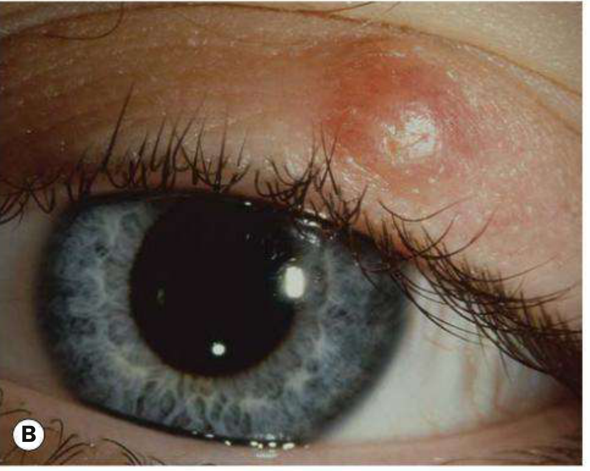
2.2 CHALAZION (Meibomian Cyst)
The mnemonic: CHALAZION = Chronic, Hard, Aseptic, Lipogranuloma, At Zeis or mIbomian gland, Over time
- A sterile chronic lipogranuloma of the meibomian gland (occasionally Zeis)
- Caused by retained sebaceous secretions blocking the duct
- NOT an infection - no pain, no acute inflammation
- Histology: lipid-laden epithelioid cells + multinucleated giant cells + lymphocytes (lipogranuloma)
Compared to Hordeolum:
HORDEOLUM CHALAZION
Acute vs. Chronic
Painful/tender vs. Painless
Infective (Staph) vs. Sterile (lipogranuloma)
Smaller, at margin vs. Larger, within tarsus
Resolves with Abx vs. Often needs I&C
Treatment:
- Most resolve spontaneously
- Hot compresses + lid massage
- Intralesional steroid injection (triamcinolone)
- Incision and curettage (I&C) - from conjunctival surface (vertical incision - perpendicular to lid margin)
🚨 CRITICAL EXAM POINT: If a chalazion recurs in the same position in an older patient → BIOPSY to exclude sebaceous gland carcinoma! This is a classic exam scenario.
- Bortezomib (for multiple myeloma) predisposes to chalazia within 3 months.
- Association: Rosacea → multiple/recurrent chalazia
👁️ PART 3: BLEPHARITIS
Definition: Chronic bilateral inflammation of the lid margins. Most common cause of ocular discomfort.
The Two Types - Easy to Remember
ANTERIOR BLEPHARITIS POSTERIOR BLEPHARITIS
(Around the lashes) (Behind the lash line)
↓ ↓
Staphylococcal or Seborrhoeic Meibomian Gland Dysfunction (MGD)
↓ ↓
- Hard crusts (Staph) - Frothy/foamy tear film
- Greasy scales (Seborrhoeic) - Dilated/blocked meibomian glands
- Collarettes = Demodex sign - Associated with ACNE ROSACEA
- Madarosis (lash loss) - Pouting/capped glands on slit lamp
Demodex blepharitis - a favourite exam topic:
- Demodex folliculorum longus → anterior blepharitis
- Demodex folliculorum brevis → posterior blepharitis
- Pathognomonic sign: COLLARETTES (cylindrical dandruff-like sleeves around lash bases)
- Treatment: tea tree oil, lid scrubs, azithromycin
Symptoms (same in all forms - poor correlation with signs!)
Burning, grittiness, mild photophobia, morning crusting, watering, mild blurring
Signs by Type
| Sign | Staphylococcal | Seborrhoeic | Posterior (MGD) |
|---|---|---|---|
| Crusts | Hard, fibrinous | Greasy/oily | Absent |
| Lash changes | Madarosis, trichiasis | Mild | Absent usually |
| Meibomian glands | Normal | Mildly blocked | Capped, inspissated |
| Cornea | Marginal infiltrates (immune) | Mild SPK | SPK, instability |
| Association | Atopic dermatitis | Seborrhoeic dermatitis | Acne rosacea |
Treatment (The 3 Cs)
- Cleaning: Lid hygiene - warm compresses + lid scrubs twice daily (lifetime)
- Control organisms: Topical antibiotics (chloramphenicol/azithromycin) ± systemic doxycycline/tetracycline (for posterior/Demodex)
- Control inflammation: Weak topical steroid short-term; artificial tears for dry eye
😴 PART 4: PTOSIS
Definition: Abnormally low position of the upper lid. Normal MRD = 4-5mm.
Measurements you MUST know for exams:
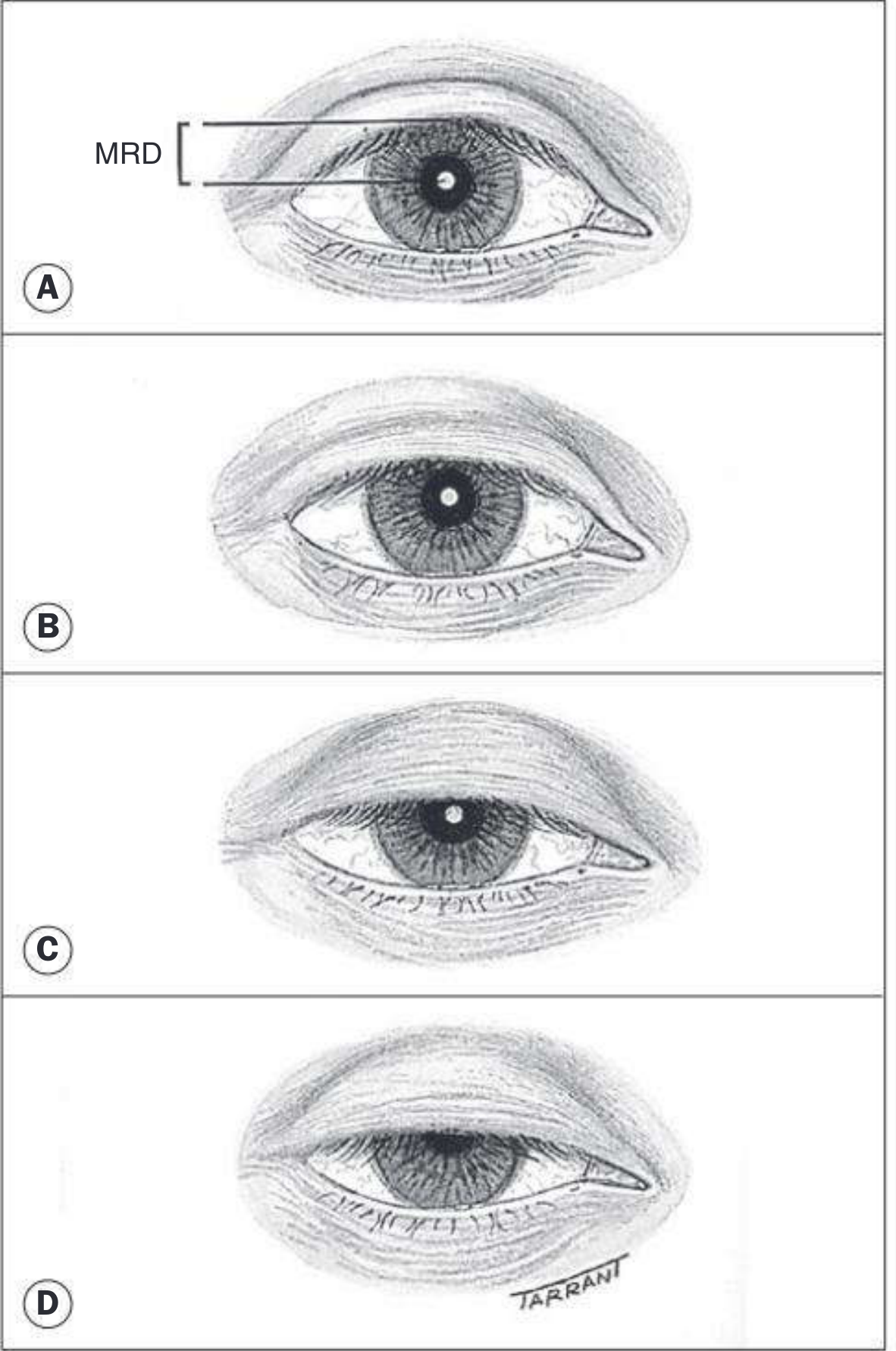
| Measurement | Normal | Grading of Ptosis |
|---|---|---|
| MRD (margin to corneal reflex) | 4-5 mm | Mild: ≤2mm drop; Mod: 3mm; Severe: ≥4mm |
| Palpebral fissure | Males 7-10mm; Females 8-12mm | - |
| Levator function | ≥15mm = Normal; 12-14 = Good; 5-11 = Fair; ≤4 = Poor | - |
| Lid crease | Males 8mm; Females 10mm | Absent = poor levator function |
Classification - "NMAM" (Neurogenic, Myogenic, Aponeurotic, Mechanical)
PTOSIS
│
┌─────────────────┼──────────────────┐─────────────────┐
▼ ▼ ▼ ▼
NEUROGENIC MYOGENIC APONEUROTIC MECHANICAL
(Involutional)
│ │ │ │
CN III palsy Myasthenia gravis Levator tendon Heavy mass
Horner syndrome Myotonic dystrophy dehiscence (tumour)
(Müller damage) CPEO Most common in Dermatochalasis
elderly Scarring
Key Features by Type
| Type | Levator function | Key feature |
|---|---|---|
| Congenital simple | Poor | Present from birth, amblyopia risk |
| Aponeurotic (involutional) | Good (≥12mm) | Deep superior sulcus, high crease |
| Myasthenia gravis | Variable | Fatigable, worse in evening, Cogan's lid twitch |
| Horner syndrome | Good | Mild ptosis + miosis + anhidrosis (classic triad) |
| CN III palsy | Variable | Ptosis + dilated pupil + "down and out" gaze |
| Marcus Gunn syndrome | Fair | Jaw-winking (lid raises on jaw movement) |
Pseudoptosis - Things That Look Like Ptosis But Aren't
- Contralateral lid retraction (making normal side look ptotic)
- Enophthalmos (sunken globe - no globe support for lid)
- Ipsilateral hypotropia (lid follows globe downward)
- Brow ptosis + dermatochalasis (excess skin hangs over)
- Microphthalmos/phthisis
Surgery
| Procedure | Indication | Principle |
|---|---|---|
| Müller (conjunctiva) resection | Mild ptosis, good LF (≥10mm) | Shortens Müller + conjunctiva, max 2-3mm lift |
| Levator advancement/resection | Any ptosis, LF ≥5mm | Shorten levator complex |
| Frontalis (brow) suspension | Severe ptosis, LF ≤4mm | Harness frontalis to open lid |
🔑 Key rule: Levator function determines the procedure. If LF ≥5mm → levator surgery. If LF <4mm (poor) → brow suspension.
📤 PART 5: ECTROPION
Definition: Eversion (outward turning) of the lid margin, away from the globe.
Memory trick: ECTropion = External turning = away from eye
Types - "ICPM"
ECTROPION
│
├── INVOLUTIONAL (most common) - Lower lid only
│ Age-related laxity of canthal tendons + lid laxity
│
├── CICATRICIAL - Any lid
│ Scarring contracts anterior lamella (skin)
│ Causes: burns, skin disease, surgery
│
├── PARALYTIC (Facial nerve palsy, CN VII)
│ Orbicularis can't hold lid to globe
│ Associated with lagophthalmos → exposure keratopathy!
│
└── MECHANICAL
Heavy lower lid (tumour, oedema)
Complications of Ectropion
- Epiphora (watering - punctum everted, can't drain tears)
- Conjunctival exposure → chemosis, keratinization
- Exposure keratopathy (corneal drying/ulceration - worst with paralytic)
- Chronic conjunctivitis
Treatment of Involutional Ectropion
- Medical (temporary): Artificial tears, lubricant ointment at night
- Surgery (definitive):
- Medial ectropion: medial canthal tendon plication, Lazy-T procedure, medial tarsoconjunctival diamond excision
- Lateral ectropion: lateral canthal sling (tarsal strip procedure)
- Total ectropion: horizontal lid shortening (pentagon excision)
🔑 The tarsal strip procedure (lateral canthal sling) is the workhorse operation for lower lid horizontal laxity - applicable to both involutional ectropion AND entropion.
📥 PART 6: ENTROPION
Definition: Inversion (inward turning) of the lid margin, towards the globe.
Memory trick: ENTropion = ENTering the eye = inward turning
Types
ENTROPION
│
├── INVOLUTIONAL (most common) - Lower lid, elderly
│ Three mechanisms:
│ 1. Horizontal lid LAXITY (canthal tendons stretch)
│ 2. Vertical INSTABILITY (lower lid retractors disinsertion)
│ 3. OVER-RIDING of preseptal over pretarsal orbicularis
│
├── CICATRICIAL - Any lid (upper > lower)
│ Conjunctival scarring pulls lid inward
│ Causes: TRACHOMA (most common worldwide!), OCP,
│ Stevens-Johnson, chemical burns
│
└── CONGENITAL
Rare, usually lower lid
The Result: Trichiasis (Lashes Rubbing Cornea)
Entropion → lashes rub cornea (pseudotrichiasis)
→ Punctate epithelial erosions
→ Pannus formation
→ Corneal ulceration (chronic cases)
Note: True trichiasis = misdirected lashes but lid position normal (e.g., from trachoma scarring). Pseudotrichiasis = lashes are normal but rubbing because the lid has turned in.
Treatment of Involutional Entropion
| Severity | Procedure | Principle |
|---|---|---|
| Temporary/frail patient | Transverse everting sutures | Quick, lasts months |
| Definitive | Wies procedure | Full-thickness lid split + everting sutures |
| Definitive | Lower lid retractor reinsertion | Direct advancement of retractors |
| With lid laxity | + Tarsal strip | Tighten horizontal laxity |
Entropion vs Ectropion - Quick Comparison
| Feature | Entropion | Ectropion |
|---|---|---|
| Direction | INWARD | OUTWARD |
| Lashes | Rub cornea | Away from cornea |
| Main complication | Corneal ulcer | Epiphora + exposure |
| Most common type | Involutional (lower lid) | Involutional (lower lid) |
| Most common cause worldwide | Trachoma (cicatricial) | - |
🏷️ PART 7: EYELID TUMOURS
Benign Tumours
| Tumour | Origin | Appearance | Notes |
|---|---|---|---|
| Papilloma | Squamous epithelium | Pedunculated/sessile fleshy tag | Most common benign lid tumour |
| Seborrhoeic keratosis | Epidermal | Greasy stuck-on appearance, pigmented | Elderly, "stuck-on" appearance |
| Cyst of Moll | Gland of Moll (sweat) | Clear fluid-filled, lid margin | Translucent on transillumination |
| Cyst of Zeis | Gland of Zeis (sebaceous) | Yellowish, lid margin, NOT transilluminant | Opaque |
| Xanthelasma | Lipid deposits | Flat/slightly raised yellow plaques, medial canthus | May indicate hypercholesterolaemia |
| Strawberry naevus | Capillary haemangioma | Bright red, grows then involutes | Treat if causing amblyopia |
| Capillary haemangioma | Endothelial | Red, present at birth | Can cause ptosis → amblyopia |
Premalignant Lesions
| Lesion | Feature | Risk |
|---|---|---|
| Actinic keratosis | Sun-exposed elderly skin, scaling erythematous patch | Transforms to SCC |
| Lentigo maligna (Hutchinson freckle) | Slowly expanding macular pigmented lesion in elderly | 3-10% transform to melanoma |
| Keratoacanthoma | Rapidly growing nodule with keratin-filled crater | May regress spontaneously but biopsy needed |
Malignant Tumours - The Big 3
7.1 BASAL CELL CARCINOMA (BCC) - Most Common (90%)
Mnemonic: BCC = Bottom lid, Commonest, Cruelly invades (no mets)
- Most common malignant eyelid tumour - 90% of all cases
- Most common location: Lower lid (50%) > medial canthus (25%) > upper lid > lateral canthus
- Risk factors: Fair skin, UV exposure, xeroderma pigmentosum, Gorlin-Goltz syndrome
- Behaviour: Locally invasive, slow growing, does NOT metastasize
- Madarosis (loss of lashes) is commonly seen
Morphological Types:
BCC Types
│
├── NODULAR - Shiny pearly nodule, telangiectasias, smooth
│
├── NODULO-ULCERATIVE (Rodent ulcer) - Classic!
│ Central ulcer + pearly rolled edges + telangiectasias
│
├── MORPHOEIC (Sclerosing) - Most dangerous for recurrence
│ Flat, indurated, ill-defined margins
│ Infiltrates along tissue planes → difficult to clear
│
└── PIGMENTED - Contains melanin
- Histology: Peripheral palisading of basal cells (the hallmark)
🔑 Medial canthal BCC = most dangerous location - invades orbit and sinuses, highest recurrence rate.
Treatment:
- Wide surgical excision with margin control (Mohs surgery = gold standard)
- Radiotherapy for inoperable cases
- Topical imiquimod/vismodegib for superficial/inoperable
7.2 SQUAMOUS CELL CARCINOMA (SCC)
- 2nd most common (but far rarer than BCC)
- More aggressive than BCC - can metastasize to regional lymph nodes
- Risk factors: UV exposure, immunosuppression, HPV, actinic keratosis, xeroderma pigmentosum
- Location: Upper or lower lid
- Appearance: Keratinizing, ulcerated, firm nodule; may have cutaneous horn
- Histology: Keratin pearls (squamous pearls), dyskeratosis
- Treatment: Wide excision + sentinel lymph node biopsy if indicated
7.3 SEBACEOUS GLAND CARCINOMA (SGC) - The Great Masquerader!
🚨 THE MOST IMPORTANT EXAM TUMOUR - it mimics everything!
- Rare but very deadly (mortality 5-10%)
- Mostly affects elderly females
- Arises from meibomian glands (most common) or glands of Zeis
- Upper lid more common (more meibomian glands there!) - contrast to BCC
Two patterns:
NODULAR SGC SPREADING SGC
│ │
Hard nodule in upper tarsal plate Diffuse lid thickening
Yellowish due to lipid Lash loss/distortion
MIMICS CHALAZION! MIMICS CHRONIC BLEPHARITIS!
Histology:
- Lobules of cells with pale foamy vacuolated (lipid-containing) cytoplasm
- Large hyperchromatic nuclei
- Pagetoid spread (intraepithelial spread) = diagnostic feature
⚠️ Key exam scenarios:
| Scenario | Diagnosis to think of |
|---|---|
| "Recurrent chalazion in elderly" | SGC masquerading as chalazion |
| "Chronic unilateral blepharitis not responding to treatment" | SGC masquerading as blepharitis |
| "Yellow mass in upper tarsus" | SGC (yellow = lipid!) |
Treatment: Wide excision with frozen section margins, exenteration if orbit invaded
Comparison Table: The Big 3 Malignancies
| Feature | BCC | SCC | SGC |
|---|---|---|---|
| Frequency | 90% | 5% | Rare |
| Location | Lower lid > medial canthus | Any | UPPER lid |
| Metastasis | NO | YES (lymph nodes) | YES (distant) |
| Mimics | - | - | Chalazion, blepharitis |
| Key histology | Peripheral palisading | Keratin pearls | Foamy vacuolated cells, pagetoid spread |
| Lash finding | Madarosis | Madarosis | Madarosis |
| Mortality | Very low | Moderate | 5-10% |
🧬 PART 8: CONGENITAL EYELID ANOMALIES
Quick-Reference Table
| Condition | What it is | Key feature |
|---|---|---|
| Epicanthic fold | Skin fold from upper lid covering medial canthus | Normal in Asians; can mimic squint |
| Blepharophimosis syndrome | Triad: blepharophimosis + ptosis + epicanthus inversus | BPES - autosomal dominant, FOXL2 gene |
| Epiblepharon | Horizontal skin fold presses lashes against cornea | Asian children; most self-resolves |
| Coloboma | Full-thickness lid defect | Superior lid; may be part of CHARGE syndrome |
| Cryptophthalmos | No eyelid development - skin covers eye | Rare, associated with Fraser syndrome |
| Ankyloblepharon | Lid margins fused by filiform bands | Can be isolated or syndromic |
| Congenital entropion | Lower lid rolls in from birth | Horizontal tarsal plate kink |
| Congenital upper lid eversion | Lid turns out at birth | Associated with Down syndrome |
| Euryblepharon | Abnormally wide palpebral fissure | Lateral lid margin displaced downward |
| Microblepharon | Abnormally small lids | May cause exposure keratopathy |
Blepharophimosis-Ptosis-Epicanthus Inversus Syndrome (BPES)
BPES Tetrad:
1. Blepharophimosis (narrow horizontal fissure)
2. Ptosis (bilateral)
3. Epicanthus INVERSUS (fold runs lower → upper lid)
4. Telecanthus (increased distance between medial canthi)
Type I: BPES + premature ovarian failure (POF)
Type II: BPES alone (without POF)
Gene: FOXL2 on chromosome 3q23
🔄 PART 9: MISCELLANEOUS ACQUIRED DISORDERS
Dermatochalasis vs Blepharochalasis
| Feature | Dermatochalasis | Blepharochalasis |
|---|---|---|
| What is it | Excess redundant upper lid skin | Recurrent episodic lid oedema causing skin atrophy |
| Age | Elderly | Young women |
| Cause | Aging + fat herniation | Idiopathic (? angioneurotic oedema) |
| Appearance | Hanging skin fold | Thin, wrinkled, atrophic skin; ptosis develops |
| Treatment | Upper lid blepharoplasty | Anti-inflammatory; surgery if stable |
Floppy Eyelid Syndrome
- Rubbery, easily everted upper lids with papillary conjunctivitis
- Associated with obstructive sleep apnoea (the key association!)
- Lid everts during sleep → chronic papillary conjunctivitis
- Treatment: lid taping at night, CPAP for OSA, surgery if severe
Lid Retraction
- Upper lid normally rests 2mm below upper limbus (covers 2mm of cornea)
- If it sits above the limbus → retraction
Causes of upper lid retraction:
- Thyroid eye disease (Graves') - most common cause; stimulation of levator + Müller (Dalrymple's sign = lid retraction; von Graefe's sign = lid lag on downgaze)
- Contralateral ptosis (Hering's law - the normal lid over-elevates to maintain binocular vision)
- Parinaud syndrome (dorsal midbrain) - bilateral lid retraction + convergence-retraction nystagmus
- Neurogenic (aberrant CN III regeneration)
Lagophthalmos
- Inability to close the eyelid completely
- Causes: CN VII palsy, proptosis, cicatricial ectropion, symblepharon
- Danger: Corneal exposure → exposure keratopathy → ulceration
- Bell's phenomenon (protective reflex - globe rolls up when closing) = partially protective
- Treatment: Lubricants, moisture chamber, taping at night, lid weights, tarsorrhaphy
⚡ PART 10: LASH ABNORMALITIES
| Condition | Definition | Cause |
|---|---|---|
| Trichiasis | Misdirected lashes (lid in normal position) | Trachoma, blepharitis, trauma |
| Distichiasis | Extra row of lashes from meibomian gland openings | Congenital or acquired (lymphoedema) |
| Madarosis | Loss of lashes | BCC, SCC, SGC, blepharitis, alopecia areata, leprosy |
| Poliosis | White lashes | Vitiligo, Vogt-Koyanagi-Harada, tuberous sclerosis |
| Trichomegaly | Abnormally long lashes | HIV, topical prostaglandins (bimatoprost), cyclosporin |
Treatment of Trichiasis:
- Epilation (temporary)
- Electrolysis/cryotherapy/argon laser (more permanent)
- Lid surgery for cicatricial cases (posterior lamellar grafting)
🎓 EXAM RAPID-FIRE SUMMARY
"The One-Liners" - Memorize These
| # | Fact |
|---|---|
| 1 | Most common eyelid malignancy = BCC (90%) |
| 2 | Most common location of BCC = lower lid |
| 3 | BCC at medial canthus = most likely to invade orbit, highest recurrence |
| 4 | BCC does NOT metastasize; SCC DOES |
| 5 | SGC occurs more on upper lid (more meibomian glands) |
| 6 | Recurrent chalazion in same site = biopsy to exclude SGC |
| 7 | SGC mimics chalazion and blepharitis = "the great masquerader" |
| 8 | Pagetoid spread = pathognomonic of SGC |
| 9 | Normal MRD = 4-5mm; Normal levator function = ≥15mm |
| 10 | Ptosis + miosis + anhidrosis = Horner syndrome |
| 11 | Ptosis + diplopia + dilated pupil = CN III palsy |
| 12 | Jaw-winking ptosis = Marcus Gunn syndrome |
| 13 | Floppy eyelid syndrome → always think obstructive sleep apnoea |
| 14 | Upper lid retraction → first think Thyroid eye disease |
| 15 | Dalrymple's sign = lid retraction; Von Graefe's sign = lid lag in Graves' |
| 16 | Demodex → collarettes (pathognomonic) |
| 17 | Posterior blepharitis → associated with acne rosacea |
| 18 | Cicatricial entropion worldwide most common cause = trachoma |
| 19 | Brow (frontalis) suspension = for ptosis with levator function <4mm |
| 20 | BPES gene = FOXL2 on chromosome 3q23 |
The Master Comparison Grid: Lid Margin Swellings
| Feature | External Hordeolum | Chalazion | Internal Hordeolum | SGC |
|---|---|---|---|---|
| Pain | Yes | No | Moderate | No (chronic) |
| Acute/Chronic | Acute | Chronic | Subacute | Chronic |
| Sterile/Infective | Infective (Staph) | Sterile | Infective (Staph) | Neoplastic |
| Location | Anterior margin (lash) | Within tarsus | Within tarsus (posterior) | Within tarsus (upper) |
| Consistency | Soft/fluctuant | Hard, rubbery | Hard | Very hard |
| Discharge | Pus | None | Pus (on conjunctiva) | None |
| Special feature | Lash at apex | Lipogranuloma | Points internally | Yellow, recurrent |
| Treatment | Abx + hot compress | I&C or steroid injection | I&C | Wide excision |
Step-by-step Logic for Exam Scenarios
Scenario 1: Old lady, "chalazion" keeps coming back in the same spot on upper lid
→ Think: Sebaceous Gland Carcinoma masquerading as chalazion → Biopsy!
Scenario 2: Patient with chronic unilateral blepharitis, not responding to treatment
→ Think: SGC spreading type → Biopsy!
Scenario 3: Elderly man, lower lid turning outward, watering eye
→ Involutional ectropion → medial punctal ectropion → epiphora → tarsal strip surgery
Scenario 4: Elderly woman, lower lid turning inward, corneal abrasion
→ Involutional entropion → pseudotrichiasis → Wies procedure or retractor reinsertion
Scenario 5: Child, unilateral ptosis, lid elevates when chewing gum
→ Marcus Gunn jaw-winking syndrome (synkinesis between CN V and CN III)
Scenario 6: Unilateral mild ptosis, small pupil, no diplopia
→ Horner syndrome → mild ptosis (Müller damage) + miosis (sympathetic loss) → look for underlying cause (lung apex, carotid, cavernous sinus)
All content sourced from Kanski's Clinical Ophthalmology: A Systematic Approach, 10th Edition - the most authoritative reference for ophthalmology exams.
This is a shared conversation. Sign in to Orris to start your own chat.