
Gerd pathophysiology
"gastroesophageal reflux disease" AND pathophysiology
GERD gastroesophageal reflux disease pathophysiology lower esophageal sphincter diagram
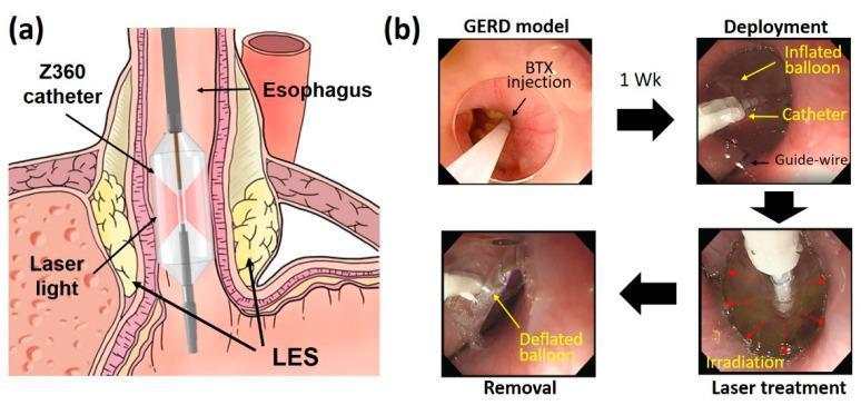
Educational figure illustrating Endoscopic Balloon-assisted Laser Treatment (EBLT) for Gastroesophageal Reflux Disease (GERD). (a) Anatomical diagram showing a Z360 balloon catheter positioned within the distal esophagus at the lower esophageal sphincter (LES). The catheter features a transparent inflated balloon and a central diffusing applicator that emits circumferential laser light to target the LES muscle layers. (b) A series of four endoscopic frames detailing the procedural stages in a porcine model: 1. GERD model induction via botulinum toxin (BTX) injection to weaken the LES; 2. Deployment stage showing the catheter and inflated balloon guided by a guide-wire; 3. Laser treatment stage showing active irradiation (indicated by red arrows); 4. Removal stage following balloon deflation. The educational focus is on the minimally invasive delivery of laser energy to the esophagogastric junction to induce tissue remodeling and increase LES pressure without mucosal injury.
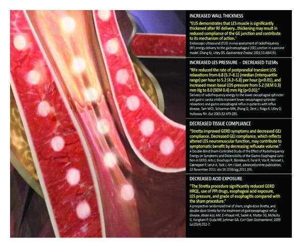
This medical illustration demonstrates the mechanism of action of the Stretta procedure, a minimally invasive treatment for gastroesophageal reflux disease (GERD). The visual depicts a cross-section of the esophageal wall near the gastroesophageal junction. Key components include multiple nickel-titanium needle electrodes delivering radiofrequency (RF) energy into the muscularis propria of the esophagus, represented by bright yellow circular glows within the red-textured tissue layers. The anatomical focus is on the lower esophageal sphincter (LES) region. Accompanying text panels summarize clinical outcomes and pathophysiology: increased wall thickness, increased LES basal pressure with decreased transient LES relaxations (TLESRs), decreased tissue compliance at the gastroesophageal junction, and significantly decreased esophageal acid exposure. The image highlights the device's ability to deliver thermal energy to induce tissue remodeling while protecting the overlying mucosa via irrigation. This illustration serves as an educational tool for gastroenterologists and surgical trainees to understand non-surgical antireflux interventions and their physiological impact on LES neuromuscular function.
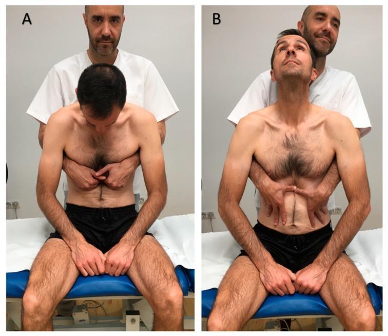
Two-panel clinical photograph demonstrating a visceral osteopathic manual technique targeting the lower esophageal sphincter, typically used for gastroesophageal reflux disease (GERD). Panel A (Initial Position) shows a male patient seated upright on a treatment table in spinal flexion with his neck flexed and head looking down. A practitioner stands behind him with hands placed horizontally in the epigastric region, specifically contacting the area just inferior to the costal margins. Panel B (Final Position) illustrates the completion of the maneuver: the patient has moved into spinal extension with his neck extended and gaze upward. Simultaneously, the practitioner has shifted hand placement to apply caudal (downward) pressure within the epigastrium. The visual demonstrates the coordination of patient respiratory/postural movement with manual tissue mobilization by the therapist. This procedural image is intended for medical students and physical therapy or osteopathic practitioners to understand the manual application and patient positioning for visceral abdominal therapy.
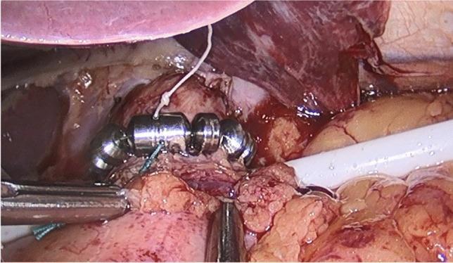
This clinical photograph captures a laparoscopic view of an implanted LINX Reflux Management System during a surgical procedure for Magnetic Sphincter Augmentation (MSA). The device, designed to treat Gastroesophageal Reflux Disease (GERD), consists of a series of cylindrical titanium beads with magnetic cores, interconnected by small wires to form an expandable annular ring. In the image, the bead chain is seen partially encircling the distal esophagus at the level of the lower esophageal sphincter (LES). Surgical instruments, including a laparoscopic grasper and a white retraction tool, are visible in the operative field, alongside a white suture tied to the device for positioning or fixation. The surrounding anatomy includes the liver retracted superiorly and the gastroesophageal junction. The photograph demonstrates the fundic-sparing nature of this anti-reflux surgery, illustrating the device's placement and the mechanical augmentation of the LES to prevent acid reflux while allowing bolus transit.
GERD Pathophysiology
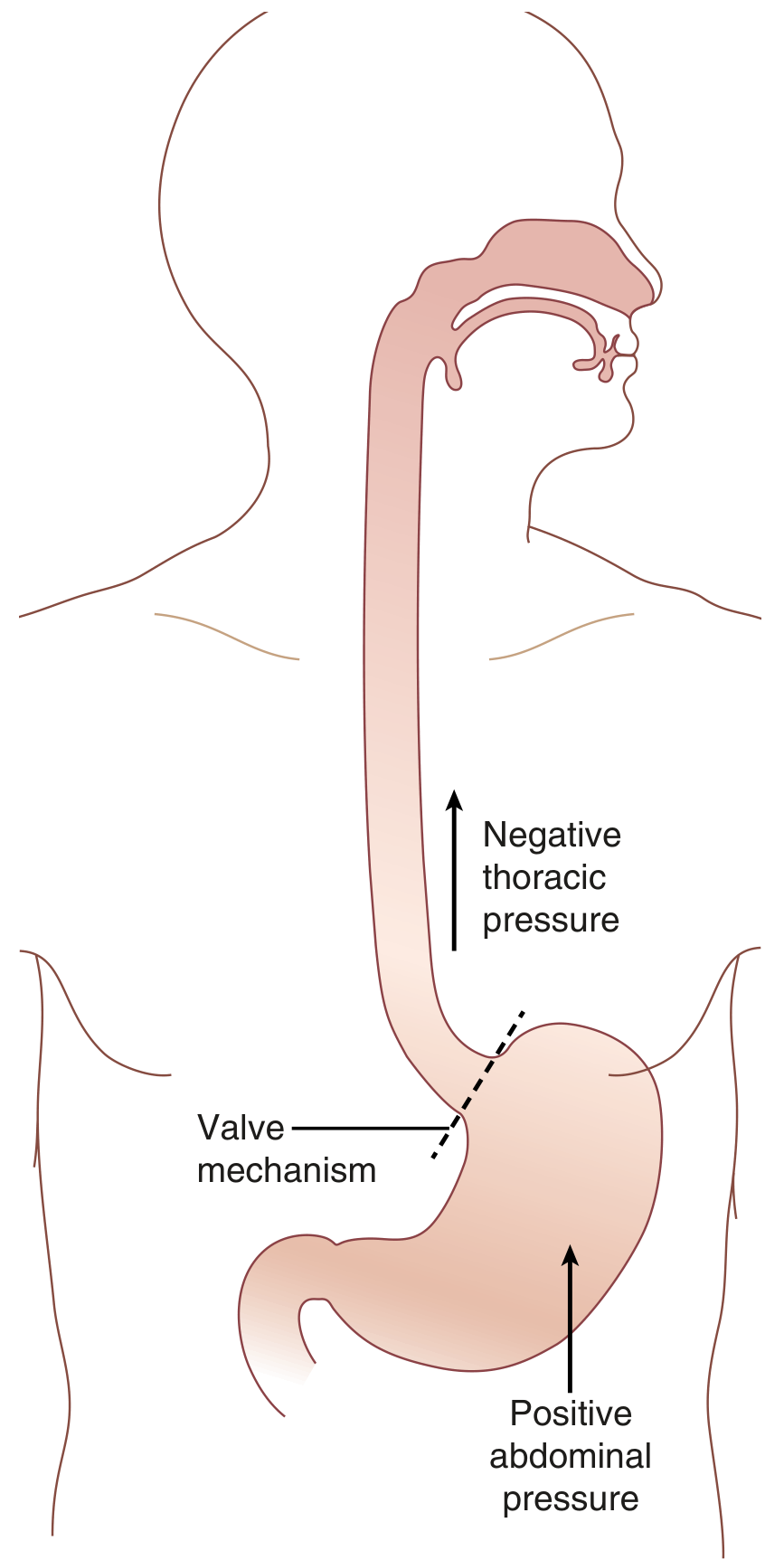
1. The Pressure Imbalance
2. The Antireflux Barrier - Five Components
a) Lower Esophageal Sphincter (LES)
| Increases LES Pressure | Decreases LES Pressure |
|---|---|
| Gastrin, motilin, substance P | CCK, secretin, somatostatin, VIP |
| α-adrenergic agonists, cholinergic agonists | β-adrenergic agonists, cholinergic antagonists |
| Protein (dietary) | Chocolate, fat, peppermint |
| Metoclopramide, domperidone, baclofen | Calcium channel blockers, diazepam, opioids, theophylline |
b) Crural Diaphragm
c) Angle of His
d) Gubaroff Valve
e) Intra-abdominal Segment of the Esophagus
3. Mechanisms of Reflux
a) Transient LES Relaxations (tLESRs) - THE dominant mechanism
- Persist for >10 seconds (vs. ~5 seconds for swallow-induced LES relaxation)
- Not accompanied by esophageal peristalsis
- Associated with inhibition of the crural diaphragm
- Triggered by proximal gastric distension (gas or food), dietary fat, stress, and subthreshold pharyngeal stimulation
b) Low Basal LES Pressure
c) Swallow-associated LES Relaxation
d) Straining During Periods of Low LES Pressure
4. Hiatal Hernia - a Key Amplifier
- Displaces the LES into the negative-pressure thoracic cavity (removing the intra-abdominal esophageal segment)
- Separates the LES from the crural diaphragm (manometric dissociation)
- Obtunds the angle of His
- Enlarges the hiatal opening
- Creates a reservoir effect - gastric contents trapped in the hernial sac reflux back into the esophagus with each subsequent LES relaxation during swallowing
- Increases frequency of tLESRs
5. Esophageal Clearance - Second Line of Defense
- Volume clearance - esophageal peristalsis propels refluxate back into the stomach
- Acid neutralization - weakly alkaline swallowed saliva neutralizes residual acid
- Peristaltic dysfunction is associated with increasing severity of esophagitis; contractions <20 mm Hg are ineffective for clearance
- Saliva production is reduced by smoking, Sjögren syndrome, and sleep (explaining why nocturnal reflux is particularly damaging)
- Goldman-Cecil Medicine
6. Mucosal Defense - Third Line of Defense
| Layer | Components |
|---|---|
| Pre-epithelial | Unstirred water layer + bicarbonate from swallowed saliva and submucosal glands |
| Epithelial | Cell membranes, tight intercellular junctions, intracellular buffers, ion transporters |
| Post-epithelial | Mucosal blood supply (delivers bicarbonate, removes acid) |
- Damage tight intercellular junctions
- Increase intercellular permeability
- Dilate intercellular spaces (a hallmark finding on electron microscopy, even before macroscopic esophagitis)
- If sufficient refluxate diffuses into the intercellular space, direct cellular damage occurs
7. Obesity and GERD
- Raised intra-abdominal pressure compresses the stomach and increases the gastroesophageal pressure gradient
- More frequent tLESRs
- Reduced LES pressure and shorter intra-abdominal LES length
- Promotes hiatal hernia formation by enhancing crural diaphragm-LES separation
- Higher gastric acid production
- Esophageal motor dysfunction
- The metabolic syndrome may have an independent contributing role
Summary Schematic
GERD = Antireflux Barrier Failure + Impaired Clearance + Mucosal Injury

Antireflux Barrier Failure:
  ├── ↑ tLESRs (vagally mediated, gastric distension trigger)
  ├── ↓ LES resting pressure (drugs, fat, hormones)
  ├── Hiatal hernia (disrupts all 5 EGJ components)
  └── ↑ Intra-abdominal pressure (obesity, pregnancy)

Impaired Clearance:
  ├── Peristaltic dysfunction
  └── ↓ Salivary bicarbonate

Mucosal Injury:
  ├── Acid + pepsin → intercellular junction disruption
  └── Chemokine-mediated inflammation → epithelial damage
explain in a very very easy language
GERD - Super Simple Explanation 🍋
What is GERD in plain English?
Imagine This...
You have a bottle of lemon juice (stomach) with a cap (LES). If the cap is loose, lemon juice spills out. If you squeeze the bottle hard (increased pressure), even more juice spills. That's GERD.
Why Does the "Cap" Fail? - 4 Simple Reasons
1. The Cap Randomly Opens - tLESR
2. The Cap is Just Too Weak
- Chocolate 🍫
- Fatty foods 🍟
- Peppermint 🍬
- Coffee ☕
- Alcohol 🍺
- Some medicines (like blood pressure pills)
3. Too Much Pressure from Below - Squeezing the Bottle
- Obesity - belly fat squeezes the stomach
- Pregnancy - baby squeezes the stomach
- Tight clothing - compresses the belly
- Bending over or straining
4. The Hernia Problem - Cap Slips Out of Place
- The cap (LES) is no longer in the right position
- The natural pressure that was keeping acid down is gone
- Acid pools in the hernia sac and shoots back up more easily
Think of it like the bottle cap getting displaced - even if the cap exists, it's no longer properly sealing the bottle.
What Happens After Acid Gets In?
- Swallowing pushes it back down (like a pump)
- Saliva washes and neutralizes the remaining acid
How Does the Acid Actually Hurt?
- Breaks the "glue" between the tiles (damages tight junctions between cells)
- Seeps into the cracks and irritates deeper tissue
- Calls in inflammatory cells which attack the lining further
- Over time, this causes redness, ulcers, and scarring
Why Do Obese People Get More GERD?
- Pushes acid upward more forcefully
- Makes the cap open more often
- Weakens the cap muscle
- Can push the stomach up into the chest (hiatal hernia)
The 3-Layer Protection System (Simplified)
| Layer | What It Does | Simple Analogy |
|---|---|---|
| Layer 1 - The Cap (LES + diaphragm) | Stops acid from entering | Security door |
| Layer 2 - Peristalsis + Saliva | Flushes and neutralizes acid | Cleaning crew |
| Layer 3 - Mucosal lining | Resists acid damage | Protective armor |
One-Line Summary
GERD = Stomach acid sneaks past a weak or faulty valve into the food pipe, burns the lining, and your body can't clean it up fast enough.
tell the pathophysiology in the easiest language possible
GERD Pathophysiology - As Simple As Possible
Start Here: Two Things You Need to Know
- Food pipe (esophagus) - the tube food travels down to reach the stomach
- Stomach - the bag that holds food and makes strong acid to digest it
The Gate
Open when food comes DOWN. Stay SHUT at all other times.
Why Does the Gate Fail?
Reason 1 - The Gate Randomly Opens on Its Own
Reason 2 - The Gate is Permanently Weak
- Chocolate, coffee, alcohol, fatty food
- Smoking
- Some medicines
- Just bad luck (genetics)
Reason 3 - Too Much Pressure Forcing the Gate Open
- Obese people - belly fat squeezes the stomach from outside
- Pregnant women - the baby pushes the stomach upward
- People who bend over, strain, or wear very tight clothing
Bonus: The Slipped Gate (Hiatal Hernia)
What Happens to the Food Pipe?
- Burns the lining of the food pipe
- Breaks the protective wall between cells
- Causes inflammation - the body sends immune cells to fight the damage
- Over time - ulcers, scarring, and even precancerous changes (Barrett's esophagus)
Why Can't the Body Just Clean the Acid Up?
- Swallowing pushes acid back down
- Saliva washes and neutralizes leftover acid
- Swallowing is weak or slow
- You swallow less saliva at night
- So acid sits in the food pipe for a long time and causes more damage
The Full Story in 5 Lines
- Your stomach makes acid to digest food.
- A gate (muscle ring) normally keeps that acid inside the stomach.
- In GERD, the gate fails - acid comes up into the food pipe.
- The food pipe burns because it isn't built to handle acid.
- The body tries to wash the acid away but can't keep up - so damage builds over time.
Visual Summary
Normal:
   Food Pipe
      ↓
   [GATE SHUT] ✅
      ↓
   Stomach (acid stays here)


GERD:
   Food Pipe ← ACID BURNS HERE 🔥
      ↑
   [GATE OPEN] ❌
      ↑
   Stomach (acid escaping upward)