
Gross easy drawing picture of leiomyomas need to be say drawing
leiomyoma gross pathology uterine fibroid cut section
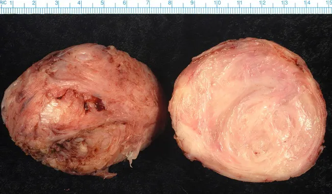
This gross pathology image shows a uterine leiomyoma (fibroid), the most common benign neoplasm of the female uterus. The specimen consists of a well-circumscribed, bulging mass with a firm, rubbery consistency. The right-hand section reveals the classic whorled tan-to-white cut surface produced by intersecting bundles of smooth muscle fibers. Degenerative changes, such as hyaline degeneration, may alter color and texture and can complicate gross evaluation. The lesion is intramural, typically arising within the myometrium and may be solitary or multiple in the uterus, sometimes enlarging the organ and producing bulk symptoms. Although histologic subtypes exist, all leiomyomas share a similar gross appearance, making differentiation from other myometial lesions primarily dependent on histology. This image is useful for gross-pathology education, surgical pathology review, and gynecologic oncology reference, illustrating tumor organization, texture, and surface characteristics. Clinically, fibroids are common in middle-aged women and can be detected by pelvic examination and imaging (ultrasound, MRI). Management depends on size, location, symptoms, and patient preference, ranging from expectant management to myomectomy or hysterectomy. The image provides context for teaching gross anatomic features, differential diagnoses, and the relevance of degenerative changes in leiomyomas. This context supports educational use in pathology laboratories and clinical rounds for learners.
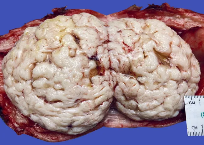
This gross pathology photograph depicts a uterus with a large intramural leiomyoma (uterine fibroid). The specimen shows a firm, well-circumscribed mass within the myometrium, with a cerebriform, gyriform cut surface produced by densely packed smooth muscle bundles arranged in a whorled pattern. The cut surface exhibits pale, whitish zones with convoluted ridges reminiscent of brain gyri, a characteristic gross feature described as cerebriform. The mass is pale to pale-tan with a slightly gleaming surface; surface vascularity is accentuated by incidental small hemorrhages at the periphery. The uterus is opened to expose the lesion, with the surrounding myometrium appearing bulky but without obvious invasion into adjacent tissue. The lesion is typically encapsulated by a fibrous capsule and shows no necrosis or overt malignant features on gross examination. The morphology is consistent with a benign smooth muscle tumor (leiomyoma), and the cerebriform cut surface is a recognized gross variant. Diagnostic significance: gross examination supports benign etiology and helps distinguish fibroids from malignant leiomyosarcoma, though histology is required for confirmation. Clinical correlation: fibroids are common in reproductive-age women and may cause menorrhagia, pelvic pressure, or infertility; management includes myomectomy or hysterectomy; imaging (ultrasound, MRI) often complements gross findings. Useful for education and research.
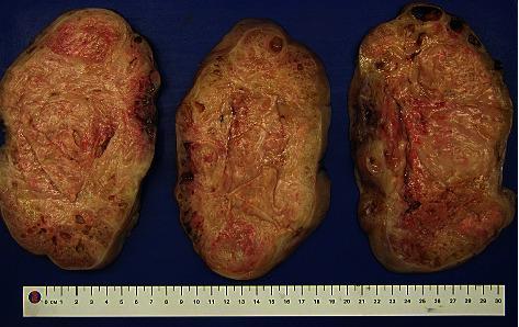
This clinical photograph displays a gross specimen of a uterine leiomyoma (uterine fibroid) that has been sectioned into three portions to reveal the internal architecture. The cut surfaces exhibit a characteristic whorled or trabecular growth pattern, consisting of interlacing bundles of smooth muscle and fibrous connective tissue. The color is predominantly a pale yellowish-tan, with notable focal areas of reddish discoloration suggesting vascularity or intramural hemorrhage. The texture appears dense and firm, although small cystic spaces or translucent regions are visible, indicating focal cystic or hydropic degeneration. The specimens are displayed against a blue background with a metric ruler for scale, demonstrating a significant size (approximately 10-12 cm in diameter per section). This image serves as an educational tool for identifying the macroscopic features of benign uterine smooth muscle tumors and their potential secondary degenerative changes.
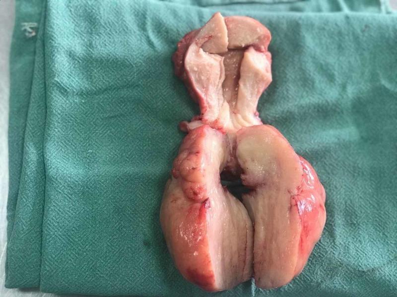
This clinical photograph displays a gross surgical specimen of a human uterus post-hysterectomy, specifically showing a longitudinal cut-section. The specimen features a bulky uterine corpus and a prominent, prolapsed fleshy fibroid. The external serosal surface is lobulated with a pinkish-red, vascular appearance. The internal cut surfaces reveal a relatively homogeneous, pale pink-to-tan tissue with a smooth and regular texture, characteristic of a uterine leiomyoma (fibroid). The lower portion of the image highlights the rounded, well-circumscribed nature of the mass. This visual is an educational example of gynecological pathology, specifically illustrating a large submucosal or pedunculated fibroid polyp that has caused significant uterine enlargement. It is relevant for medical training in surgical pathology and clinical gynecology, demonstrating the morphological characteristics that differentiate benign leiomyomas from other myometrial lesions.
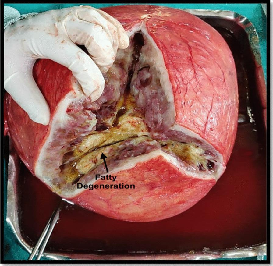
This clinical photograph shows a gross pathological specimen of a large uterine leiomyoma (fibroid) following a total abdominal hysterectomy. The specimen is presented in a surgical tray, with a gloved hand retracting a deep Y-shaped incision to reveal the internal structure. The external surface of the mass is reddish-pink with visible vascularity. The cut section demonstrates a heterogeneous internal composition characteristic of secondary degeneration in a benign smooth muscle tumor. A central region, marked by an arrow and labeled 'Fatty Degeneration,' exhibits a distinct yellowish, oily, and somewhat greasy appearance. This area is surrounded by mottled reddish-brown, off-white, and greyish tissue, with some darker regions suggestive of hemorrhagic or cystic changes. This visual is an educational example of macroscopic degenerative changes in chronic uterine leiomyomas, illustrating the loss of the typical whorled appearance and its replacement by fibro-fatty tissue. It is relevant for pathology and gynecology learners studying benign uterine neoplasms and their potential for massive enlargement and structural transformation.
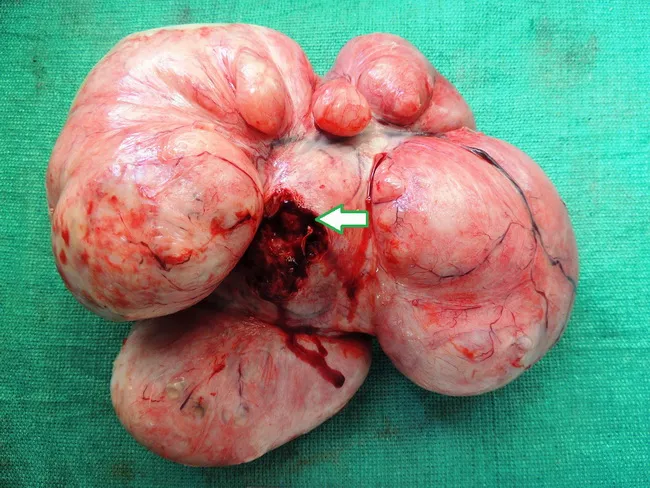
This is a gross pathology photograph of a giant uterine leiomyoma (fibroid) arising from the uterus, with a thick pedicle anchored to the right uterine cornual region. Imaging modality was not used for this capture; the image represents intraoperative gross anatomy of a large subserosal mass extending into the pelvic and abdominal cavities. The mass measures approximately 36 x 34 x 30 cm and weighs about 9.8 kg. On cut surface, the specimen shows a predominantly fleshy, whorled appearance with focal cystic degeneration containing serous fluid, and a pale, firm capsule surrounding lobulated nodules. The external surface is smooth to lobulated, with some congestion and serosal adherence. Histologically, samples show classical benign smooth muscle fibers arranged in intersecting fascicles (leiomyomatous tumor) with hyaline degeneration; no overt atypia or high mitotic activity is evident. Clinically, giant subserosal fibroids can mimic ovarian neoplasms and cause mass effect including abdominal distension and urinary frequency. Diagnostic significance lies in distinguishing benign leiomyoma from malignant sarcoma or ovarian malignancy; intraoperative frozen section can aid immediate surgical decision-making. Differential diagnoses include ovarian fibroma/thecoma, serous cystadenoma, broad ligament fibroid, or pedunculated ovarian tumor. This image is relevant for education on gynecologic pathology, surgical anatomy, and gross morphologic features of giant fibroids.
1. Classic Whorled Cut Surface

2. Cerebriform (Brain-like) Cut Surface
3. Multiple Sectioned Fibroids with Degeneration
4. Submucosal/Pedunculated Fibroid - Hysterectomy Specimen
5. Fatty Degeneration in a Large Leiomyoma
6. Giant Subserosal Leiomyoma
Summary of Gross Features to Remember
| Feature | Description |
|---|---|
| Shape | Well-circumscribed, round/ovoid |
| Capsule | Pseudo-capsule of compressed myometrium |
| Cut surface | White-to-tan, whorled/trabecular pattern |
| Texture | Firm, rubbery |
| Degeneration | Hyaline (most common), cystic, red (carneous), fatty, calcific |
| Malignant risk | Low; suspect sarcoma if necrosis, hemorrhage, soft texture |
Gross drawing picture
Medical gross pathology labeled diagram drawing of uterine leiomyoma (fibroid). Clean educational illustration showing: a uterus cut open in cross-section revealing multiple leiomyomas in different locations. Labels pointing to: 1) Subserosal leiomyoma (on outer surface), 2) Intramural leiomyoma (within myometrium wall), 3) Submucosal leiomyoma (bulging into uterine cavity), 4) Pedunculated leiomyoma (on a stalk). Each fibroid shown with classic whorled white cut surface, firm rubbery texture, well-circumscribed borders with pseudo-capsule. Uterine cavity, endometrium, myometrium, and cervix clearly labeled. White background, clean medical textbook illustration style with clear arrows and labels.
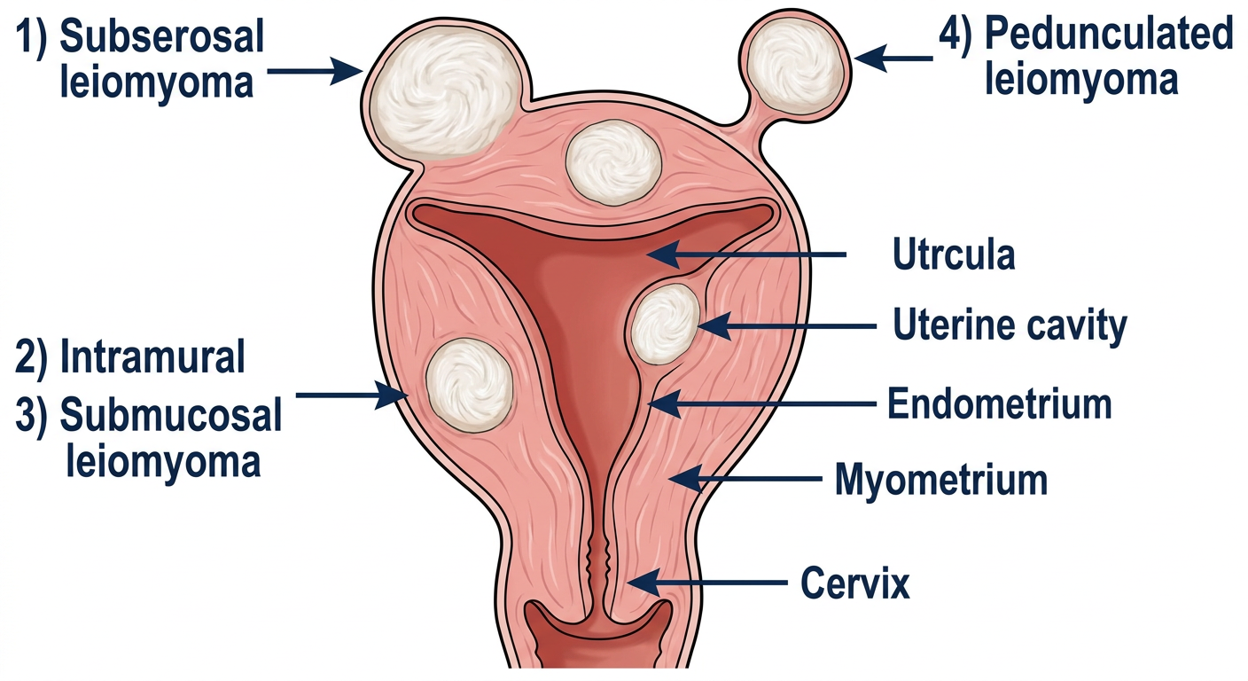
What This Diagram Shows
| Type | Location | Key Gross Feature |
|---|---|---|
| Intramural | Within the myometrium wall | Most common type; whorled white cut surface |
| Subserosal | Outer surface (under serosa) | May become pedunculated; can grow very large |
| Submucosal | Bulges into uterine cavity | Causes heavy bleeding; visible on hysteroscopy |
| Pedunculated | On a stalk (can be subserosal or submucosal) | Risk of torsion |
Gross Appearance (Key Points for Exams)
- Color: White to grayish-white on cut surface
- Pattern: Classic whorled/trabecular arrangement
- Capsule: Well-circumscribed with pseudo-capsule of compressed myometrium
- Texture: Firm, rubbery
- Degeneration: Can undergo hyaline (most common), cystic, red/carneous (pregnancy), calcific, or fatty degeneration