
What is anterior opercular syndrome
anterior opercular syndrome Foix-Chavany-Marie syndrome clinical features
"anterior opercular syndrome" OR "Foix-Chavany-Marie"
anterior opercular syndrome Foix-Chavany-Marie MRI brain lesion
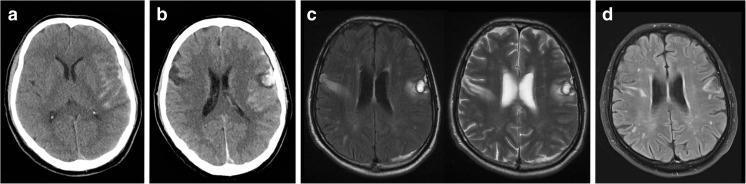
This neuroradiological composite displays the progression of traumatic brain injury leading to Foix-Chavany-Marie syndrome (FCMS). Panel (a) is an initial non-contrast axial CT scan showing traumatic subarachnoid hemorrhage within the left Sylvian fissure and over the left cerebral convexity. Panel (b), a follow-up CT at 48 hours, reveals a new hyperdense hemorrhagic contusion in the left frontal operculum and a corresponding hypodense lesion in the right inferolateral frontal region. Panels (c) and (d) provide axial MRI views. Panel (c) utilizes FLAIR and T2-weighted sequences at 2 weeks post-injury, clearly demonstrating hyperintense bilateral lesions localized to the frontal opercula, which are characteristic of FCMS. Panel (d) shows a progress FLAIR MRI at 13 months, illustrating the chronic persistence of these bilateral opercular signal abnormalities. The series highlights the superior sensitivity of MRI over CT in characterizing parenchymal opercular damage and its long-term sequelae in patients with persistent bulbar dysfunction.
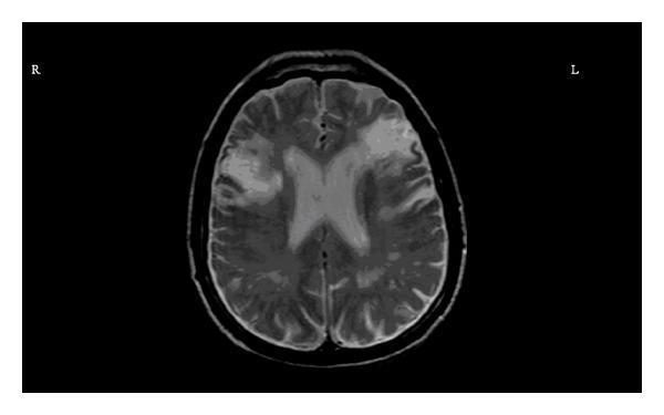
This diagnostic image is an axial T2-weighted magnetic resonance imaging (MRI) scan of the brain, specifically at the level of the lateral ventricles. The image demonstrates bilateral, slightly asymmetrical areas of high signal intensity (hyperintensities) located within the inferior frontal gyri (opercular regions). On the right side (labeled 'R'), the hyperintensity appears more irregular and prominent compared to the left side (labeled 'L'). These findings are consistent with chronic bilateral cortical infarcts involving the frontal opercula. The surrounding anatomy, including the ventricular system and posterior cortical structures, is visible, though the primary pathology is localized to the anterior cerebral parenchyma. This clinical imaging is educationally significant for illustrating the radiological presentation of chronic ischemic strokes and their potential association with clinical syndromes such as Foix-Chavany-Marie syndrome (anterior opercular syndrome), which presents with facio-glosso-pharyngo-masticatory diplegia.
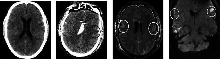
A multi-panel series of neuroimaging studies demonstrating post-traumatic and ischemic brain lesions. Panel (a) is an initial non-contrast axial CT scan showing acute traumatic changes. Panel (b) is a postoperative axial CT scan highlighting a subacute ischemic lesion in the left operculum (black circle) alongside residual post-traumatic changes and generalized cerebral edema. Panel (c) is an axial T2-weighted MRI showing bilateral hyperintense lesions in the opercula (white circles); the right-sided lesion represents parenchymal damage from traumatic contusion, while the left-sided lesion indicates an ischemic event. Panel (d) is a coronal diffusion-weighted image (DWI) confirming restricted diffusion (hyperintensity) within both opercula (white circles). This imaging set illustrates the diagnostic findings for Foix-Chavany-Marie Syndrome (FCMS), characterized by bilateral anterior opercular lesions of mixed traumatic and ischemic etiology.
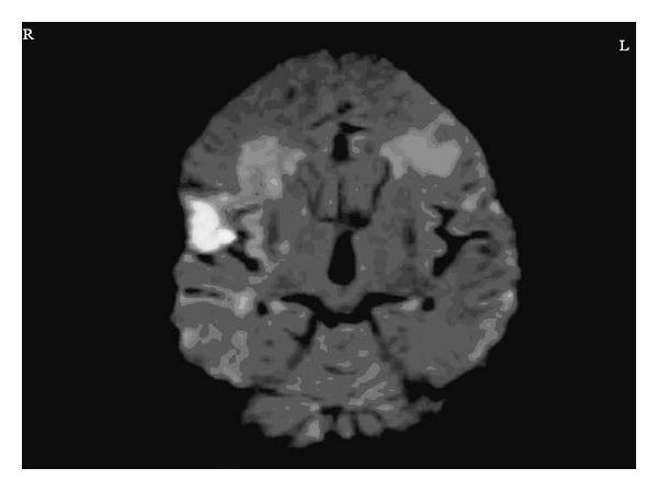
This diagnostic image is a coronal Diffusion-Weighted MRI (DWI) of the human brain. The image displays a prominent, hyperintense (bright white) region in the right frontal opercular area, indicating acute restricted diffusion. This finding is characteristic of an acute infarct, measuring approximately 3.3 x 2.6 cm. Additionally, there are bilateral areas of altered signal intensity in the inferior frontal regions, which correspond to chronic ischemic changes. The ventricular system and midline structures are visible in this section, and orientation markers 'R' and 'L' are provided to denote the anatomical right and left hemispheres. This imaging is clinically significant for diagnosing Foix-Chavany-Marie syndrome (anterior opercular syndrome) when presenting with the described clinical features of aphemia and loss of voluntary facio-pharyngo-glosso-masticatory control. It is an essential resource for neuroradiology and neurology education regarding ischemic stroke localization.
Anterior Opercular Syndrome (Foix-Chavany-Marie Syndrome)
Definition and Names
Anatomy
Pathophysiology
Classic Clinical Features
| Feature | Description |
|---|---|
| Anarthria / severe dysarthria | Complete or near-complete loss of voluntary speech production |
| Dysphagia | Severely impaired swallowing; aspiration risk |
| Bilateral facial paresis (voluntary) | Cannot open or close mouth, show teeth, or smile on command |
| Lingual paresis | Cannot protrude or move tongue voluntarily |
| Masticatory muscle weakness | Difficulty chewing voluntarily |
| Absent/reduced gag reflex | Decreased palatal movement |
| Drooling | Saliva accumulates due to loss of oral motor control |
| Mouth held half open | Characteristic appearance |
| Automatic-voluntary dissociation | Patient can smile when amused, yawn spontaneously, cry in pain - but CANNOT do so on command |
| Limb commands preserved | Comprehension intact; limb movements follow commands normally |
Etiology / Causes
- Ischemic stroke (most common) - bilateral MCA territory infarcts, especially with staggered timing
- Hemorrhagic stroke
- Traumatic brain injury - bilateral frontal opercular contusions
- Epilepsy / cortical dysplasia - especially in children (developmental variant)
- Chronic herpes simplex encephalitis
- Tumors
- Multiple sclerosis
- Neurodegenerative diseases (e.g., corticobasal syndrome can mimic FCMS)
- Rarely, arachnoid cysts involving the perisylvian region
Classification / Forms
- Bilateral anterior opercular syndrome - classic; lesions in both frontal opercula
- Opercular-subopercular syndrome - opercular cortex on one side + subcortical corticobulbar disruption contralaterally
- Subopercular syndrome - pure subcortical corticobulbar projections bilaterally
- Unilateral anterior opercular syndrome - rare, incomplete form
Neuroimaging
Differential Diagnosis
| Condition | Key Distinguishing Feature |
|---|---|
| Classical pseudobulbar palsy | Has emotional lability; more diffuse UMN signs |
| Bulbar palsy | Lower motor neuron; fasciculations, atrophy; no automatic-voluntary dissociation |
| Broca's aphasia | Language comprehension impaired; aphasic, not just dysarthric |
| Apraxia of speech | No facial paralysis; motor planning disorder |
| Oral-buccal apraxia | No actual paresis; can move muscles reflexively AND automatically |
| Locked-in syndrome | Involves limb paralysis; total deefferentation below pons |
Diagnosis
- Presence of bilateral facio-pharyngo-glosso-masticatory paresis
- Automatic-voluntary dissociation - the key test
- Preserved limb commands (excluding aphasia)
- MRI confirming bilateral anterior opercular or perisylvian lesions
Treatment and Prognosis
- Speech and language therapy - communication aids, AAC devices
- Dysphagia management - modified diet, nasogastric/PEG feeding if severe aspiration risk
- Physiotherapy
- Treatment of the underlying cause (thrombolysis/thrombectomy if acute stroke, antiepileptics if seizure-related)
- Bradley and Daroff's Neurology in Clinical Practice, Opercular Syndrome, p. 151
- Localization in Clinical Neurology, 8e, Opercular Syndrome / Pseudobulbar Palsy, p. 1272
- NORD - Foix-Chavany-Marie Syndrome
- Recent case reports: PMID 35855258, 37228548, 38173897