

IMG-20260706-WA0038(1).jpg
Diagnosis life cycle epidemiology pathophysiology differential diagnosis signs and symptoms treatment prevention conclusion
Reading File
The case clearly points to hepatic amoebiasis (Entamoeba histolytica liver abscess) - a Fijian soldier with fever, leukocytosis, interstitial shadowing, multiple liver abscesses, negative bacterial cultures, parasite detected by PCR, and treated with metronidazole + paromomycin. Now let me search the medical library for a thorough answer.
Finding Sources
Finding Sources
Searching PubMed
"Entamoeba histolytica" AND "liver abscess"
Good results. Now let me read the key sections from Bailey & Love, Park's, and Yamada's for full content.
Reading File
Reading File
Reading File
Excellent content. Now let me read the treatment section, differential diagnosis, and more epidemiology from Bailey & Love and Park's.
Reading File
Reading File
Reading File
I now have comprehensive material. Let me fetch the key microscopy images and read the signs/symptoms and prevention sections from Park's.
Loading Image
Reading File
Reading File
I now have all the content needed. Let me read the signs/symptoms and diagnosis section for amoebiasis from Bailey & Love and Yamada's specifically.
Reading File
Finding Sources
I now have comprehensive material from multiple authoritative textbooks. Let me compile the full, structured answer.
Case Study 2 - Diagnosis: Amoebic Liver Abscess (Hepatic Amoebiasis)
Causative agent: Entamoeba histolytica
The clues in the case are: Fijian soldier (endemic region traveler), fever unresponsive to antibiotics, multiple liver abscesses on CT, negative bacterial cultures, parasite detected by PCR of abscess material, clinical response to metronidazole + paromomycin, and negative post-treatment stool antigen/PCR confirming eradication.
1. Diagnosis
The definitive diagnosis is amoebic liver abscess (ALA), the most common extraintestinal manifestation of Entamoeba histolytica infection.
Diagnostic workup:
-
Imaging: Ultrasound or CT scan - shows single (80%) or multiple hypoechoic lesions, predominantly in the right lobe. In this case, CT confirmed multiple liver abscesses.
-
Serology: Indirect haemagglutination (IHA) and ELISA are highly sensitive (>90%) for extraintestinal amoebiasis. Serology is often negative in purely intestinal disease.
-
PCR of abscess material: As in this case, PCR is the most sensitive method when cultures are negative and serology inconclusive - it confirmed the diagnosis here.
-
Stool antigen detection: ELISA-based antigen kits are specific for E. histolytica (not the non-pathogenic E. dispar). Post-treatment stool antigen and PCR were negative, confirming eradication.
-
Aspiration: Yields the classic "anchovy sauce" (chocolate-brown, odourless) fluid - a mixture of necrotic liver and blood. Trophozoites are found in the abscess wall (not the pus) in only a minority of cases.
-
Blood cultures: Negative (as in this patient) - distinguishing ALA from pyogenic liver abscess.
-
Yamada's Textbook of Gastroenterology, 7th ed.
-
Bailey and Love's Short Practice of Surgery, 28th ed.
2. Life Cycle of Entamoeba histolytica
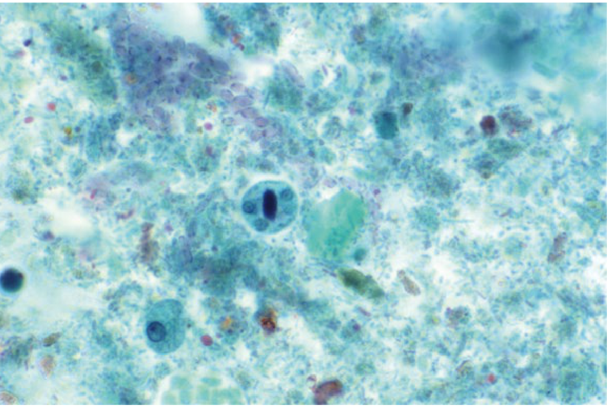
The life cycle consists of two stages:
- Cyst (infective stage): Quadrinucleate cysts are excreted in faeces and are stable in the environment - resistant to chlorine at normal water disinfection doses, but killed by heat (>55°C), drying, or freezing.
- Excystation: Ingested cysts pass through the stomach (resistant to gastric acid) and excyst in the small intestine, undergoing nuclear and cytoplasmic division to release 8 trophozoites.
- Trophozoite (tissue-invasive stage): The motile feeding form with a single nucleus and pseudopodia. Trophozoites colonize the large intestine, invade the submucosa to form flask-shaped ulcers, multiply by binary fission, and produce uninucleate cysts.
- Portal dissemination: Trophozoites enter the portal circulation, become trapped in interlobular veins of the liver, multiply in portal triads, and cause focal hepatocyte infarction and liquefactive necrosis via proteolytic enzymes - forming the abscess.
- Transmission: Only cysts (not trophozoites) transmit the disease. Trophozoites rapidly degenerate outside the body and are destroyed by gastric acid. Humans are the only reservoir.
- Yamada's Textbook of Gastroenterology, 7th ed., p. 3091
- Bailey and Love's Short Practice of Surgery, 28th ed., p. 88
- Park's Textbook of Preventive and Social Medicine
3. Epidemiology
- Global burden: ~500 million people infected with Entamoeba spp. worldwide; ~40 million develop invasive colitis or extraintestinal abscesses; ~50,000 deaths annually. Among parasitic diseases, only cryptosporidiosis, malaria, and schistosomiasis cause comparable mortality.
- Geographic distribution: Most prevalent in Central and South America (especially Mexico), the Indian subcontinent, Indonesia, sub-Saharan and tropical Africa, and South East Asia. Prevalence of E. histolytica in stool samples in high-endemic zones averages ~10%; incidence of ALA can reach 21 per 100,000 population.
- High-risk groups in non-endemic countries: Travelers, immigrants, institutionalized individuals, and men who have sex with men (MSM).
- Sex difference: The prevalence of colonic disease is equal between sexes, but ALA and other extraintestinal disease is 3-10 times more common in men. This case (48-year-old male) fits this pattern exactly.
- Other risk factors for severe disease: Children (especially neonates), pregnant women, malignancy, malnutrition, corticosteroid therapy.
- Socioeconomic link: More closely tied to poor sanitation and low socioeconomic status than to climate. In areas with wet-dry seasons, transmission increases during rains due to cyst survival in water.
A 2025 systematic review and meta-analysis (PMID: 39979548) from India confirmed the epidemiological profile, risk factors, and clinical characteristics of ALA.
- Yamada's Textbook of Gastroenterology, 7th ed.
- Park's Textbook of Preventive and Social Medicine, p. 283-284
4. Pathophysiology
- Ingestion: Contaminated food/water introduces cysts via the faecal-oral route.
- Intestinal invasion: Trophozoites invade the colonic submucosa, producing flask-shaped ulcers (wide base, narrow neck) - predominantly in the caecum and ascending colon, then rectum and sigmoid. This stage may be asymptomatic in up to 90% of infected individuals.
- Portal entry: Trophozoites gain access to the mesenteric venules and are carried via the portal vein to the liver.
- Hepatic trapping: Trophozoites are filtered and trapped in the interlobular veins and multiply in portal triads.
- Liquefactive necrosis: Trophozoites produce proteolytic enzymes (cysteine proteases, amoebapores) causing focal infarction and coalescent liquefactive necrosis. The necrotic cavity fills with the characteristic anchovy-sauce fluid.
- Right lobe predominance (80%): Blood from the superior mesenteric vein flows more directly into the larger right lobe via the portal vein, explaining this anatomical predilection. Left lobe involvement accounts for ~10%; multiple abscesses (as in this case) are the remainder.
- Chest complications: Abscesses high on the diaphragmatic surface of the right lobe can cause reactive pleural effusion, basal pneumonitis, and even transdiaphragmatic rupture into the pleural space - explaining this patient's dyspnoea and pulmonary infiltrates on initial presentation.
- Secondary infection: Sterile unless secondarily infected (which causes the pus to become malodorous).
- Bailey and Love's Short Practice of Surgery, 28th ed., p. 88
5. Differential Diagnosis
| Condition | Key Distinguishing Features |
|---|---|
| Pyogenic liver abscess | Positive bacterial blood/pus cultures, often Gram-negative rods (Klebsiella, E. coli), older patients, biliary/GI source, septate lesions |
| Hydatid cyst (Echinococcus) | Cystic lesion with daughter cysts on imaging, serology positive, eosinophilia, history of sheep/dog contact |
| Hepatocellular carcinoma | AFP elevated, cirrhotic background, solid mass with arterial enhancement on CT |
| Pyogenic pneumonia / lung abscess | Productive purulent cough, sputum cultures positive, no liver lesion |
| Typhoid fever | Rose spots, positive blood/bone marrow culture for Salmonella typhi, splenomegaly, relative bradycardia |
| Visceral leishmaniasis | Massive splenomegaly, pancytopenia, positive rK39 antigen, bone marrow aspirate |
| Hepatic tuberculosis | Calcified granulomas, positive Mantoux/IGRA, chronic course, constitutional symptoms |
In this case, negative bacterial cultures of both blood and abscess, combined with positive PCR for the parasite, excluded pyogenic abscess and confirmed amoebic aetiology.
6. Signs and Symptoms
Typical patient: Young adult male with insidious onset progressing over days to weeks.
Systemic symptoms:
- Fever (often swinging, as in this patient - 39.5°C)
- Night sweats and rigors
- Anorexia, malaise, weight loss
Localizing symptoms:
- Right upper quadrant / right hypochondrial pain (progresses to right shoulder tip pain - diaphragmatic irritation)
- Non-productive cough and dyspnoea (from diaphragmatic elevation/reactive effusion)
- Hiccoughs (diaphragmatic involvement)
Signs on examination:
- Toxic, ill-appearing, anaemic patient
- Tender hepatomegaly (most consistent finding)
- Tender and bulging intercostal spaces with overlying skin oedema
- Upper abdominal rigidity
- Pleural effusion and basal pneumonitis (late)
- Occasionally jaundice or ascites
Complications:
- Rupture into pleural cavity (empyema), peritoneal cavity (peritonitis), or pericardial cavity (tamponade - most feared)
- Secondary infection of the abscess
- Amoeboma (chronic granuloma in large bowel from incomplete treatment)
Laboratory findings:
-
Leukocytosis with neutrophilia (as in this patient: 25,000/mm³, 84%)
-
Elevated ESR and CRP
-
Mildly elevated liver enzymes (ALP most consistently raised)
-
Anaemia in chronic cases
-
Bailey and Love's Short Practice of Surgery, 28th ed.
7. Treatment
Medical (first-line)
- Metronidazole 400-800 mg three times daily for 5-10 days (or tinidazole as an alternative) - this is the tissue amoebicide that kills trophozoites.
- Followed by a luminal amoebicide to eradicate residual cysts in the gut: Paromomycin (500 mg three times daily for 7 days) or diloxanide furoate (500 mg three times daily for 10 days). This step prevents relapse and ongoing transmission.
- In this case: metronidazole + paromomycin - the textbook regimen. Ceftriaxone was added initially for presumed secondary bacterial infection and was discontinued once cultures were negative.
Drainage indications
- Large abscesses (>5 cm): percutaneous needle aspiration or pigtail catheter drainage combined with drug treatment.
- Drainage is recommended when: the abscess is large and at high risk of rupture; there is inadequate response to medical therapy within 72 hours; left lobe abscess (risk of pericardial rupture); suspected secondary infection.
- In this patient, a drain was placed and removed on day 21 as the discharge transitioned from purulent to serosanguinous.
Surgical treatment
Reserved for complications:
-
Rupture into pleural, peritoneal, or pericardial cavity
-
Acute toxic megacolon (intestinal amoebiasis)
-
Severe haemorrhage
-
Bailey and Love's Short Practice of Surgery, 28th ed.
-
Maingot's Abdominal Operations
8. Prevention
Primary Prevention
- Sanitation: Safe disposal of human excreta; hand washing after defecation and before eating. This is the single most effective measure.
- Water supply: Protection from faecal contamination. Note - chlorine at normal doses does NOT kill amoebic cysts. Sand filtration and boiling are more effective. Iodine-based purification tablets (used in military settings) are also effective.
- Food hygiene: Thorough washing of raw vegetables with detergent in running water; alternatively, soaking in 5-10% acetic acid or full-strength vinegar removes cysts. Uncooked vegetables from fields irrigated with sewage water are a major risk. Food handlers should be screened, treated, and educated.
- Vector control: Control of flies, cockroaches, and rodents that mechanically transmit cysts.
- Health education: Long-term community education on personal hygiene.
- Military-specific: Soldiers deployed to endemic regions should receive pre-deployment health briefings, use water purification beyond chlorination alone, and practice strict food/hand hygiene.
Secondary Prevention
-
Early diagnosis of symptomatic and carrier individuals.
-
Treatment of asymptomatic carriers (luminal agents like paromomycin) to interrupt transmission.
-
Periodic screening of food handlers in endemic areas.
-
Contact tracing within households (amoebiasis is frequently a household infection - when one member is infected, others may also be affected).
-
Park's Textbook of Preventive and Social Medicine, p. 284
9. Conclusion
This case of a 48-year-old Fijian soldier illustrates several teaching points about amoebic liver abscess:
- Delayed diagnosis is common because initial presentation with fever, dyspnoea, and interstitial infiltrates mimics community-acquired pneumonia. ALA should be considered in any patient from or traveling to an endemic region who does not respond to standard antibiotics for CAP.
- PCR of abscess material is the key diagnostic tool when cultures are negative - it was diagnostic in this case.
- Medical therapy is highly effective: metronidazole as the tissue agent followed by paromomycin as the luminal agent is the standard regimen. This patient was successfully treated without surgery.
- Drainage is needed for large/complex abscesses - this patient required percutaneous drainage with drain removal on day 21.
- Post-treatment negative stool antigen and PCR confirmed microbiological cure.
- Prevention in military/travel settings hinges on water boiling/filtration (not chlorination alone), food hygiene, and sanitation - all critical for deployed personnel in endemic zones.
Sources: Bailey and Love's Short Practice of Surgery, 28th ed. | Yamada's Textbook of Gastroenterology, 7th ed. | Park's Textbook of Preventive and Social Medicine | Maingot's Abdominal Operations | Ghalib et al. 2025, Int Microbiol [PMID: 39979548]
This is a shared conversation. Sign in to Orris to start your own chat.