
Duodenal peptic ulcer
"duodenal ulcer" AND treatment
duodenal peptic ulcer endoscopy image
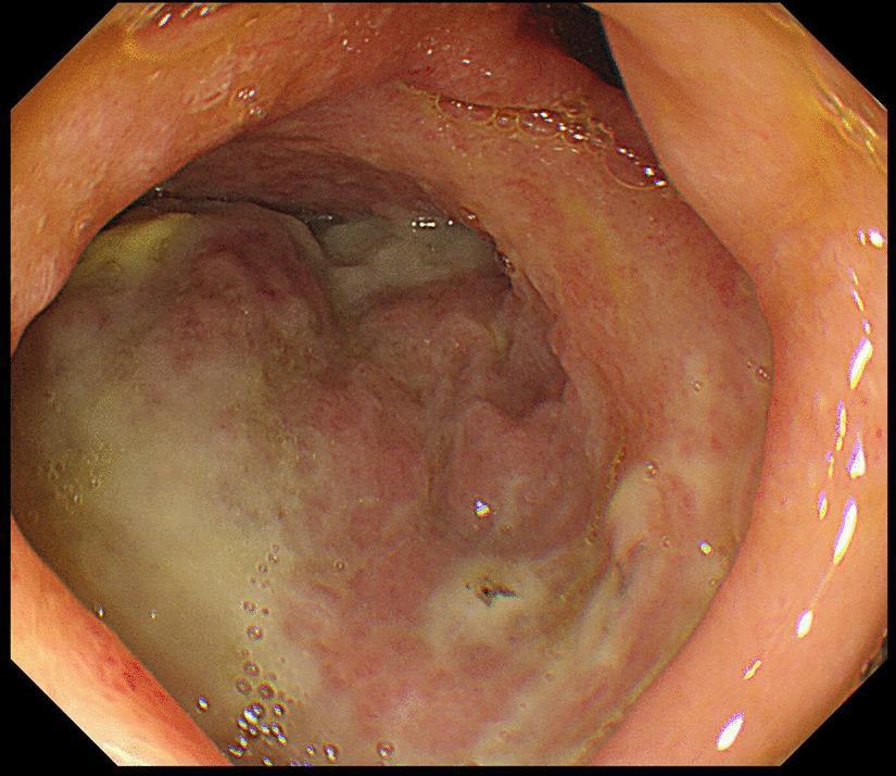
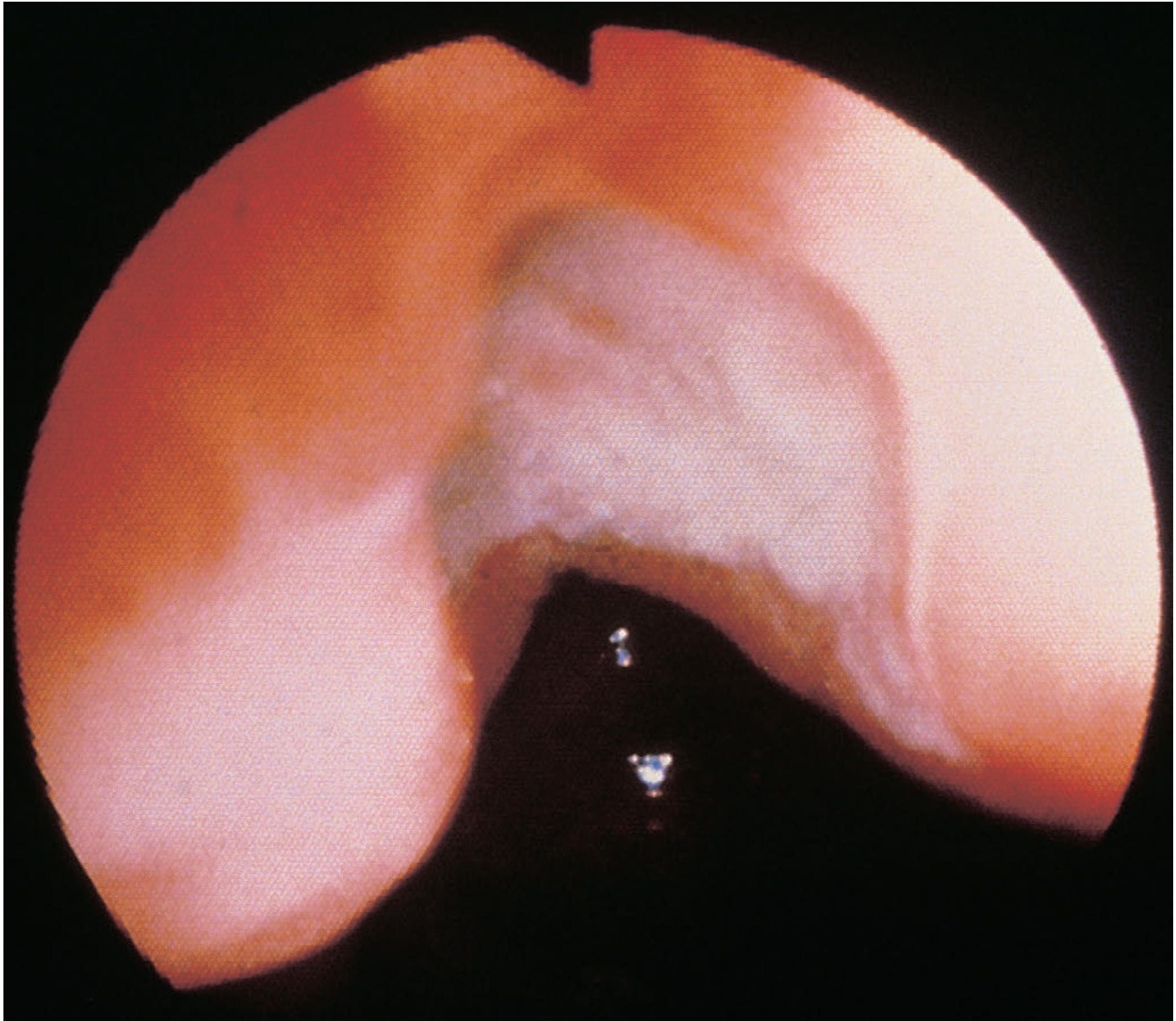
Gastrointestinal fiberscopy image of the duodenal bulb demonstrating a large, irregular ulcer on the posterior wall. The ulcer is characterized by a prominent white-to-yellowish fibrin base, consistent with an active stage (A1 according to the Sakita-Miwa classification). The lesion stands in sharp contrast to the surrounding duodenal mucosa, which appears erythematous and edematous with irregular contours and loss of normal mucosal texture. Visible clear fluid bubbles are scattered across the mucosal surface, likely representing gastric or duodenal secretions. The image illustrates a significant peptic ulcer in a clinical case complicated by perforation and subsequent intra-abdominal abscess formation. This visual is suitable for educational purposes regarding upper gastrointestinal endoscopy, peptic ulcer disease (PUD) morphology, and Sakita-Miwa staging.
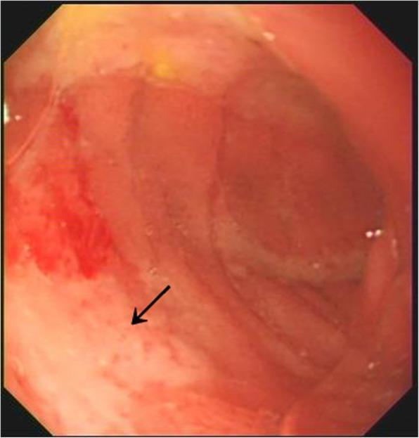
This gastroscopy image displays an active peptic ulcer located within the duodenal bulb. The endoscopic view reveals a prominent, irregular ulcerated lesion characterized by a thick, fibrinous white-yellowish exudate, colloquially referred to as 'white moss.' The surrounding duodenal mucosa exhibits significant pathological changes, including intense erythema (redness) and a granular, edematous texture, indicating localized inflammatory response. An arrow highlights the specific area of mucosal disruption and exudative deposition. This finding is clinically significant for diagnosing peptic ulcer disease and assessing the stage of ulcer healing or activity. The image serves as a clear educational example of upper gastrointestinal pathology for medical students and gastroenterology trainees focusing on diagnostic endoscopy.
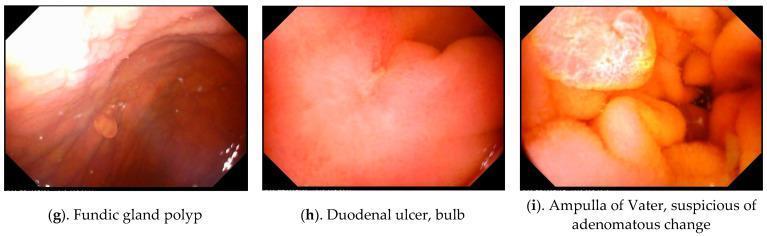
This composite clinical diagnostic image features three endoscopic frames (g, h, i) obtained via Magnetically Controlled Capsule Endoscopy (MACE), demonstrating various upper gastrointestinal organic lesions. Image (g) depicts a fundic gland polyp within the stomach, appearing as a small, smooth, rounded sessile protrusion from the reddish-brown mucosal wall. Image (h) shows the duodenal bulb containing a duodenal ulcer, characterized by a localized area of mucosal depression and pale fibrinoid coating amidst surrounding erythematous tissue. Image (i) focuses on the Ampulla of Vater showing suspicious adenomatous changes, visualized as a complex, multi-lobulated, and nodular structure with variegated yellow-red coloration and irregular surface texture compared to the surrounding duodenal mucosa. These images serve as educational examples of common and significant gastric and duodenal findings in geriatric patients, emphasizing the utility of capsule endoscopy in identifying benign polyps, peptic ulcer disease, and potentially premalignant anatomical changes in the periampullary region.
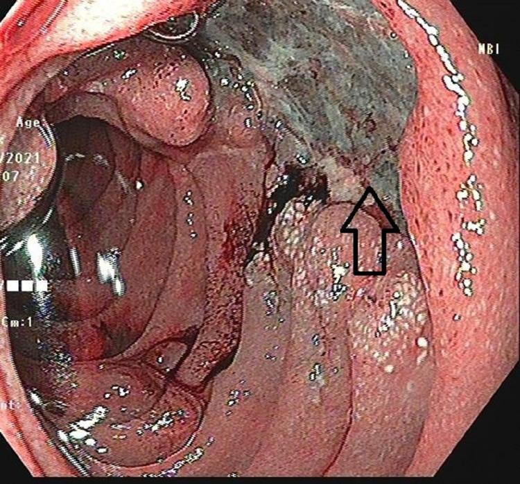
This clinical photograph displays an upper gastrointestinal endoscopy (UGIE) view of the second part of the duodenum. The primary finding is a large, irregularly shaped ulcer approximately 2x2 cm in size, situated on the medial wall. The ulcer features prominent nodular margins and a base covered with dark, necrotic-appearing material and focal areas of hemorrhage. An arrow indicates the close anatomical proximity of the lesion to the duodenal papilla. The surrounding duodenal mucosa is erythematous and inflamed, with thickened, irregular folds and a granular texture suggestive of significant tissue reaction. This endoscopic view is critical for the differential diagnosis of duodenal pathologies, including malignancy (duodenal adenocarcinoma), complex peptic ulcer disease, or atypical presentations of granulomatous diseases like tuberculosis or groove pancreatitis. The image illustrates key markers for malignancy or chronic inflammation, such as nodularity and mucosal distortion.
Duodenal Peptic Ulcer
Definition
Epidemiology
Pathogenesis
- H. pylori infection: present in up to 90% of duodenal ulcer patients and found in over 70% of PUD overall. It sits on a background of chronic gastritis; only 5-10% of infected people actually develop an ulcer, implying host and bacterial strain factors matter.
- NSAIDs: an increasingly common cause as H. pylori prevalence declines; impair mucosal prostaglandin-mediated defenses.
- Hyperacidity: from parietal cell hyperplasia, excess secretory drive, or loss of inhibitory signals. Gastrinoma (Zollinger-Ellison syndrome) causes ulcers through pure acid hypersecretion and should be considered in refractory or atypical cases.
- Cofactors: smoking (reduces mucosal blood flow/healing), high-dose corticosteroids (suppress prostaglandin synthesis), alcohol-related cirrhosis, COPD, chronic renal failure, and hyperparathyroidism (hypercalcemia stimulates gastrin release)
- Duodenal ulcer patients as a group have higher gastric acid output than normal, though there is considerable overlap with the general population
Clinical Features
- Relieved by food or antacids (unlike gastric ulcer pain, which food can worsen)
- Occurs a few hours after meals, often nocturnal
- Chronic, relapsing-remitting course
- Constant pain -> suggests deeper penetration
- Pain radiating to the back -> penetration into the pancreas
- Sudden, diffuse peritoneal pain/rigidity -> free perforation
Diagnosis
- Esophagogastroduodenoscopy (EGD) is the diagnostic test of choice. Biopsy of a duodenal ulcer itself is not routinely necessary (malignancy is extremely rare here, unlike gastric ulcers), but gastric biopsies are taken to test for H. pylori.
- H. pylori testing: urea breath test, fecal antigen test, rapid urease test, or biopsy/histology at endoscopy - all positive patients should be treated.
- Upper GI barium radiography can also demonstrate the ulcer crater but is less sensitive than endoscopy.
- Fasting serum gastrin should be checked in refractory, recurrent, or multiple ulcers to screen for Zollinger-Ellison syndrome.
Management
- Acid suppression with a PPI
- Test for and eradicate H. pylori if positive
- Discontinue NSAIDs; smoking cessation
- Long-term PPI is not routinely needed unless the patient required hospitalization for a complication, or needs ongoing NSAIDs/aspirin/anticoagulants, in which case PPI prophylaxis is continued indefinitely
- Low-dose aspirin for cardiovascular indications can generally continue safely if a daily PPI is co-administered
| Complication | Key Features |
|---|---|
| Perforation | Most common indication for surgery today. Sudden severe pain, tachycardia progressing to peritonitis, rigid abdomen, leukocytosis. Erect CXR shows pneumoperitoneum in 85-90%; CT nearly always shows free air/fluid. Mortality up to 30% in modern series, worsened by delay to treatment. Anemia should raise suspicion for a second "kissing" posterior bleeding ulcer. |
| Bleeding | Most common overall complication; posterior duodenal ulcers can erode into the gastroduodenal artery |
| Gastric outlet obstruction | From chronic scarring/edema at the pylorus/duodenal bulb |
| Intractability | Rare now with modern medical therapy; requires confirmation of H. pylori eradication and NSAID cessation before being labelled "refractory" |
Recent Evidence Note
Gastric peptic ulcer
"gastric ulcer" AND "Helicobacter pylori"
Gastric Peptic Ulcer
Definition
Pathogenesis
- H. pylori infection: causes chronic mucosal inflammation that primes the lamina propria with inflammatory cells, impairing local defenses; it also affects acid/gastrin secretion. Gastric ulcer from H. pylori can occur with normal or reduced acid output - the injury is largely from the inflammatory response to the infection itself.
- NSAIDs/aspirin: block prostaglandin synthesis, the key mediator of mucus secretion, bicarbonate secretion, and mucosal blood flow. Increasingly the dominant cause as H. pylori rates fall.
- Smoking: reduces mucosal blood flow and healing.
- Physiologic/psychological stress, alcohol, cocaine, radiation, long-term corticosteroids: additional risk factors.
- Normal healing depends on rapid restitution of surface epithelial cells, mucus "bandaging," augmented mucosal blood flow, and prostaglandins - all of which the above insults disrupt.
Classification (Modified Johnson Types I-V)
| Type | Location | Acid Level | Notes |
|---|---|---|---|
| I | Lesser curve at the incisura (transition between body and antral mucosa) | Low to normal | Most common (~50-60%); not associated with acid hypersecurity; vagotomy traditionally not needed |
| II | Gastric body, combined with a duodenal ulcer (active or healed) | Increased | ~15-20%; behaves like a duodenal ulcer |
| III | Prepyloric | Increased | ~20%; resembles duodenal ulcer pathophysiology |
| IV | High on lesser curve, near the gastroesophageal junction | Normal | <10%; excision can require Roux reconstruction (Csendes operation) due to proximity to the esophagus |
| V | Anywhere (often greater curvature) | Normal | NSAID-induced; amenable to simple wedge resection |
Clinical Features
The Critical Issue: Malignancy
- A truly benign chronic ulcer undergoing malignant transformation (rare)
- A gastric cancer presenting as an "ulcerated" lesion that looks benign endoscopically or radiographically but is malignant on biopsy (more common)
Diagnosis
- Upper endoscopy (EGD) with multiple biopsies is mandatory - this is the fundamental difference in workup versus duodenal ulcer.
- H. pylori testing (biopsy-based rapid urease test/histology, or noninvasive urea breath/fecal antigen test).
- Serum gastrin in refractory or atypical cases.
- Repeat endoscopy after 6-10 weeks of treatment to confirm complete healing and re-biopsy if not fully healed.
Management
- PPI acid suppression
- H. pylori eradication if positive
- Discontinue NSAIDs/aspirin, smoking cessation
- Mandatory endoscopic surveillance until healing is confirmed and malignancy excluded
- Confirmed or suspected malignancy
- Complications: perforation, uncontrolled bleeding, obstruction
- Non-healing/intractable ulcers despite adequate medical therapy and confirmed H. pylori eradication
Duodenal vs Gastric Ulcer - Key Differences
| Feature | Duodenal Ulcer | Gastric Ulcer |
|---|---|---|
| Malignancy risk | Extremely rare | Must always be excluded by biopsy |
| Acid secretion | Usually elevated | Variable, often normal/low |
| Pain-food relationship | Relieved by food | Food may not relieve or can worsen pain |
| Biopsy of ulcer itself | Not routinely needed | Mandatory, multiple biopsies |
| Age | Younger patients | Older (55-65 peak) |
Recent Evidence Note
Treatment modality for gastric ulcer
"gastric ulcer" AND vonoprazan
Treatment Modality for Gastric Ulcer
1. Medical Therapy (First-line for essentially all uncomplicated cases)
- PPI is the mainstay (e.g., omeprazole, lansoprazole, pantoprazole). Lansoprazole dosing example: prophylaxis 15 mg once daily; treatment 30 mg once daily for a defined course.
- Typical treatment course is around 8 weeks of PPI therapy; recurrence/non-healing is unusual if H. pylori is eradicated and NSAIDs/smoking are stopped, but recurs if these persist - Current Surgical Therapy 14e, p. 2959-2961.
- H2-receptor antagonists are an alternative but generally less effective than PPIs for healing and are mainly used for prophylaxis in lower-risk settings.
- Standard triple or quadruple antibiotic + PPI regimens. Eradication significantly reduces symptom recurrence and re-ulceration - Textbook of Family Medicine 9e; Maingot's Abdominal Operations.
- Discontinue NSAIDs/aspirin where possible; if they must continue, co-prescribe a PPI.
- Smoking cessation (impairs mucosal blood flow and healing).
- Avoid alcohol.
2. Endoscopic Therapy (for bleeding gastric ulcers)
3. Surgery (reserved for complications, non-healing, or malignancy)
- Perforation
- Uncontrolled/recurrent bleeding
- Gastric outlet obstruction
- Confirmed or suspected malignancy
- True intractability despite confirmed H. pylori eradication and NSAID cessation
| Type | Operation |
|---|---|
| I (lesser curve, low acid) | Distal gastrectomy including the ulcer, without vagotomy (Billroth I reconstruction preferred) |
| II (body + duodenal ulcer, high acid) | Distal gastrectomy with truncal vagotomy; Billroth II often preferred given concomitant duodenal disease |
| III (prepyloric, high acid) | Distal gastrectomy with truncal vagotomy |
| IV (high lesser curve/near GE junction) | Distal subtotal gastrectomy with in-continuity excision; Roux-en-Y esophagogastrojejunostomy (Csendes procedure) if resection encroaches on the cardia; Pauchet procedure for more distal type 4/proximal type 1 lesions |
| V (NSAID-related, often greater curvature) | Simple wedge resection |
| Operation | Operative mortality | Significant side effects | Recurrence |
|---|---|---|---|
| Gastrectomy | 1-2% | 20-40% | 1-4% |
| Gastroenterostomy alone | <1% | 10-20% | 50% |
| Truncal vagotomy + drainage | <1% | 10-20% | 2-7% |
| Selective vagotomy + drainage | <1% | 10-20% | 5-10% |
| Highly selective vagotomy | <0.2% | <5% | 2-10% |
| Truncal vagotomy + antrectomy | 1% | 10-20% | 1% |
Emerging Evidence: Vonoprazan vs PPI
Bottom Line
Pylori eradication
"Helicobacter pylori" AND eradication AND resistance
H. Pylori Eradication
Why eradicate
Who should be treated
- All patients with documented peptic ulcer disease (gastric or duodenal), regardless of symptom severity, first episode vs recurrent, or NSAID use
- Uninvestigated dyspepsia in patients under 60 (test-and-treat), or where local H. pylori prevalence is >20%
- Patients starting long-term NSAIDs/low-dose aspirin, especially with a prior ulcer history (note: eradication alone does not eliminate ulcer risk in these patients - continued PPI is still needed)
- Gastric MALT lymphoma (60-90% achieve complete remission with eradication alone)
- After resection of early gastric cancer; first-degree relatives of gastric cancer patients; severe atrophic gastritis/pangastritis; long-term acid inhibition >1 year; heavy occupational/environmental carcinogen exposure
- Unexplained iron-deficiency anemia, idiopathic thrombocytopenic purpura, unexplained vitamin B12 deficiency
Regimens
| Regimen | Drugs | Duration | Notes |
|---|---|---|---|
| Clarithromycin triple therapy (PAC) | PPI bid + amoxicillin 1g bid + clarithromycin 500mg bid | 10-14 days | Acceptable first-line ONLY in low clarithromycin-resistance areas (<15%) |
| PCM (penicillin-allergic) | PPI bid + clarithromycin 250-500mg bid + metronidazole 400mg bid | 10-14 days | Substitute for PAC in penicillin allergy |
| Bismuth quadruple therapy | PPI + tetracycline 500mg qid + metronidazole 250mg qid + bismuth 525mg qid | 14 days | First-line, especially in high clarithromycin-resistance regions or after prior macrolide exposure |
| Concomitant (nonbismuth quadruple) therapy | PPI bid + amoxicillin 1g bid + clarithromycin 500mg bid + metronidazole 500mg bid | 14 days | First-line, effective even against some clarithromycin resistance |
| Sequential therapy | Amoxicillin + PPI for 5-7 days, then clarithromycin + metronidazole + PPI for another 5-7 days | 10-14 days total | First-line alternative |
| Hybrid therapy | Amoxicillin + PPI for 7 days, then add clarithromycin + metronidazole for 7 more days | 14 days | First-line |
| Levofloxacin triple therapy | Levofloxacin 250mg bid + amoxicillin 1g bid + PPI | - | First-line alternative where fluoroquinolone resistance is low |
- Bismuth quadruple with levofloxacin, tetracycline, or metronidazole substitutions
- Rifabutin-based salvage regimen (amoxicillin + rifabutin + PPI) for 10 days
- Culture-guided therapy: two antibiotics selected by susceptibility testing + bismuth + PPI
Key Practical Points
- Clarithromycin resistance is the single biggest determinant of triple therapy failure. Empiric clarithromycin triple therapy should NOT be used first-line in high-resistance areas - cure rates below 80%, sometimes below 50%, have been widely reported (Greece, India, Italy, Japan, Mexico, Spain), though still acceptable in Korea, Singapore, Thailand.
- Longer courses (14 days) outperform shorter ones (7-10 days); dual therapy and simpler regimens are inferior to triple/quadruple therapy.
- If triple therapy fails in a compliant patient, this is usually due to a resistant organism, inadequate acid suppression, or inadequate compliance/duration.
- Confirm eradication with a urea breath test or stool antigen test at least 4 weeks after completing therapy and at least 1-2 weeks off PPI (to avoid false negatives).
- Concerns exist about growing antibiotic resistance and effects on the microbiome from widespread empiric antibiotic use, reinforcing the move toward regional resistance-guided or culture-guided regimen selection.
Recent Evidence Check
- PMID 38898622 - global primary antibiotic resistance rates over the last decade (2024)
- PMID 37016679 / 37436391 - global clarithromycin resistance prevalence meta-analyses (2023)
- PMID 38477847 - high resistance rates reported in Turkey, with implications for regimen selection