

Explqin me this topic in easy exam orinted ponit of view my exam is near and im unable to understand with this video detailed explanation easy way exam orinted to get full marks with diagram
TENS mechanism gate control theory segmental inhibition diagram
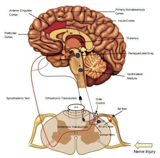
This pathophysiology diagram illustrates the neural pathways of nociceptive processing and the mechanisms of spinal cord stimulation (SCS). The graphic features a sagittal cutaway of the human brain and a cross-section of the spinal cord. Key intracranial structures labeled include the prefrontal cortex, anterior cingulate cortex (highlighted in red), primary somatosensory cortex, insula, thalamus, and amygdala. The brainstem is detailed with the periaqueductal gray and ventrolateral medulla, indicating the descending inhibitory system. At the spinal level, the diagram shows the dorsal horn and its interaction with Aβ and Aδ/C nerve fibers following a 'nerve injury'. The visual highlights the ascending spinothalamic tract and descending pathways that facilitate segmental spinal inhibition. Key physiological concepts depicted include gate control theory, orthodromic and antidromic transduction, and cortical modulation. This educational material is designed to demonstrate how neuromodulation through SCS impacts pain signaling and inhibitory feedback loops between the peripheral nervous system and the central nervous system.
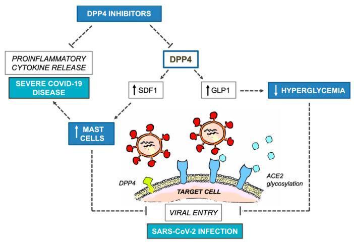
This pathophysiology diagram illustrates the complex relationship between DPP4 inhibitors, glycemic control, and SARS-CoV-2 infection outcomes. The central mechanism depicts SARS-CoV-2 virions utilizing two cell-surface pathways for 'Viral Entry' into a 'Target Cell': the DPP4 protease (green) and ACE2 glycosylation sites (blue). The diagram outlines the systemic effects of DPP4 inhibition through two primary branches. First, the inhibition of DPP4 leads to increased Glucagon-like Peptide 1 (GLP1), which reduces hyperglycemia, potentially mitigating SARS-CoV-2 infection. Second, DPP4 inhibition increases Stromal Derived Factor 1 (SDF1), which elevates mast cell levels. This increase in mast cells presents a dual clinical implication: it may inhibit viral entry but simultaneously promote proinflammatory cytokine release, contributing to 'Severe COVID-19 Disease.' The chart effectively summarizes the therapeutic paradox where glycemic benefits and viral entry inhibition must be weighed against the risk of exacerbated inflammatory responses in COVID-19 management.
Educational figure illustrating the molecular mechanism and experimental model of retinal detachment and Rho-kinase (ROCK) inhibition. Panel A is a pathophysiology diagram showing that Fasudil inhibits ROCK, which normally activates LIMK and Myosin Light Chain. This inhibition prevents cofilin phosphorylation and actomyosin contraction, theoretically stopping photoreceptor axon retraction. Panels B and C are clinical specimen photographs of porcine eyecups (right and left eyes) used for in vivo studies. The images show the posterior segment with anatomical markers: S (Superior), I (Inferior), T (Temporal), N (Nasal), and OD (Optic Disc). Yellow squares indicate sample collection areas for morphological analysis: 'BC' and 'FC' represent attached retinal control areas, while 'BD' (control BSS) and 'FD' (Fasudil-treated) indicate areas of iatrogenic retinal detachment created in the inferior-nasal quadrant. The visual contrast between attached and detached retinal folds is evident. This material is designed for advanced ophthalmic research on neuroprotection and synaptic remodeling following retinal injury.
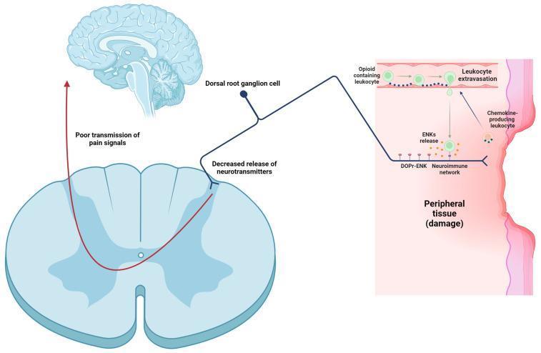
A pathophysiology diagram illustrating the mechanism of peripheral pain control via leukocyte-derived enkephalins (ENKs). The visual depicts three primary anatomical regions: damaged peripheral tissue, the spinal cord (axial cross-section), and the brain. In the periphery, opioid-containing leukocytes undergo extravasation from blood vessels, attracted by chemokines produced at the site of tissue damage. These leukocytes enter the DOPr-ENK neuroimmune network and release enkephalins. These ENKs bind to the peripheral endings of primary nociceptors (dorsal root ganglion cell axons). This interaction leads to a functional inhibition shown at the spinal level: there is a 'decreased release of neurotransmitters' from the primary afferent into the dorsal horn. The resulting effect is depicted by a red arrow labeled 'poor transmission of pain signals' ascending toward the brain, illustrating the analgesic effect of immune-mediated opioid release in response to inflammatory peripheral damage.
TENS transcutaneous electrical nerve stimulation nerve fiber types pain pathway
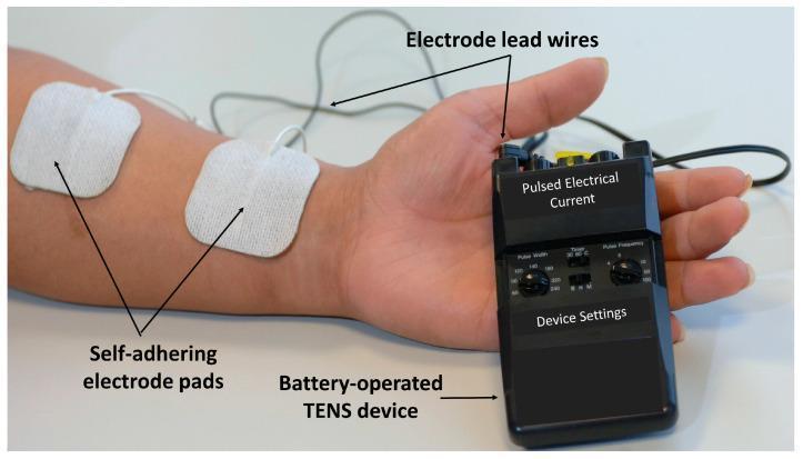
A clinical photograph illustrating a Transcutaneous Electrical Nerve Stimulation (TENS) setup for pain management. The image displays a portable, handheld, battery-operated TENS device held in a patient's hand. The device is black with physical control knobs for adjusting 'Pulse Width' and 'Pulse Frequency,' alongside a 'Timer' switch and a yellow intensity dial. Labeled electrode lead wires extend from the top of the device to two square, white, self-adhering electrode pads placed on the patient's anterior forearm. This procedural image demonstrates the correct positioning for delivering pulsed electrical currents across the skin's surface to stimulate peripheral nerves (A-beta fibers) for symptomatic pain relief. The visual focuses on the integration of hardware, skin interface, and user-adjustable settings in physical therapy and rehabilitation medicine.
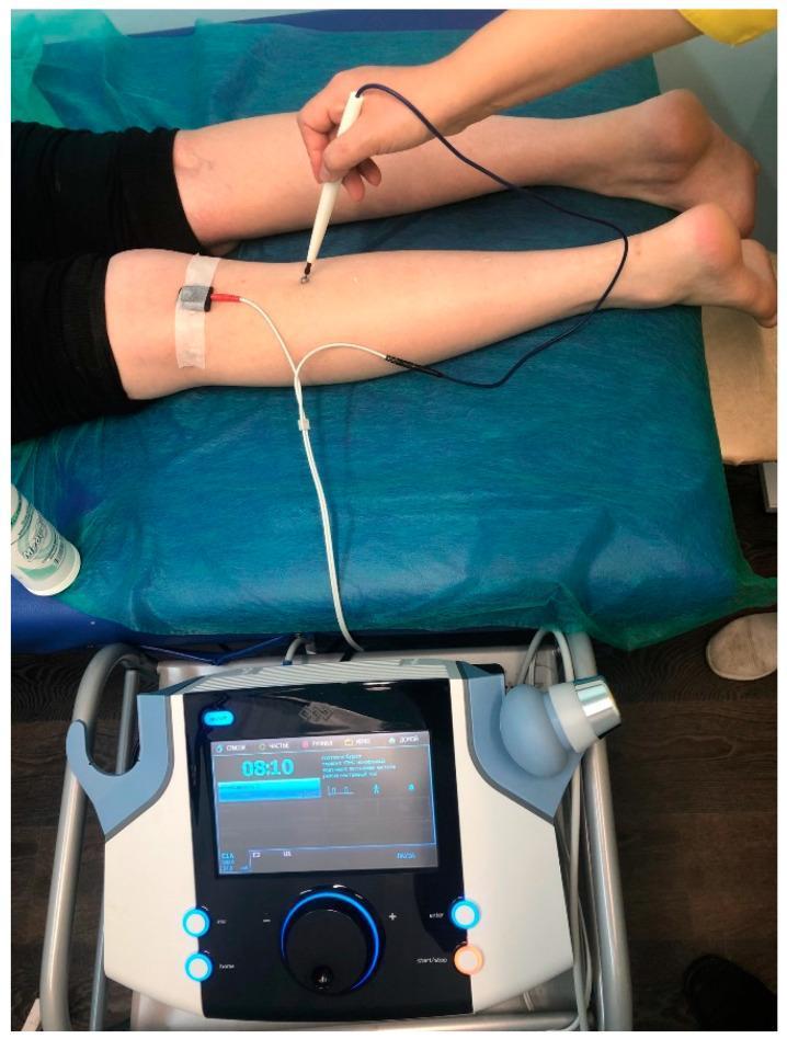
This clinical photograph illustrates a procedural setup for Low-Frequency Transcutaneous Electrical Nerve Stimulation (LF TENS) of the tibial nerve. The subject is positioned prone on a medical table. A stationary rectangular cathode is fixed with white adhesive tape over the nerve in the popliteal fossa of the lower leg. A clinician is shown manually applying a mobile, pen-like anode electrode, moving it along the posterior calf in a proximal-to-distal direction to target the nerve pathway. Both electrodes are connected to a digital medical stimulation device positioned in the foreground. The device interface displays a timer (08:10), wave parameters, and clinical settings regulated by a large central rotary dial and illuminated functional buttons. This procedure is commonly utilized in physical therapy and neurology for managing neuropathic pain, such as diabetic painful distal axonal neuropathy, by inducing painless muscle contractions to modulate pain perception.
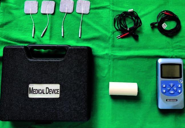
Clinical photograph of a transcutaneous electrical nerve stimulation (TENS) device kit, commonly used in physical therapy, pain management, and experimental medicine—such as parotid gland stimulation to increase salivary flow. The components are arranged on a green sterile field and include a dual-channel TENS control unit (model MH8000), four square self-adhesive electrode pads with integrated lead wires, two coiled black electrical cables with dual-terminal connectors, a roll of micropore medical tape for securing electrodes, and a black hard-shell carrying case labeled 'MEDICAL DEVICE'. The blue control unit features a digital LCD screen and tactile interface buttons for adjusting pulse rate, pulse width, and intensity (0-80mA). This visual serves to illustrate the equipment required for non-invasive neurostimulation procedures in both clinical and research settings.
⚡ Mechanism of TENS - Complete Exam Notes
(Peripheral + Segmental + Extra-Segmental Mechanisms)
🔷 What is TENS? (Start here - 2 marks intro)
- A non-invasive electrotherapy modality
- Electrodes placed on skin deliver low-voltage electrical current
- Used for pain relief in arthritis, tendonitis, chronic pain, etc.
- Patient feels a tingling sensation but no pain
One-line definition for exam: "TENS is a safe, cutaneous electrical stimulation method that delivers low-intensity current through skin electrodes to activate sensory nerve fibers for pain relief." (Campbell-Walsh Urology)
🔷 Key Nerve Fibers - MUST KNOW TABLE
| Fiber Type | Diameter | Myelination | Carries | Role in TENS |
|---|---|---|---|---|
| Aβ (A-beta) | Large | Heavily myelinated | Touch, pressure | ACTIVATED by TENS - closes pain gate |
| Aδ (A-delta) | Medium | Thinly myelinated | Sharp/fast pain | Blocked by conventional TENS |
| C fiber | Small | Unmyelinated | Slow/dull pain | Pain signals - INHIBITED by TENS |
Exam tip: TENS selectively activates large-diameter Aβ fibers without activating small nociceptive fibers.
🔷 MECHANISM 1: Peripheral Mechanism
How it works:
- TENS electrodes placed over/around the painful area
- Electrical current activates Aβ (large diameter) sensory fibers
- These fibers carry non-painful touch/tingling sensations
- This blocks the transmission of pain signals from C fibers and Aδ fibers
- Antidromic conduction - impulse travels backwards toward the periphery, causing local effect
Key point for exam:
- Conventional TENS (high frequency, low intensity) works mainly via this mechanism
- The pain relief is localized to the area of stimulation
- Video note: "TENS-induced activity in large diameter afferents (Aβ fibers) can block afferent activity in large diameter fibres"
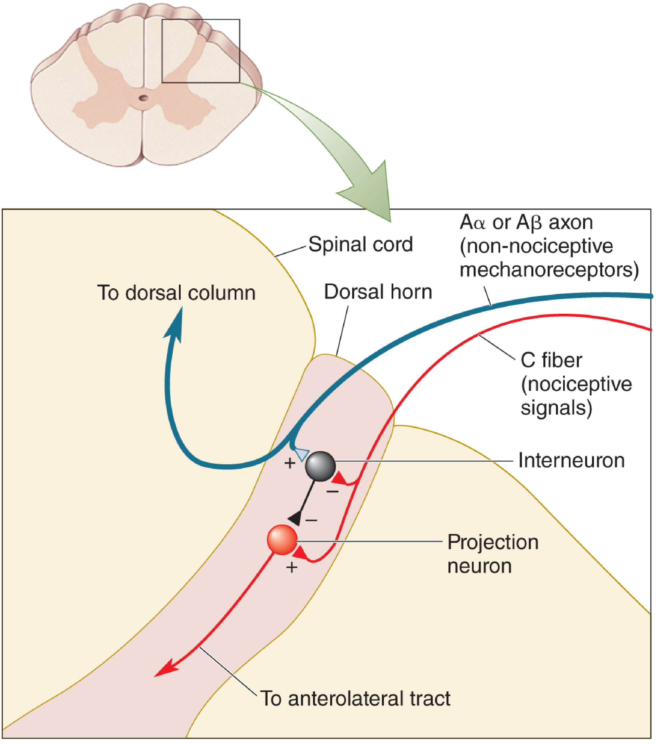
🔷 MECHANISM 2: Segmental Mechanism (Gate Control Theory)
Gate Control Theory (Melzack & Wall, 1965)
The Gate - How it Works:
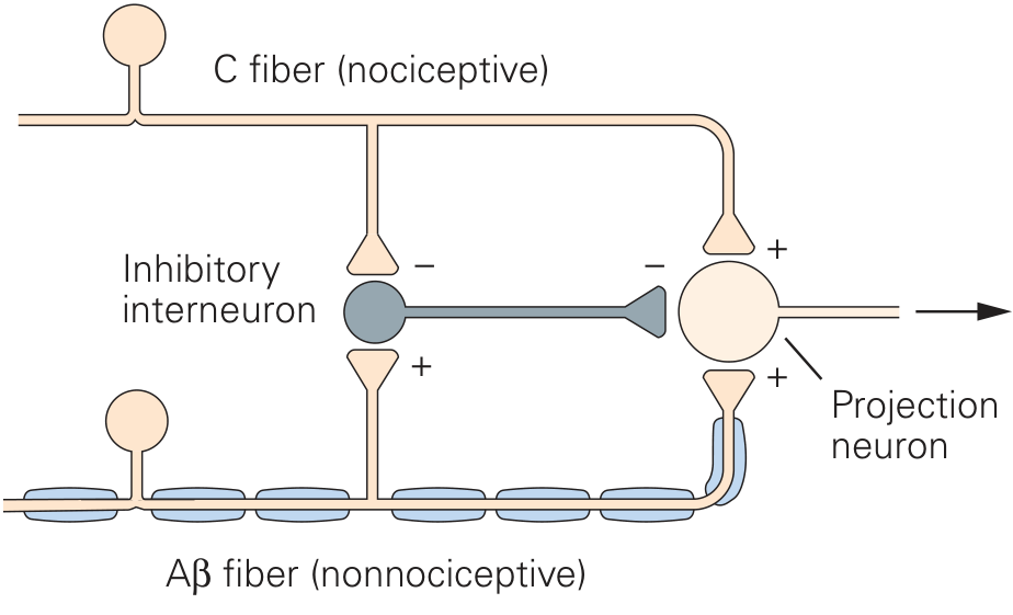
- C fiber (nociceptive/pain fiber) - wants to OPEN the gate
- Aβ fiber (non-nociceptive/touch fiber) - wants to CLOSE the gate
- Inhibitory Interneuron (the gatekeeper)
- Projection Neuron (the signal sender - sends pain to brain)
Step-by-step (write this in exams):
| Situation | What Happens | Result |
|---|---|---|
| Only C fibers active (pain) | C fiber INHIBITS interneuron + excites projection neuron | Gate OPENS → Pain felt |
| Aβ fibers active (TENS/touch) | Aβ EXCITES interneuron → interneuron INHIBITS projection neuron | Gate CLOSES → Pain blocked |
| Both active | Competition - if Aβ > C → gate closes | Pain reduced |
TENS and Segmental Mechanism:
- TENS activates Aβ fibers in the painful segment
- Aβ fibers excite inhibitory interneurons in the dorsal horn
- Interneurons INHIBIT the projection neuron (T-cell/wide dynamic range neuron)
- Pain signals from C fibers cannot get through = Gate is CLOSED
- This effect is segmental - works only for the spinal cord segment corresponding to the treated area
"The region of the body in which pain is reduced maps to those segments of the spinal cord in which nociceptive and non-nociceptive afferents from that body region terminate." (Eric Kandel, Principles of Neural Science)
🔷 MECHANISM 3: Extra-Segmental Mechanism (Descending Pain Inhibition / Opiate Mechanism)
How it works:
- Low-frequency TENS (acupuncture-like TENS) activates deeper nerve fibers including Aδ fibers
- This activates supraspinal (brain) pain-modulating centers:
- Periaqueductal Gray (PAG) in midbrain
- Nucleus Raphe Magnus in brainstem
- Ventrolateral Medulla
- These centers release endogenous opioids:
- Endorphins (beta-endorphin)
- Enkephalins
- Dynorphins
- These opioids travel DOWN the spinal cord via descending inhibitory pathways
- They inhibit pain transmission at multiple spinal cord levels
Key difference from segmental mechanism:
- Works beyond the stimulation segment (hence "extra-segmental")
- Uses endogenous opioids as the mediator
- Can be blocked by naloxone (opioid antagonist) - important exam fact!
- Involved in acupuncture-like TENS (low frequency, high intensity)
"Another described mechanism is the release of endorphins and their precursors into the cerebrospinal fluid." (Campbell-Walsh Urology)
🔷 Comparison Table - All 3 Mechanisms (High-value exam table)
| Feature | Peripheral | Segmental | Extra-Segmental |
|---|---|---|---|
| Level | Peripheral nerve | Spinal cord dorsal horn | Brain + descending pathways |
| Fiber activated | Aβ fibers | Aβ fibers | Aδ fibers |
| TENS type | Conventional (HF) | Conventional (HF) | Acupuncture-like (LF) |
| Mediator | Direct nerve block | Inhibitory interneuron | Endorphins/Enkephalins |
| Effect area | Local/peripheral | Same spinal segment | Beyond segment (widespread) |
| Blocked by naloxone? | No | No | Yes |
| Onset | Fast | Fast | Slower |
| Duration after TENS | Short | Short | Longer |
🔷 TENS Parameters - Quick Reference
| Parameter | Conventional TENS | Acupuncture-Like TENS |
|---|---|---|
| Frequency | High (80-150 Hz) | Low (1-4 Hz) |
| Intensity | Low (tingling) | High (muscle twitch) |
| Mechanism | Segmental + Peripheral | Extra-segmental |
| Fiber | Aβ | Aδ |
| Onset | Rapid | Delayed |
🔷 Clinical Pearls for Exam (Short Answer Points)
- Why shake your hand after hitting it? - Activates Aβ fibers → closes pain gate (same principle as TENS)
- Why does TENS not relieve pain elsewhere? - Segmental mechanism is spinal segment-specific
- Levin and Chan finding: Healthy individuals cannot tolerate direct Aδ activation, so intense TENS is given for only brief periods in clinical practice
- Naloxone blocks extra-segmental (opioid) mechanism but NOT segmental mechanism
- TENS is contraindicated: Over eyes, carotid sinus, transcerebrally, over pacemaker, during pregnancy (abdomen/lower back)
🔷 Draw This Diagram in Your Exam (Gate Control Theory)
                    DORSAL HORN (Spinal Cord)

PERIPHERAL NERVE          DORSAL HORN           TO BRAIN

Aβ fiber ─────────►  Inhibitory    ─(-)→  Projection
(large, touch)       Interneuron           Neuron ───► Pain signal
         ↑(+)excites                  ↑(+)
                              ↑(+)
C fiber ──────────────────────────────┘
(small, pain)        ↓(-)inhibits
                  Interneuron

TENS activates Aβ → Excites interneuron → Inhibits projection neuron
→ GATE CLOSED = No Pain Signal reaches brain