
Cartilaginous joint
I'll look this up in the medical library for a thorough answer.
Reading File
Searching sources...
Finding Sources
Finding Sources
Good - found relevant sources. Let me now read the detailed sections from the best anatomy sources.
Reading File
Reading File
Finding Sources
Now let me get more detail from the imaging anatomy textbook and search for any images of cartilaginous joints.
Reading File
Finding Sources
Finding Sources
Let me read the rest of the cartilaginous joints section and fetch the relevant images.
Reading File
Loading Image
Now let me fetch the key cartilaginous joint images (pubic symphysis and synchondrosis).
Loading Image
Loading Image
I now have comprehensive information. Let me compile the full answer.
Cartilaginous Joints
Cartilaginous joints are joints in which two bones are united by cartilage (either hyaline cartilage or fibrocartilage). They have no joint cavity and permit little to no movement compared to synovial joints. They are structurally intermediate between fibrous joints and synovial joints.
Classification
Cartilaginous joints are divided into two types based on the type of cartilage present and the degree of movement allowed:
| Feature | Primary (Synchondrosis) | Secondary (Symphysis) |
|---|---|---|
| Cartilage type | Hyaline cartilage | Fibrocartilage |
| Movement | None (except bone growth) | Slight (amphiarthrosis) |
| Location | Not necessarily midline | Midline of the body |
| Tendency to ossify | Yes (temporary) or no (permanent) | Less likely to ossify |
1. Synchondrosis (Primary Cartilaginous Joint)
- Bones are joined by hyaline cartilage
- Functionally classified as a synarthrosis (immobile)
- Subclassified as temporary or permanent
Temporary Synchondroses
These exist during growth and are eventually replaced by bone (synostosis):
- Epiphyseal growth plates (physis) between the diaphysis and epiphysis of long bones - injury to these in a growing child can cause shortening or deformity of the bone
- Y-shaped triradiate cartilage connecting the ilium, ischium, and pubis - completely ossified by ~age 25
- Spheno-occipital synchondrosis at the base of the skull
Permanent Synchondroses
These persist throughout life:
- Costochondral junctions (ribs to costal cartilage)
- First sternocostal joint (1st rib to manubrium)
- Note: The 2nd through 7th sternocostal joints are synovial joints
2. Symphysis (Secondary Cartilaginous Joint)
- Bones are united by a fibrocartilage disk and supported by periarticular ligaments
- Functionally classified as amphiarthrosis (slightly mobile)
- Located in the midline of the body
- Fibrocartilage is much stronger than hyaline cartilage due to thick collagen fibers, making symphyses efficient at firmly uniting adjacent bones
- Fibrocartilage of a symphysis is less likely to ossify than hyaline cartilage of a synchondrosis
Examples:
- Pubic symphysis - narrow fibrocartilaginous joint connecting the two pubic bones; ends of pubic bones are covered by a thin layer of hyaline cartilage attached to the fibrocartilage disk; reinforced by several ligaments (anterior and posterior pubic ligaments)
- Intervertebral discs - classified as wide symphyses; fibrocartilaginous joints between vertebral bodies
- Manubriosternal joint - symphysis at the sternal angle (angle of Louis); filled with fibrocartilage (note: this particular symphysis is synarthrotic)
- Sacrococcygeal articulation - amphiarthrodial symphysis homologous with intervertebral discs; may be partially or completely obliterated in old age
- Mandibular symphysis (in early life; fuses by ~age 2)
Pubic Symphysis - Anatomy
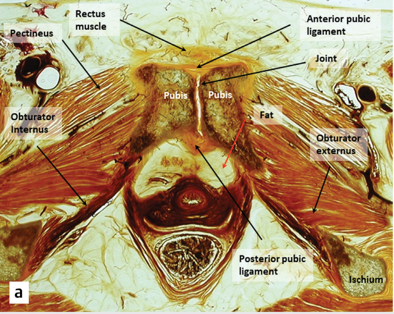
Fig. 1.41a - Axial cadaveric section of the pubic symphysis showing the fibrocartilage joint space and surrounding reinforcing ligaments.
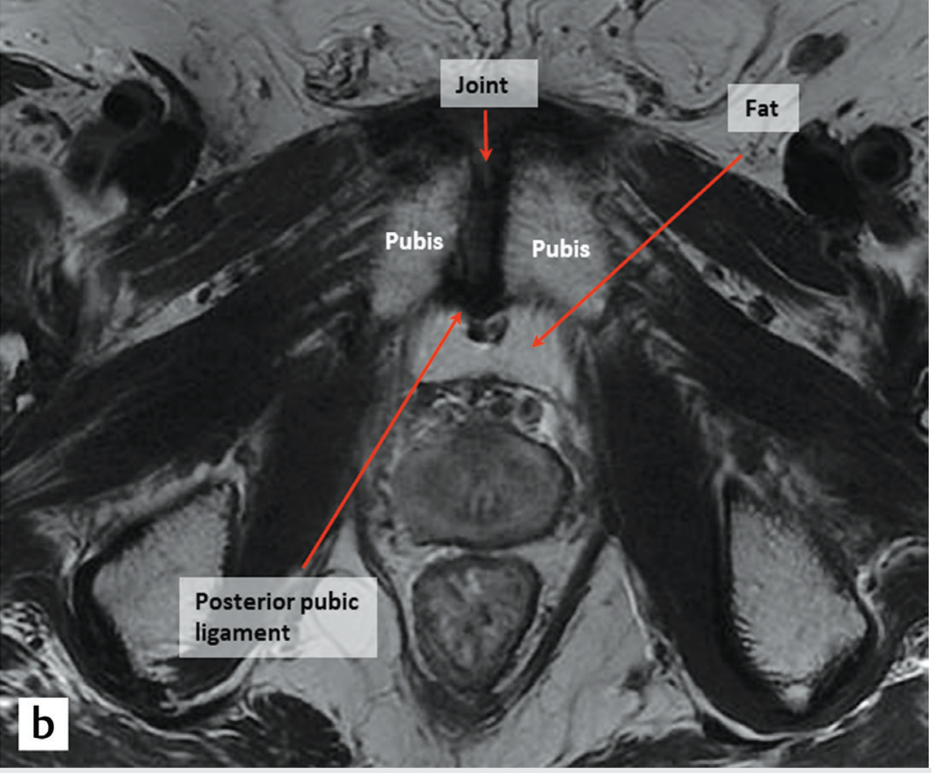
Fig. 1.41b - Axial MRI of the pubic symphysis. The gap between the two pubic bones is bridged by the fibrocartilage disc; a small amount of fluid may normally be present.
Functional Classification Summary
| Functional Class | Movement | Structural Type | Examples |
|---|---|---|---|
| Synarthrosis | Immobile | Synchondrosis (hyaline) | Epiphyseal plates, 1st sternocostal joint, costochondral |
| Amphiarthrosis | Slightly mobile | Symphysis (fibrocartilage) | Pubic symphysis, intervertebral discs, sacrococcygeal |
Comparison with Other Joint Types
- Fibrous joints - united by dense fibrous connective tissue (e.g., skull sutures, tibiofibular syndesmosis, gomphosis of teeth)
- Cartilaginous joints - united by cartilage, no joint cavity
- Synovial joints - freely mobile, have a fluid-filled joint cavity lined by synovial membrane; bones do not fuse
Clinical Points
- Growth plate injuries in children (fracturing through a temporary synchondrosis) are classified by the Salter-Harris system and can disrupt normal longitudinal bone growth
- Symphysis pubis dysfunction (commonly in pregnancy due to relaxin-mediated ligament laxity) causes pain and widening of the pubic symphysis
- Intervertebral disc degeneration involves the fibrocartilage of symphyseal joints and is a major cause of back pain
- The manubriosternal joint can be used as a surface landmark (sternal angle / angle of Louis) to identify the 2nd rib and the level of T4/T5 vertebra
Sources: Imaging Anatomy: Bones, Joints, Vessels and Nerves, Vol. 3; Forensic Anthropology: A Comprehensive Introduction, 2nd Ed.
This is a shared conversation. Sign in to Orris to start your own chat.