


Explain visually
aminoglycoside mechanism of action protein synthesis inhibition ribosome
aminoglycoside transport bacterial cell membrane entry EDP
aminoglycoside resistance mechanisms bacterial enzymes
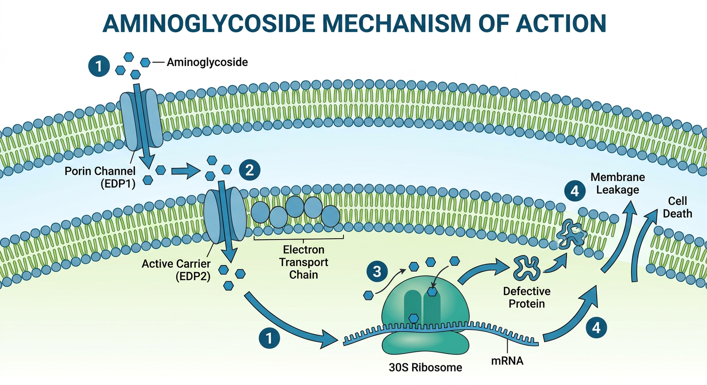
Medical pharmacology diagram showing aminoglycoside antibiotic mechanism of action: step-by-step transport through gram-negative bacterial outer membrane via porin channels (EDP1), then active carrier-mediated transport across cytoplasmic membrane (EDP2) linked to electron transport chain, then binding to 30S ribosome causing mRNA misreading and wrong amino acid incorporation, leading to defective protein inserted into cell membrane, causing membrane leakage and bacterial cell death. Clean educational diagram with labeled arrows and clear steps numbered 1-4.
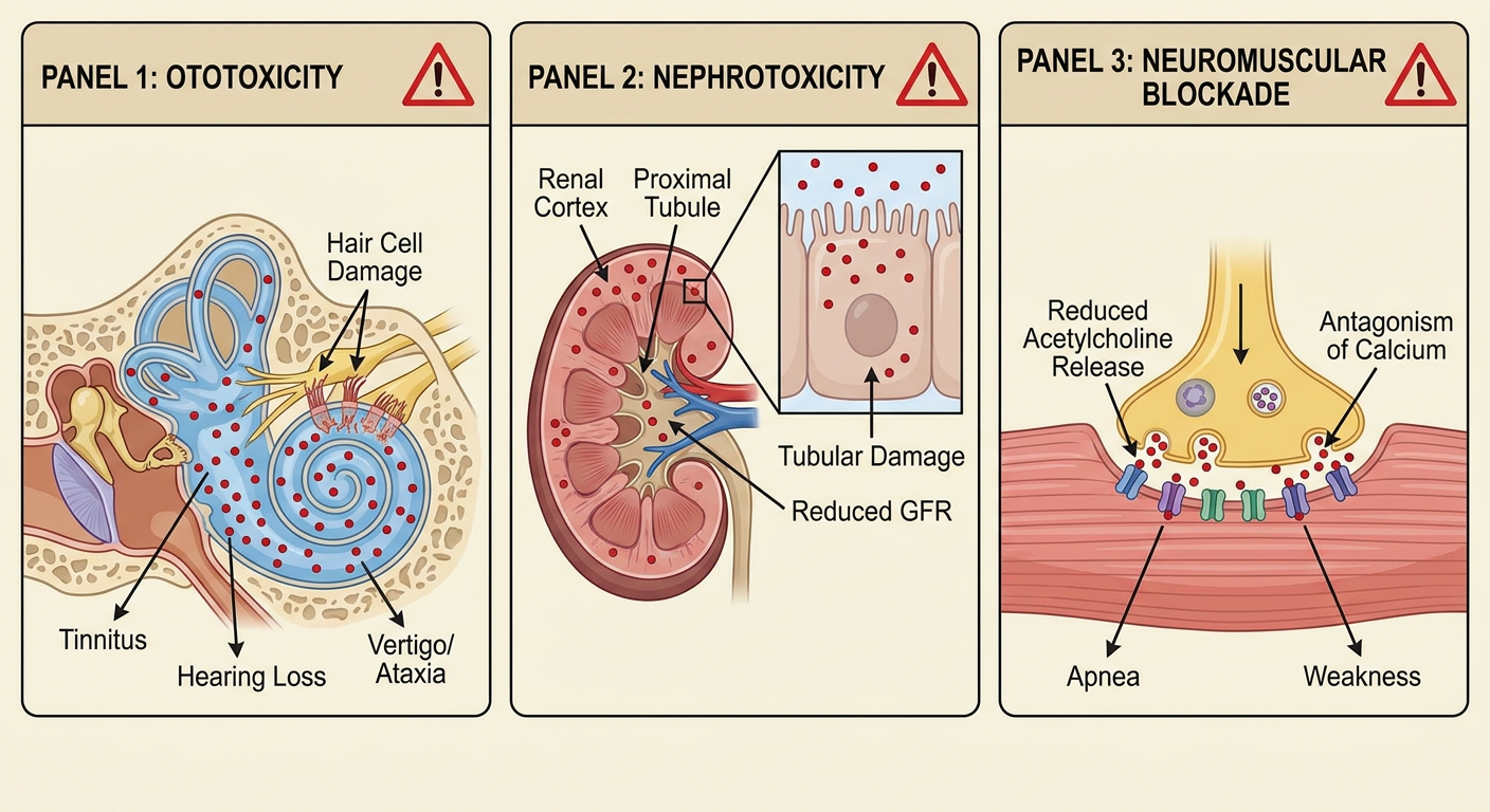
Medical diagram showing aminoglycoside toxicity: three panels side by side. Panel 1 - Ototoxicity showing inner ear cochlea and vestibular apparatus with drug concentrating in labyrinthine fluid, hair cell damage, tinnitus progressing to hearing loss and vertigo/ataxia. Panel 2 - Nephrotoxicity showing kidney tubular cell with drug accumulating in renal cortex proximal tubule, causing tubular damage and reduced GFR. Panel 3 - Neuromuscular blockade showing nerve-muscle junction with reduced acetylcholine release, antagonism of calcium, causing apnea and weakness. Each panel has clear labels and warning symbols.
🔬 What Are Aminoglycosides?
⚙️ Mechanism of Action (Step-by-Step)
| Step | What Happens |
|---|---|
| ① EDP₁ (Passive) | Drug diffuses through porin channels in the outer membrane of gram-negative bacteria into the periplasmic space |
| ② EDP₂ (Active) | Drug crosses the cytoplasmic membrane via carrier-mediated active transport linked to the electron transport chain — requires O₂ (so anaerobes are resistant) |
| ③ Ribosome Binding | Binds 30S subunit (and 30S-50S junction); freezes initiation, causes mRNA misreading → wrong amino acids incorporated |
| ④ Membrane Disruption | Defective proteins insert into cell membrane → membrane becomes leaky → ions/proteins escape → cell death (bactericidal, not just static) |
Key insight: The membrane damage increases EDP₂ entry even further — a self-reinforcing lethal cycle. Alkaline pH favors penetration (~20× more active in alkaline vs. acidic medium).
🧬 Ribosome Action in Detail
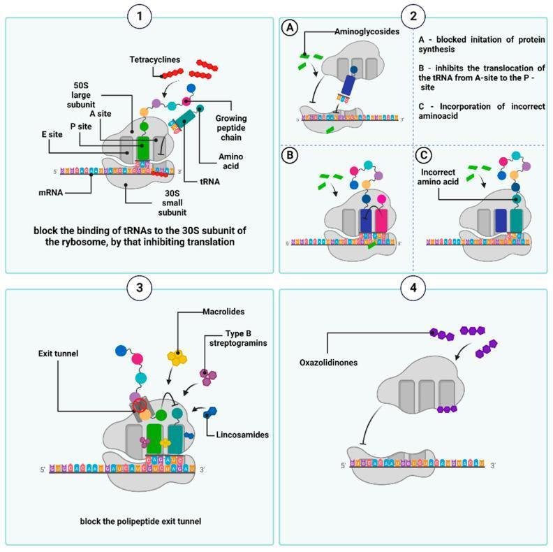
- (A) Block initiation of protein synthesis
- (B) Inhibit tRNA translocation from A-site to P-site
- (C) Cause mRNA misreading → incorrect amino acids incorporated
🛡️ Mechanisms of Resistance
| Mechanism | How It Works |
|---|---|
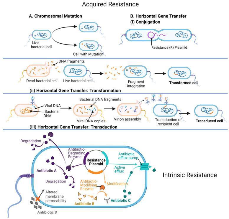
| Modifying enzymes (most important) | Phosphorylation, adenylation, or acetylation of the antibiotic → can't bind ribosome. Enzymes are encoded on plasmids transferred by conjugation (nosocomial spread!) |
| Ribosomal mutation | Decreased affinity of ribosomal proteins for the drug → high-level resistance, but limited extent |
| Reduced uptake | Porin loss or impaired active transport → less drug enters the cell |
☠️ Shared Toxicities
1. Ototoxicity
- Drug concentrates in labyrinthine fluid and is slowly cleared
- Cochlear damage: starts at base (high-frequency hearing loss first) → progresses to lower frequencies → permanent deafness (no regeneration). Kanamycin & amikacin are worst.
- Vestibular damage: headache → nausea → vertigo → ataxia → chronic phase with difficulty walking. Streptomycin & gentamicin are worst.
- Threshold for gentamicin: keep trough plasma < 1 µg/mL to avoid toxicity.
2. Nephrotoxicity
- Accumulates in renal cortex proximal tubules
- Tubular damage → ↓ urinary concentrating power → nitrogen retention → albuminuria
- Reversible if drug stopped promptly; worse with pre-existing kidney disease
- Streptomycin is the least nephrotoxic. Single daily dosing causes less nephrotoxicity than thrice-daily dosing.
3. Neuromuscular Blockade
- Reduces ACh release from motor nerve endings (antagonizes Ca²⁺ at terminal membrane)
- Can cause apnea + fatalities especially after peritoneal/pleural use post-surgery when a curare-like relaxant was used
- Partially reversed by IV calcium salt; neostigmine is inconsistent
⚠️ Comparative Toxicity at a Glance
| Drug | Vestibular | Cochlear | Nephrotoxic |
|---|---|---|---|
| Streptomycin | ++ | ± | + |
| Gentamicin | ++ | + | ++ |
| Kanamycin | + | ++ | ++ |
| Tobramycin | +± | + | +± |
| Amikacin | + | +± | +± |
| Sisomicin | +± | + | +± |
| Netilmicin | +± | + | +± |
🚫 Key Precautions
- Avoid in pregnancy — risk of fetal ototoxicity
- Avoid with other nephrotoxic drugs: NSAIDs, amphotericin B, vancomycin, cyclosporine, cisplatin
- Caution with ototoxic drugs: vancomycin, minocycline, furosemide
- Caution in patients >60 years or those with pre-existing kidney damage
Explain first week of development in embryology visually
first week embryo development fertilization cleavage morula blastocyst implantation
zygote cleavage morula blastocyst formation embryology diagram
blastocyst implantation uterus endometrium trophoblast inner cell mass embryology
human fertilization sperm egg oocyte zona pellucida acrosome reaction embryology
Medical embryology timeline diagram showing the first week of human development as a journey through the fallopian tube to uterus. Day 0: fertilization in ampulla of fallopian tube - sperm penetrates egg, zygote forms with 2 pronuclei. Day 1-2: cleavage - 2 cell then 4 cell stage, cells called blastomeres, zona pellucida intact. Day 3: morula - 16 cell compact ball entering uterus. Day 4: early blastocyst - fluid cavity (blastocoel) forms, zona pellucida thinning. Day 5-6: late blastocyst hatches from zona pellucida, inner cell mass (embryoblast) and outer trophoblast visible. Day 6-7: implantation begins in posterior wall of uterine endometrium, trophoblast invades decidua. Clean labeled educational diagram with fallopian tube anatomy showing progression from ampulla to uterus.
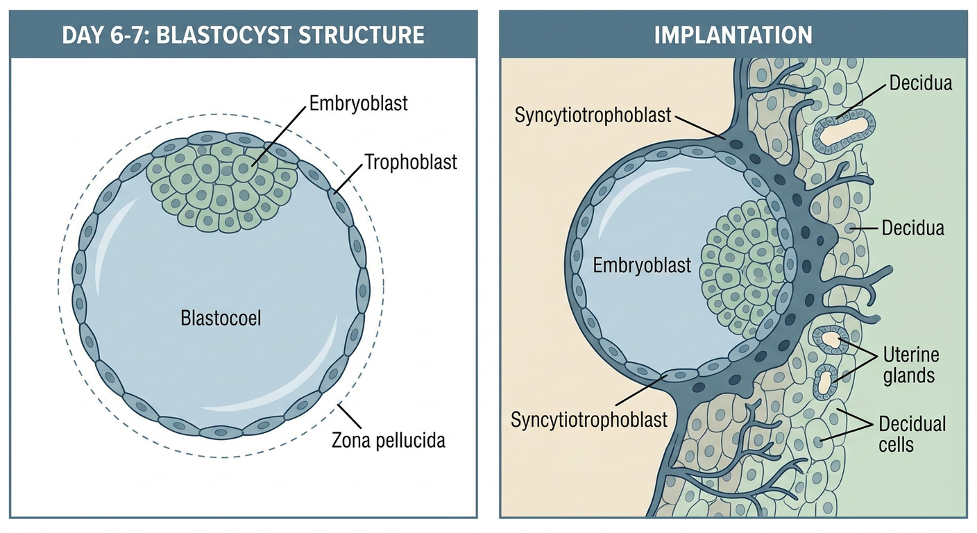
Medical embryology diagram showing blastocyst structure and implantation at day 6-7. Left panel: cross-section of blastocyst showing inner cell mass (embryoblast) at one pole, outer layer of trophoblast cells, fluid-filled blastocoel cavity, zona pellucida (hatching away). Right panel: implantation into uterine wall showing trophoblast differentiating into syncytiotrophoblast (invading, multinucleated) and cytotrophoblast (inner layer), decidua of endometrium with decidual cells, and the embryoblast oriented toward endometrium. Labels: embryoblast, trophoblast, blastocoel, syncytiotrophoblast, cytotrophoblast, decidua, uterine glands. Clear anatomical educational diagram.
acrosome reaction cortical reaction zona pellucida sperm penetration block polyspermy
Medical embryology diagram showing fertilization events in detail. Step 1: capacitation of sperm in female reproductive tract. Step 2: sperm binds ZP3 glycoprotein of zona pellucida, triggering acrosome reaction releasing hyaluronidase and acrosin enzymes. Step 3: sperm penetrates zona pellucida and fuses with oocyte plasma membrane. Step 4: cortical reaction - cortical granules release enzymes that harden zona into fertilization membrane, preventing polyspermy. Step 5: oocyte completes meiosis II, forms female pronucleus. Step 6: sperm nucleus decondenses into male pronucleus. Step 7: syngamy - two pronuclei fuse, forming zygote with 46 chromosomes (2n). Clear numbered steps with labeled diagrams of sperm, oocyte, zona pellucida, pronuclei.
🧬 First Week of Human Embryogenesis
📍 The Big Picture: Journey Through the Fallopian Tube to Uterus
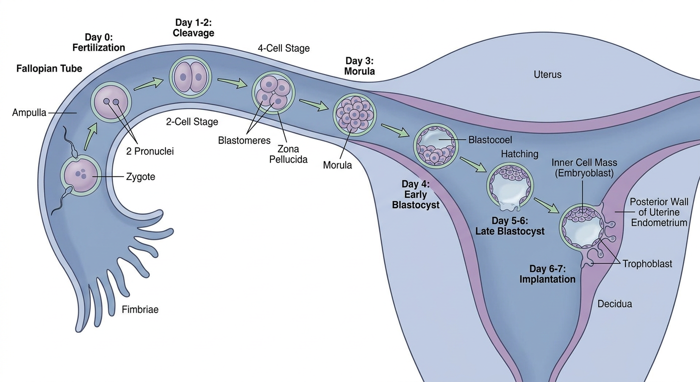
Day 0 — Fertilization (Syngamy)
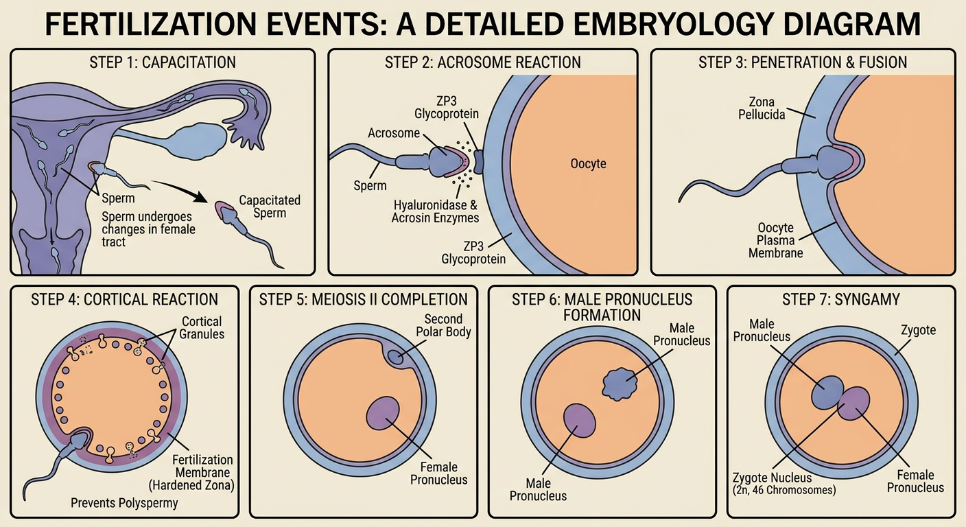
| Step | Event | Key Detail |
|---|---|---|
| 1 | Capacitation | Sperm undergoes changes in female tract (removal of cholesterol from membrane) that enable fertilization |
| 2 | Acrosome reaction | Sperm binds ZP3 glycoprotein on zona pellucida → acrosome releases hyaluronidase + acrosin to penetrate zona |
| 3 | Membrane fusion | Sperm head fuses with oocyte plasma membrane |
| 4 | Cortical reaction | Cortical granules release enzymes → zona hardens into fertilization membrane → blocks polyspermy |
| 5 | Meiosis II completes | Secondary oocyte (arrested at MII) now completes division → ejects 2nd polar body, forms female pronucleus |
| 6 | Male pronucleus forms | Sperm nucleus decondenses |
| 7 | Syngamy | Two pronuclei fuse → zygote with 46 chromosomes (2n) |
Important: The oocyte at ovulation is a secondary oocyte arrested in metaphase II. It only completes meiosis after sperm penetration.
Days 1–3 — Cleavage
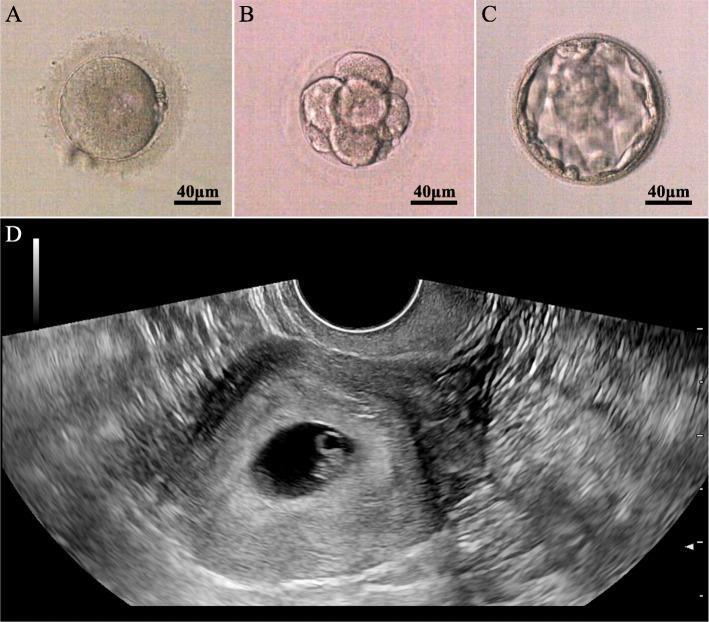
| Day | Stage | Key Features |
|---|---|---|
| Day 1 | 2-cell | First cleavage division (~30 hrs after fertilization) |
| Day 2 | 4-cell | Cells called blastomeres; totipotent at this stage |
| Day 3 | 8–16 cells (Morula) | Enters uterine cavity; undergoes compaction — cells flatten and maximize contact |
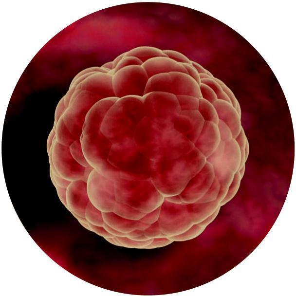
The morula resembles a mulberry (Latin: morus) — hence the name. The zona pellucida is still intact throughout cleavage. Cell size decreases with each division (no growth between divisions), so overall embryo size stays ~same as zygote.
Days 4–5 — Blastocyst Formation (Cavitation)
| Cell Type | Location | Fate |
|---|---|---|
| Embryoblast (Inner Cell Mass, ICM) | Eccentric pole inside blastocoel | Gives rise to the embryo proper + amnion + yolk sac |
| Trophoblast | Outer shell | Forms placenta, chorion, and all extraembryonic membranes |
At Day 5–6, the blastocyst "hatches" out of the zona pellucida — a prerequisite for implantation.
Days 6–7 — Implantation
| Layer | Type | Action |
|---|---|---|
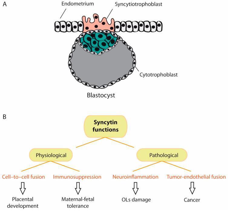
| Syncytiotrophoblast | Outer invasive layer | Multinucleated; invades decidua (maternal endometrium); produces hCG |
| Cytotrophoblast | Inner cellular layer | Mitotically active; feeds cells into syncytium |
Implantation window: Days 20–24 of the menstrual cycle (days 6–10 post-fertilization). The endometrium must be in the secretory phase (progesterone-primed) and express pinopodes on its surface.
📊 First Week — Summary Timeline
| Day | Event | Location |
|---|---|---|
| 0 | Fertilization → Zygote (2n) | Ampulla of fallopian tube |
| 1 | 2-cell stage | Fallopian tube |
| 2 | 4-cell stage | Fallopian tube |
| 3 | Morula (16 cells) | Entering uterus |
| 4 | Early blastocyst (cavitation begins) | Uterine cavity |
| 5–6 | Late blastocyst hatches from zona pellucida | Uterine cavity |
| 6–7 | Implantation begins | Posterior uterine wall |
⚠️ Key Clinical Points
| Concept | Significance |
|---|---|
| Ectopic pregnancy | If implantation occurs in the fallopian tube (95% in ampulla) — life-threatening; needs urgent treatment |
| hCG production | Begins at implantation (day 6–7) by syncytiotrophoblast → basis of pregnancy tests |
| Identical twins | If the embryo splits before day 4 → dichorionic; days 4–8 → monochorionic diamniotic; day 8–12 → monochorionic monoamniotic |
| IVF timing | Embryo transfer typically done at blastocyst stage (day 5) for best implantation rates |
| Zona pellucida | Must hatch for implantation; IVF uses "assisted hatching" if zona is thick |
Create a visual summary document on week 1 embryology
ls /home/daytona/skills/docx/