
it femur facture defination and classification and signs symptoms and investigations and treatment with appropriate diagrams and complete details regarding it
Reading File
Finding Sources
Finding Sources
Reading File
Reading File
Finding Sources
Reading File
Finding Sources
Finding Sources
Reading File
Reading File
Reading File
Reading File
Reading File
Reading File
Searching Images
femur fracture classification diagram anatomy
Reading File
Searching Images
femoral shaft fracture signs symptoms intramedullary nail treatment
Reading File
I now have comprehensive information. Let me also fetch the key classification and anatomy diagram images before writing the final response.
Loading Image
Loading Image
Loading Image
Loading Image
Here is a complete, structured overview of femur fractures.
Femur Fracture — Complete Clinical Review
1. Definition
A femur fracture is a break in the continuity of the femur (thigh bone), the longest, strongest, and heaviest bone in the human body. Because the femur requires significant force to fracture in healthy bone, these injuries carry substantial morbidity and mortality due to associated injuries and blood loss. The incidence varies from 1–9% of all fractures and is a major source of morbidity in trauma patients.
— Rockwood and Green's Fractures in Adults, 10th ed.
2. Anatomy Overview
The femur extends from the hip joint (proximal) to the knee joint (distal) and is divided into distinct anatomical regions, each associated with specific fracture patterns:
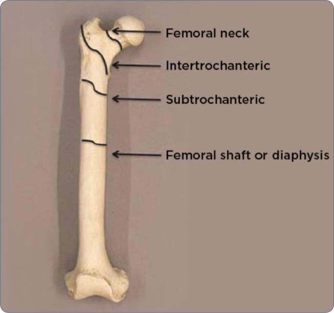
| Region | Location |
|---|---|
| Femoral head | Articulates with acetabulum |
| Femoral neck | Between head and trochanters (intracapsular) |
| Intertrochanteric | Between greater and lesser trochanter |
| Subtrochanteric | 5 cm distal to lesser trochanter |
| Femoral shaft (diaphysis) | Long central portion |
| Distal femur | Supracondylar / condylar region |
3. Epidemiology & Mechanism of Injury
Age & Gender Distribution (Bimodal)
- Young males (15–25 years): High-energy trauma — MVAs, motorcycle accidents, falls from heights, gunshot wounds, pedestrian injuries
- Elderly females (>75 years): Low-energy trauma — fall from standing height on osteoporotic bone
- 50–70 years: Atypical femoral fractures associated with long-term bisphosphonate use
Fracture Pattern by Force
| Force Type | Fracture Pattern |
|---|---|
| Bending | Transverse ± butterfly fragment |
| Axial loading | Associated hip/knee injuries |
| Rotational | Spiral or oblique |
| High energy | Comminuted, displaced |
4. Classification
A. By Anatomical Region
1. Femoral Neck Fractures
Garden Classification (most widely used for femoral neck fractures):
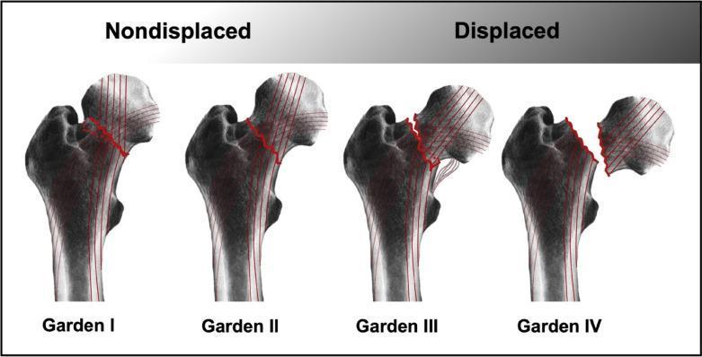
| Type | Description | Displacement |
|---|---|---|
| Garden I | Incomplete/impacted fracture, valgus angulation | Non-displaced |
| Garden II | Complete fracture, no displacement | Non-displaced |
| Garden III | Complete, partial displacement, femoral head tilted | Displaced |
| Garden IV | Complete, full displacement, no bony contact | Displaced |
Clinical significance: Types I–II have lower risk of avascular necrosis (AVN). Types III–IV carry high AVN risk due to disruption of the retinacular vessels supplying the femoral head.
Pauwels Classification (by fracture line angle to horizontal):
- Type I: 0–30° — stable, compressive forces → good prognosis
- Type II: 30–70° — increasing shear forces
- Type III: >70° — highly unstable, high pseudarthrosis risk
2. Intertrochanteric Fractures
- Extracapsular fractures between greater and lesser trochanters
- Blood supply intact — AVN rare
- AO/OTA Classification: A1 (simple, stable) → A2 (multifragment) → A3 (reverse oblique/transverse — most unstable)
3. Subtrochanteric Fractures
- Within 5 cm distal to lesser trochanter
- High biomechanical stress zone — prone to implant failure
- Seinsheimer Classification: Types I–V based on number of fragments and fracture pattern
4. Femoral Shaft (Diaphyseal) Fractures
- Winquist and Hansen Classification (degree of comminution):
| Grade | Description |
|---|---|
| 0 | Simple fracture, no comminution |
| I | Small butterfly fragment (<25% diameter) |
| II | Butterfly fragment 25–50% diameter |
| III | Butterfly fragment >50% diameter |
| IV | Segmental, no cortical contact |
- AO/OTA Classification: Type A (simple), Type B (wedge), Type C (complex/comminuted)
5. Distal Femur (Supracondylar/Condylar) Fractures
- AO/OTA Classification:
- A1–A3: Extra-articular
- B1–B3: Partial articular
- C1–C3: Complete articular (most complex)
B. By Fracture Characteristics
| Type | Description |
|---|---|
| Open (compound) | Bone penetrates or communicates with skin |
| Closed | No skin breach |
| Displaced | Fracture ends separated/malaligned |
| Non-displaced | Fracture line present but position maintained |
| Comminuted | Shattered into >2 fragments |
| Stress | Fatigue fracture from repetitive loading |
| Pathological | Through diseased bone (tumor, osteoporosis) |
| Periprosthetic | Around a hip/knee prosthesis — Vancouver Classification |
5. Signs and Symptoms
Subjective (Symptoms)
- Severe thigh/hip pain — often immediate and incapacitating
- Pain worsens with any attempted movement
- History of trauma (or trivial fall in elderly/osteoporotic patients)
- Inability to bear weight or walk
Objective (Signs)
| Sign | Description |
|---|---|
| Deformity | Visible angulation or shortening of the limb |
| Shortening | Affected limb appears shorter (muscle spasm pulls fragments proximally) |
| External rotation | Especially with femoral neck/intertrochanteric fractures (psoas spasm rotates distal fragment) |
| Swelling/bruising | Significant thigh swelling and echymosis — up to 1–2 L blood loss into thigh |
| Tenderness | Point tenderness over fracture site |
| Crepitus | Grating sensation on palpation or passive movement |
| Abnormal mobility | Movement at non-joint site |
| Loss of function | Unable to lift leg or bear weight |
| Neurovascular compromise | Distal pulses checked — popliteal/femoral artery injury possible |
In Femoral Neck Fractures Specifically:
- Leg shortened AND externally rotated (psoas pulls lesser trochanter proximally and rotates)
- Hip held in slight flexion
- Patient unable to straight-leg raise
Systemic Signs (with shaft fractures):
- Hemorrhagic shock — 1–2 L blood loss typical; up to 3 L with high-energy injuries
- Tachycardia, hypotension
- Fat embolism syndrome: petechiae, confusion, respiratory compromise (typically 24–72 hrs post-injury)
6. Investigations
A. Imaging
| Investigation | Details |
|---|---|
| Plain X-ray (AP + Lateral) | First-line investigation. Must include AP pelvis, full AP and lateral femur, joint above (hip) and below (knee). Identifies fracture line, pattern, displacement, comminution |
| CT scan | For complex fractures, intra-articular extension (distal femur), occult femoral neck fracture with shaft fracture, acetabular involvement, preoperative planning |
| MRI | Gold standard for occult/stress fractures — detects fracture-related edema invisible on X-ray. Used when X-ray is negative but clinical suspicion high |
| Bone scan (isotope) | Alternative for stress fractures when MRI unavailable |
| Angiography/CTA | If vascular injury suspected (distal ischaemia, expanding haematoma, no distal pulse) |
"Both undisplaced femoral fractures and stress fractures often appear in conventional X-rays only as slight abnormalities of the trabecular bone structure, so an MRI is needed to identify a fracture because fracture-related edema can be identified through the lesser signals it emits." — THIEME Atlas of Anatomy
B. Laboratory Tests
| Test | Purpose |
|---|---|
| FBC / CBC | Assess blood loss (Hb, Hct), baseline |
| Blood group & crossmatch | Prepare for surgery and transfusion |
| Coagulation screen (PT, APTT, INR) | Pre-operative |
| U&E, renal function, LFTs | Pre-operative baseline |
| Bone profile (Ca²⁺, phosphate, ALP) | If pathological fracture suspected |
| Blood glucose, ECG, CXR | Pre-operative fitness for anaesthesia |
C. Special Investigations
- DEXA scan: Post-acute assessment of bone mineral density if osteoporosis suspected
- CT of femoral neck: Mandatory in femoral shaft fractures to exclude occult concurrent neck fracture (up to 5.8% incidence)
7. Treatment
Initial (Emergency) Management — ABCDE Approach
- Airway, Breathing, Circulation (hemorrhage control is priority)
- IV access × 2, fluid resuscitation, blood products if shocked
- Traction splint (e.g., Thomas splint) to reduce blood loss and pain
- Analgesia — IV morphine, femoral nerve block or fascia iliaca block
- Open fractures: wound cover, IV antibiotics (cefazolin), tetanus prophylaxis
- Imaging as above
A. Non-Operative Treatment
Indications:
- Minimally displaced, stable fractures (Garden I neck fractures in elderly low-demand patients)
- Patients unfit for surgery
- Impacted, non-displaced neck fractures managed conservatively in some settings
Methods:
- Skeletal traction (Steinmann/Kirschner pin traction) — historically used; still used where surgery unavailable, or as temporary stabilization
- Cast bracing — limited role in adults
- Protected weight bearing with crutches — select stable patterns
B. Operative Treatment
1. Femoral Neck Fractures
| Fracture Type | Treatment |
|---|---|
| Non-displaced (Garden I–II), young patient | Internal fixation — cannulated screws or sliding hip screw |
| Displaced (Garden III–IV), elderly | Hemiarthroplasty (femoral head replacement; acetabulum preserved) |
| Displaced + arthritic hip or active elderly | Total Hip Arthroplasty (THA) |
| Young patient with displaced fracture | Attempt reduction and internal fixation to preserve femoral head |
In elderly patients, the sole blood supply to the femoral head via retinacular vessels is transected at fracture — making arthroplasty necessary. — Gray's Anatomy for Students
2. Intertrochanteric Fractures
- Dynamic Hip Screw (DHS) / Sliding Hip Screw: Standard for stable (A1, A2) fractures — allows controlled collapse at fracture site
- Cephalomedullary Nail (CMN/Gamma nail): Preferred for unstable (A3), reverse oblique, or subtrochanteric extension — load-sharing, reduced bending moment
- Blade plate / 95° condylar plate: Largely replaced by CMN due to high failure rates
3. Subtrochanteric Fractures
- Cephalomedullary intramedullary nail — treatment of choice
- High failure rates with plates due to extreme mechanical stress at this site
- Cerclage wires may supplement for comminuted patterns
4. Femoral Shaft Fractures
Gold standard: Reamed Intramedullary Nailing (IMN)
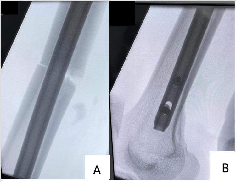
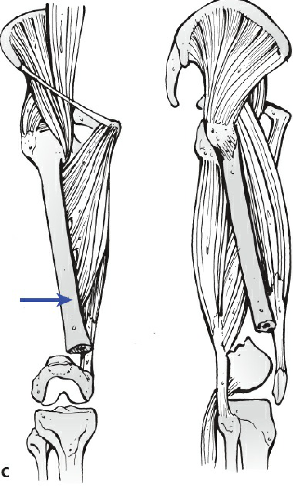
The above illustration shows how muscles deform the fracture fragments:
- Proximal fragment: Flexed by iliopsoas, abducted by glutei
- Distal fragment: Pulled proximally by quadriceps and hamstrings, adducted by adductors
| Method | Details |
|---|---|
| Antegrade IMN | Entry via piriformis fossa or greater trochanter tip; most common; proximal interlocking in femoral neck optional for added stability |
| Retrograde IMN | Entry via knee (intercondylar notch); used in obese patients, ipsilateral acetabular fracture, or bilateral femur fractures |
| External fixation | Temporizing measure in hemodynamically unstable patients, polytrauma, damage control |
| Plate (ORIF) | Used when IMN contraindicated (e.g., narrow canal, implant in situ), or for distal/proximal fractures extending near joints |
"Reaming has been shown to improve union rates compared to unreamed nails and is now the preferred method." — Rockwood and Green's, 10th ed.
Timing: Stabilization within 24 hours of injury reduces pulmonary complications in multiply injured patients.
5. Distal Femur (Supracondylar/Condylar) Fractures
- Retrograde IMN: For extra-articular and simple articular fractures
- Lateral locking plate (LISS / distal femoral locking plate): Minimally invasive; especially useful with osteoporotic bone and comminuted patterns
- Dual plating: For highly comminuted bicondylar fractures
- Total Knee Arthroplasty: For elderly patients with pre-existing arthritis + distal femur fracture
C. Post-Operative Care
| Component | Details |
|---|---|
| DVT prophylaxis | LMWH, mechanical compression stockings — high risk for VTE |
| Physiotherapy | Early mobilization as tolerated; quadriceps strengthening |
| Weight bearing | Dependent on fixation stability — toe-touch to full weight bearing |
| Analgesia | Multimodal — NSAIDs, opioids, nerve blocks |
| Nutritional support | Especially elderly patients |
| Monitoring for complications | Neurovascular observations, wound check |
8. Complications
Early
| Complication | Details |
|---|---|
| Hemorrhagic shock | 1–3 L blood loss into thigh; life-threatening |
| Neurovascular injury | Femoral/popliteal artery, sciatic/femoral nerve |
| Fat embolism syndrome | Petechiae, hypoxia, altered consciousness |
| Compartment syndrome | Raised pressure in thigh compartments; emergency fasciotomy |
| Open fracture infection | Higher risk with contaminated wounds |
| DVT / Pulmonary embolism | High risk — prophylaxis mandatory |
Late
| Complication | Details |
|---|---|
| Avascular necrosis (AVN) | Femoral head — especially displaced neck fractures |
| Non-union / Delayed union | More common with comminuted, open, or poorly fixed fractures |
| Malunion | Angular or rotational deformity |
| Post-traumatic arthritis | Especially after articular fractures |
| Implant failure | Broken nails/screws, loosening |
| Knee/hip stiffness | Especially after prolonged immobilization |
| Heterotopic ossification | Ectopic bone formation in muscle |
| Leg length discrepancy | From shortening or malunion |
| Refracture | After implant removal |
9. Summary Table
| Region | Key Classification | Preferred Treatment |
|---|---|---|
| Femoral neck | Garden I–IV / Pauwels | Screws (undisplaced) → Hemiarthroplasty/THA (displaced) |
| Intertrochanteric | AO/OTA A1–A3 | DHS (stable), CMN (unstable/A3) |
| Subtrochanteric | Seinsheimer I–V | Cephalomedullary nail |
| Femoral shaft | Winquist 0–IV / AO A-C | Reamed intramedullary nail (gold standard) |
| Distal femur | AO/OTA A1–C3 | Retrograde nail / Locking plate |
Sources:
- Rockwood and Green's Fractures in Adults, 10th Edition (2025)
- Miller's Review of Orthopaedics, 9th Edition
- Gray's Anatomy for Students
- THIEME Atlas of Anatomy — General Anatomy and Musculoskeletal System
- Rosen's Emergency Medicine: Concepts and Clinical PracticeThe response above covers everything you asked for. Here is a quick orientation to the sections:
What's covered:
- Definition — what a femur fracture is, its clinical significance
- Anatomy — the 6 anatomical regions of the femur with a labeled diagram
- Epidemiology & Mechanism — bimodal age distribution, force-pattern relationship
- Classification — all major systems:
- Garden Classification (neck fractures) with diagram
- Pauwels Classification (neck angle)
- AO/OTA Classification (intertrochanteric, shaft, distal)
- Winquist–Hansen Classification (shaft comminution)
- Seinsheimer (subtrochanteric)
- Vancouver (periprosthetic)
- Signs & Symptoms — both local (deformity, shortening, external rotation, crepitus) and systemic (hemorrhagic shock, fat embolism)
- Investigations — X-ray (AP + lateral), CT, MRI (for occult fractures), bloods, crossmatch
- Treatment — emergency management, non-operative, and operative by region (DHS, CMN, IMN, arthroplasty, locking plates) with intraoperative fluoroscopy images
- Complications — early (shock, fat embolism, compartment syndrome) and late (AVN, non-union, malunion, arthritis)
This is a shared conversation. Sign in to Orris to start your own chat.