
Tuberculosis bhms 3rd year
Reading File
Tuberculosis is a chronic granulomatous infection caused by Mycobacterium tuberculosis, spread by airborne droplets. It primarily affects the lungs (pulmonary TB) but can affect any organ. Key features include caseating granulomas histologically, a cell-mediated immune response, and the classic triad of fever, night sweats, and weight loss.
Searching sources...
Finding Sources
Finding Sources
Let me search specifically in respiratory/pathology/community medicine textbooks which are most relevant for BHMS 3rd year TB content:
Finding Sources
Finding Sources
Finding Sources
Excellent! Found Robbins Pathology, Microbiology, Park's (Community Medicine), and Murray & Nadel's Respiratory. Let me now get the TB sections from these key books simultaneously:
Finding Sources
Finding Sources
Now let me read the key TB sections from Robbins Pathology, Microbiology, and Park's simultaneously:
Reading File
Reading File
Reading File
Now let me read more from Robbins (primary + secondary TB, pathology) and Park's TB section:
Reading File
Reading File
I have a great diagram image URL. Let me fetch it and also read the remaining Robbins content on secondary TB, morphology, and the TB bone section:
Loading Image
Reading File
I now have rich content. Let me get the secondary TB, treatment, and DOTS content sections:
Reading File
Finding Sources
I have all the essential content. Let me fetch one more image and compile the comprehensive answer:
Loading Image
I now have comprehensive content from all major sources. Here is the complete BHMS 3rd year-level note on Tuberculosis:
Tuberculosis - BHMS 3rd Year
1. Definition
Tuberculosis (TB) is a communicable chronic granulomatous disease caused by Mycobacterium tuberculosis. It usually involves the lungs but may affect any organ or tissue in the body.
- Robbins & Kumar Basic Pathology
2. Causative Organism
| Property | Detail |
|---|---|
| Organism | Mycobacterium tuberculosis hominis (most common) |
| Type | Weakly gram-positive, strongly acid-fast, aerobic rods |
| Cell wall | Lipid-rich (mycolic acids) - resists disinfectants, detergents, and common antibiotics |
| Key property | Capable of intracellular growth in alveolar macrophages |
| Stain | Ziehl-Neelsen (carbol fuchsin) - appears red on blue background |
| Other species | M. bovis (bovine TB, from unpasteurized milk); M. avium complex (in AIDS patients) |
Humans are the only natural reservoir. Spread is by person-to-person aerosol inhalation.
- Medical Microbiology 9e
3. Epidemiology
-
One of the leading infectious causes of death globally; WHO estimated 10 million new cases and 1.3 million deaths in 2021
-
One-third of the global population is asymptomatically infected; only 5-10% develop clinical disease during their lifetime
-
An infectious TB patient can infect 10-15 persons per year
-
High burden countries: India, Pakistan, sub-Saharan Africa, South Africa, China, Eastern Europe
-
MDR-TB: 3.3% of new cases; 18% of previously treated cases had MDR/RR-TB
-
Risk factors: poverty, overcrowding, malnutrition, HIV/AIDS, diabetes, silicosis, chronic renal failure, Hodgkin lymphoma, alcohol use disorder, immunosuppression
-
Park's Textbook of Preventive and Social Medicine; Robbins & Kumar Basic Pathology
4. Pathogenesis
Key Concept: Infection vs. Disease
- Infection = seeding of a focus with organisms (may not cause disease)
- Disease = clinically significant tissue damage
Sequence of Events (see diagram below):
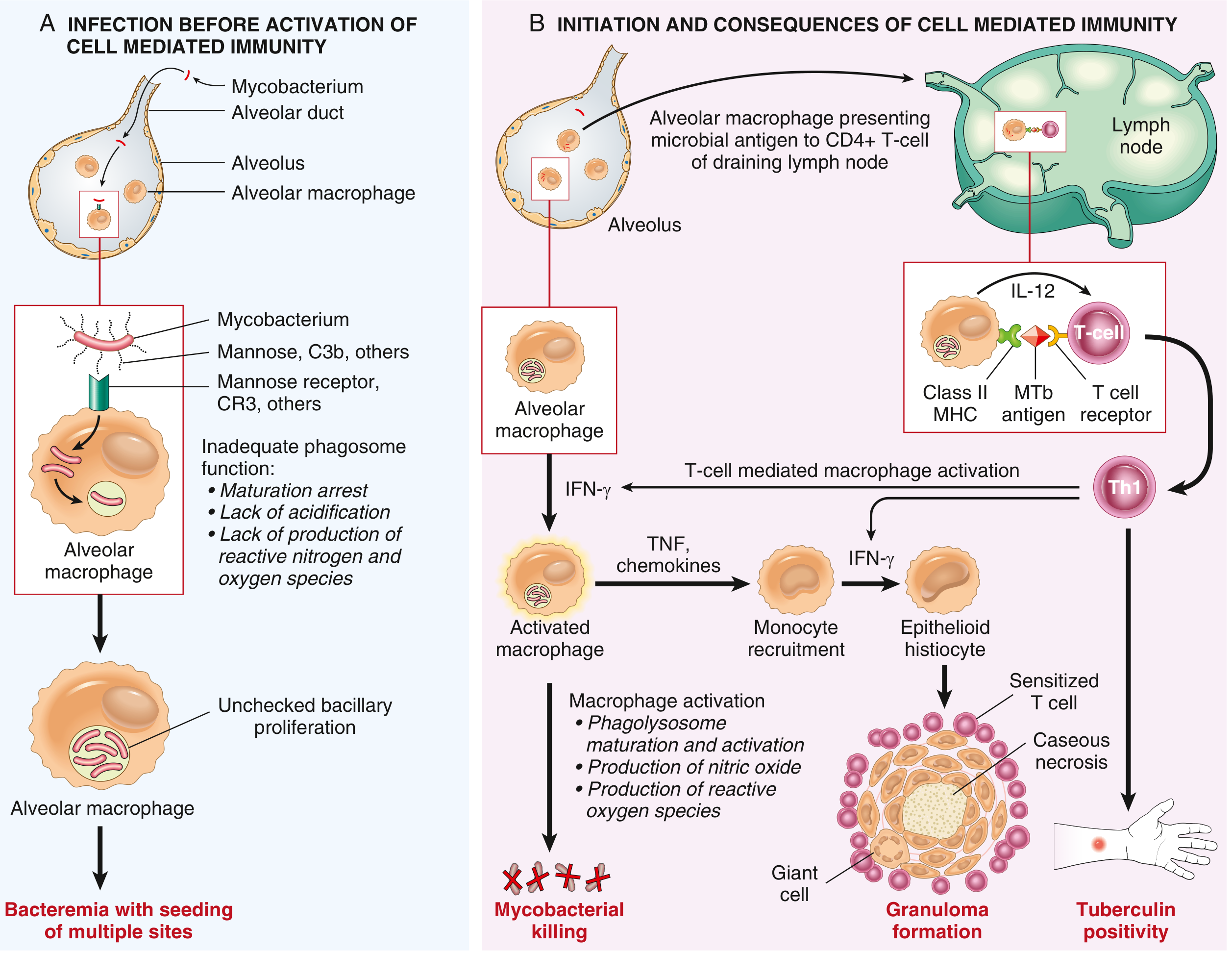
Phase A - First 3 weeks (Before cell-mediated immunity):
- Mycobacteria inhaled → enter alveolar macrophages via mannose receptors and complement receptors (CR3)
- Organisms inhibit phagolysosome fusion → prevent acidification and killing → proliferate unchecked in macrophages
- Bacteremia occurs → organisms seed multiple body sites
- Patient is usually asymptomatic or has mild flu-like illness
Phase B - After 3 weeks (Cell-mediated immunity develops):
- Macrophages present mycobacterial antigens to CD4+ T cells via MHC Class II
- Under macrophage-secreted IL-12, CD4+ T cells differentiate into Th1 cells
- Th1 cells secrete IFN-γ → activates macrophages
- Activated macrophages produce: nitric oxide, reactive oxygen species → kill mycobacteria
- TNF secreted → recruits more monocytes
- Macrophages differentiate into epithelioid histiocytes → form granulomas
- Some epithelioid cells fuse → Langhans giant cells
- Outcome: granuloma formation + caseous necrosis + tuberculin skin test positivity
Important: Immunity and hypersensitivity are two sides of the same coin - the same Th1 cells that mediate protection also cause tissue destruction (caseation).
- Robbins & Kumar Basic Pathology, p. 475-476
5. Primary Tuberculosis
- Occurs in a previously unexposed, unsensitized host
- ~5% of newly infected individuals develop significant disease
Ghon Complex (Morphology):
- Bacilli implant in distal air spaces of lower upper lobe or upper lower lobe, close to pleura
- A 1-1.5 cm gray-white area of consolidation forms = Ghon focus (often with central caseous necrosis)
- Bacilli travel via lymphatics to regional hilar lymph nodes (also caseate)
- Ghon complex = Ghon focus + affected regional lymph nodes (parenchymal + nodal lesion)
- Hematogenous dissemination occurs in first few weeks
- In ~95% of cases, cell-mediated immunity controls the infection → Ghon complex undergoes progressive fibro-calcification
Progressive Primary TB:
- Occurs in immunocompromised (especially HIV+ with CD4 < 200 cells/µL), severely malnourished
- No granuloma formation (cannot mount CD4+ T cell response)
6. Secondary (Reactivation) Tuberculosis
- Arises in a previously sensitized host
- May follow primary TB (reactivation of dormant lesions, even decades later) or reinfection
- Only < 5% of primary disease progresses to secondary TB
Key Features:
| Feature | Primary TB | Secondary TB |
|---|---|---|
| Host | Unsensitized | Previously sensitized |
| Location | Lower upper lobe / upper lower lobe (subpleural) | Apex of upper lobe (bilateral) |
| Lymph node involvement | Prominent (Ghon complex) | Less prominent |
| Cavitation | Uncommon | Common (erodes airways) |
| Granulomas | Yes (once CMI develops) | Yes, more vigorous |
| Infectivity | Low | High (sputum contains bacilli) |
| Tuberculin test | Converts positive | Already positive |
Morphology of Secondary TB:
- Initial lesion: small (<2 cm) consolidation, 1-2 cm from apical pleura
- Sharply circumscribed, firm, gray-yellow areas with central caseation + peripheral fibrosis
- Active lesions: coalescent granulomas with central caseation (histologically)
- Resolution leaves fibrocalcific scars
Histological appearances of TB:
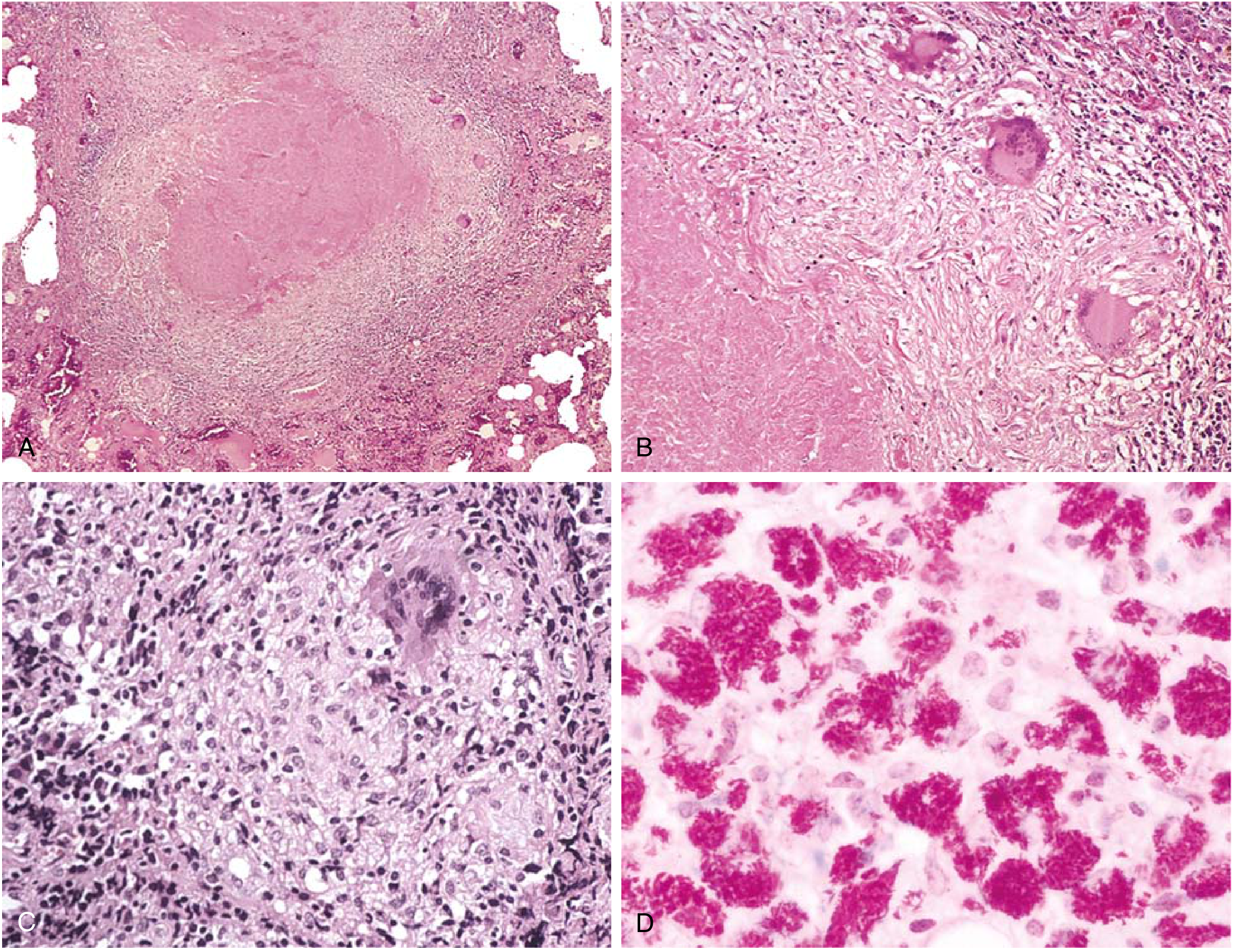
- A: Low-power - central caseating necrosis surrounded by granulomatous inflammation
- B: Fibrous wall with caseous necrosis
- C: High-power - epithelioid histiocytes + Langhans giant cell
- D: Acid-fast bacilli (red rods on ZN stain)
7. Complications / Forms of Progressive TB
| Complication | Mechanism / Notes |
|---|---|
| Miliary TB | Hematogenous dissemination → millet seed-sized granulomas in lungs, liver, spleen, bone marrow, meninges |
| Caseous pneumonia | Liquefied caseum spills into airways → consolidation of an entire lobe |
| Cavitary TB | Drainage of caseum into bronchus → cavity formation (most infectious) |
| Tuberculous pleuritis | Pleural effusion from rupture of a subpleural focus |
| TB meningitis | Spread from miliary TB to meninges |
| Pott's disease | Vertebral TB - most common form of skeletal TB |
| Scrofula | Cervical lymphadenopathy from TB |
| TB in HIV | Extrapulmonary TB >50% when CD4 <200; atypical presentation |
8. Diagnosis
| Test | Details |
|---|---|
| Tuberculin (Mantoux) test | Intradermal PPD (purified protein derivative); induration read at 48-72 hrs; positive = delayed hypersensitivity (Th1 mediated) |
| IGRA (IFN-γ release assay) | In vitro; T cells stimulated with MTb antigens; IFN-γ measured; less affected by BCG |
| Sputum smear (ZN stain) | Acid-fast bacilli (AFB); rapid, cheap, but low sensitivity |
| Sputum culture (gold standard) | Lowenstein-Jensen medium; slow (4-8 weeks); confirms diagnosis |
| CBNAAT/GeneXpert MTB/RIF | Rapid molecular test; detects MTb + rifampicin resistance simultaneously |
| Chest X-ray | Upper lobe infiltrates, cavitation (secondary); hilar adenopathy + lower lobe consolidation (primary) |
False-negatives for tuberculin test: viral infections, sarcoidosis, malnutrition, Hodgkin lymphoma, overwhelming TB, immunosuppression (anergy)
False-positives: atypical mycobacterial infections
- Medical Microbiology 9e; Robbins & Kumar Basic Pathology, p. 476
9. Treatment
Standard Regimen (RNTCP/NTEP - India):
| Phase | Duration | Drugs | Mnemonic |
|---|---|---|---|
| Intensive phase | 2 months | Isoniazid (H) + Rifampicin (R) + Pyrazinamide (Z) + Ethambutol (E) | 2 HRZE |
| Continuation phase | 4 months | Isoniazid (H) + Rifampicin (R) | 4 HR |
Total duration: 6 months for new pulmonary TB
Drug Mnemonics:
- RIPE = Rifampicin, Isoniazid, Pyrazinamide, Ethambutol (first-line drugs)
- SHREK = Streptomycin, isoniazid (H), Rifampicin, Ethambutol, piraZinamide (second-line and reserve)
Key Side Effects:
| Drug | Important Side Effect |
|---|---|
| Isoniazid (INH) | Peripheral neuropathy (→ give pyridoxine/B6), hepatotoxicity |
| Rifampicin | Orange-red discoloration of urine/secretions, hepatotoxicity, enzyme inducer |
| Pyrazinamide | Hyperuricemia (gout), hepatotoxicity |
| Ethambutol | Optic neuritis (color blindness, visual acuity loss) |
| Streptomycin | Ototoxicity, nephrotoxicity |
Drug-Resistant TB:
-
MDR-TB: resistant to at least Isoniazid + Rifampicin (the two most effective drugs)
-
XDR-TB: MDR-TB + resistance to any fluoroquinolone + at least one of bedaquiline/linezolid
-
Treatment: 18-20 months with second-line agents
-
Medical Microbiology 9e; Sherris & Ryan's Medical Microbiology
10. Prevention and Control
BCG Vaccine:
- Bacillus Calmette-Guérin (BCG) - live attenuated M. bovis
- Given at birth (part of UIP in India)
- Protective efficacy: 28-60% (mainly prevents disseminated/miliary TB and TB meningitis in children)
- Does not prevent adult pulmonary TB reliably
DOTS (Directly Observed Treatment Short-course):
- 5 components of DOTS strategy:
- Political commitment
- Case detection by sputum smear microscopy
- Standardized short-course chemotherapy under direct observation
- Regular, uninterrupted drug supply
- Standardized recording and reporting system
RNTCP / NTEP (India):
- India's National TB Elimination Programme (NTEP) aims to eliminate TB by 2025 (5 years ahead of the global target of 2030)
- Nikshay portal for TB case notification
- Ni-kshay Poshan Yojana - nutritional support Rs 500/month to TB patients
Chemoprophylaxis:
-
Isoniazid preventive therapy (IPT) for 6-9 months for high-risk contacts (household contacts of smear-positive cases, HIV-positive patients)
-
Park's Textbook of Preventive and Social Medicine
11. Extrapulmonary TB - Key Points for Exams
| Site | Clinical Features |
|---|---|
| Lymph nodes (most common extrapulmonary) | Cervical scrofula; matted, painless nodes; collar-stud abscess |
| Pleura | Exudative pleural effusion; lymphocytic; ADA elevated |
| Spine (Pott's disease) | Gibbus deformity; paraplegia possible |
| Kidney | Sterile pyuria (WBCs in urine, negative routine culture) |
| Meninges | TB meningitis; basal meningitis; CN palsies |
| Intestine | Ileocaecal region most common; malabsorption, obstruction |
| Miliary | Multiple organ involvement; fever, hepatosplenomegaly, choroidal tubercles on fundus |
12. Quick Revision Points (High-yield for BHMS Exams)
- Causative organism: M. tuberculosis - acid-fast bacillus
- Stain: Ziehl-Neelsen (ZN stain) - red on blue background
- Ghon complex = Ghon focus + hilar lymph node involvement (primary TB)
- Secondary TB: apex of upper lobe, cavitation, high infectivity
- Granuloma components: Epithelioid histiocytes + Langhans giant cells + caseous necrosis + lymphocytes
- Immunity: Th1 / CD4+ T cells (cell-mediated immunity); IFN-γ is the key cytokine
- Tuberculin test: delayed hypersensitivity (Type IV) - read at 48-72 hours
- Treatment: 2 HRZE + 4 HR (6 months total for new cases)
- Most common site of extrapulmonary TB: lymph nodes (cervical)
- Most common site of skeletal TB: vertebral column (Pott's disease)
- MDR-TB = resistant to INH + Rifampicin
- BCG: live attenuated M. bovis
- Key side effect to remember: Ethambutol → optic neuritis; INH → peripheral neuropathy; Rifampicin → orange urine
Sources: Robbins & Kumar Basic Pathology; Robbins, Cotran & Kumar Pathologic Basis of Disease; Medical Microbiology 9e; Sherris & Ryan's Medical Microbiology; Park's Textbook of Preventive and Social Medicine
This is a shared conversation. Sign in to Orris to start your own chat.