
Tell me about cell injury mechanisms
Reading File
Now let me search the medical library for comprehensive content on cell injury mechanisms.
Finding Sources
Finding Sources
Excellent results. Let me read the key sections from both Robbins textbooks simultaneously.
Reading File
Reading File
Now let me read the detailed mechanisms sections from both books in parallel.
Reading File
Reading File
Good - I now have comprehensive core content. Let me read more on the specific mechanisms (oxidative stress, calcium, membrane damage) and the reversible vs. irreversible injury section.
Reading File
Reading File
I now have very rich content. Let me fetch the key diagram images and read the ischemia/reperfusion section.
Reading File
Loading Image
Let me fetch the ER stress diagram and the mechanisms of cell injury figure to include in the answer.
Loading Image
Reading File
I now have comprehensive information to write a detailed, well-structured answer. Let me compile everything.
Cell Injury Mechanisms
Cell injury occurs when cells are exposed to stresses that exceed their adaptive capacity. The outcome ranges from reversible injury (full recovery if the stimulus is removed) to irreversible injury and cell death - by either necrosis or apoptosis.
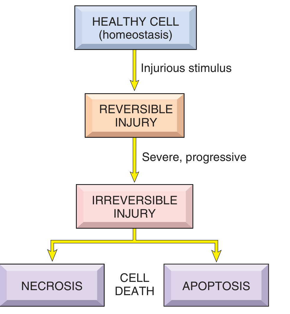
1. Causes of Cell Injury
Before examining mechanisms, it helps to know the major categories of injurious stimuli:
| Category | Examples |
|---|---|
| Hypoxia / ischemia | Arterial blockage, respiratory failure, severe anemia |
| Physical agents | Mechanical trauma, extremes of temperature, radiation, electric shock |
| Chemical agents / drugs | Glucose/salt in hypertonic concentrations, arsenic, cyanide, ethanol, CCl4 |
| Infectious agents | Viruses, bacteria, fungi, parasites (via toxins and immune responses) |
| Immunologic reactions | Autoimmunity, allergic reactions, chronic inflammation |
| Genetic abnormalities | Chromosomal defects, single-gene mutations (e.g., sickle cell anemia) |
| Nutritional imbalances | Protein-calorie deficiency, vitamin deficiencies, obesity |
- Robbins & Kumar Basic Pathology, p. 10-11
2. General Principles
Before individual pathways are discussed, several principles apply to all forms of cell injury:
- Dose- and time-dependence: Low doses of a toxin or brief ischemia cause reversible injury; larger doses or prolonged ischemia lead to irreversible injury and necrosis.
- Cell-type specificity: Skeletal muscle survives complete ischemia for 2-3 hours; cardiac muscle dies after only 20-30 minutes. Neurons are even more sensitive.
- Multiple overlapping pathways: Any single injurious stimulus often triggers several biochemical mechanisms simultaneously, which is why targeting a single pathway rarely prevents cell death completely.
- Genetic variation matters: Individuals with polymorphisms in cytochrome P-450 genes, for example, metabolize the same toxin at different rates, yielding different outcomes.
- Robbins & Kumar Basic Pathology, p. 11-12; Robbins, Cotran & Kumar Pathologic Basis of Disease, p. 62
3. Key Biochemical Mechanisms
3a. Mitochondrial Dysfunction and ATP Depletion
Mitochondria are the primary targets in most forms of injury. They are damaged by hypoxia, reactive oxygen species (ROS), and elevated cytosolic Ca2+, making them sensitive to virtually all injurious stimuli.
Consequences of mitochondrial damage:
-
ATP depletion: Failure of oxidative phosphorylation reduces ATP, which is required for virtually every enzymatic and biosynthetic activity in the cell.
- Na+/K+-ATPase pump fails → intracellular Na+ accumulates → osmotic water influx → cell swelling and ER dilation
- Anaerobic glycolysis increases → lactic acid accumulates → intracellular pH drops → enzymatic activity impaired
- Ribosomes detach from rough ER → protein synthesis falls
- Lipid deposition occurs (fatty change) due to defective lipoprotein assembly
-
Mitochondrial permeability transition pore (MPTP): Opens under injury conditions → loss of mitochondrial membrane potential → failure of oxidative phosphorylation. MPTP opening is a key step in both necrosis and the intrinsic (mitochondrial) apoptosis pathway.
-
Release of pro-apoptotic proteins: Cytochrome c, Smac/DIABLO, and apoptosis-inducing factor (AIF) leak into the cytoplasm when the outer mitochondrial membrane is permeabilized, activating caspases and triggering apoptosis.
-
Robbins & Kumar Basic Pathology, p. 10; Pathologic Basis of Disease, p. 62-63
3b. Oxidative Stress and Reactive Oxygen Species (ROS)
Oxidative stress refers to the accumulation of ROS - superoxide (O2•-), hydrogen peroxide (H2O2), and the hydroxyl radical (•OH) - which overwhelm the cell's antioxidant defenses.
Sources of ROS:
- Normal mitochondrial respiration (low-level "leak")
- Ischemia-reperfusion injury (major source - see below)
- Activated phagocytes (respiratory burst)
- Ionizing radiation
- Reactions with redox-active metals (Fe2+ + H2O2 → •OH, Fenton reaction)
- Metabolism of drugs/chemicals (e.g., CCl4)
Cellular damage caused by ROS:
- Lipid peroxidation of membrane phospholipids → membrane disruption
- Protein oxidation → cross-linking, fragmentation, enzymatic inactivation
- DNA damage → strand breaks, base modifications
Antioxidant defenses include: Superoxide dismutase (SOD), catalase, glutathione peroxidase, and vitamins E and C. When these are overwhelmed, injury escalates.
- Robbins & Kumar Basic Pathology, p. 11-12
3c. Membrane Damage
Plasma membrane integrity is critical for cell viability. Damage to membranes occurs through:
- Direct injury: Toxins (e.g., complement, bacterial toxins) form pores or disrupt bilayer structure
- Phospholipase activation: Elevated cytosolic Ca2+ activates phospholipases that degrade membrane phospholipids
- Lipid peroxidation by ROS: Oxidative attack on polyunsaturated fatty acids
- Cytoskeletal damage: Protease activation detaches the plasma membrane from the cytoskeleton, making cells fragile and prone to rupture under mechanical stress
- Lysosomal membrane rupture: Leakage of acid hydrolases into the cytoplasm causes digestion of cellular components, contributing to irreversible damage and necrosis
Membrane damage to mitochondria (as above) has additional consequences for the apoptotic pathway.
- Robbins & Kumar Basic Pathology, p. 13
3d. Disturbance in Calcium Homeostasis
Normally, free cytosolic Ca2+ is kept at very low levels (~0.1 μmol), while extracellular Ca2+ is ~1.3 mmol. Most intracellular Ca2+ is sequestered in mitochondria and the ER.
Ischemia and toxins raise cytosolic Ca2+ by:
- Releasing it from intracellular stores (ER and mitochondria) early on
- Increasing influx across the dysfunctional plasma membrane later
Downstream effects of elevated cytosolic Ca2+:
-
Protease activation → cytoskeletal and membrane protein degradation
-
Phospholipase activation → membrane phospholipid breakdown
-
ATPase activation → accelerated ATP depletion
-
Endonuclease activation → chromatin fragmentation
-
MPTP opening → mitochondrial dysfunction and apoptosis
-
Robbins & Kumar Basic Pathology, p. 13; Pathologic Basis of Disease, p. 64
3e. Endoplasmic Reticulum (ER) Stress and the Unfolded Protein Response
The ER is responsible for folding newly synthesized proteins. When misfolded proteins accumulate (due to mutations, toxins, ischemia, or viral infection), the cell mounts an Unfolded Protein Response (UPR):
- Mild ER stress (adaptive UPR): Sensors like IRE1 detect misfolded proteins → ↑ chaperone synthesis, ↓ protein translation, ↑ misfolded protein degradation → reduces the load of misfolded proteins
- Severe ER stress (terminal UPR): If the load cannot be cleared → activation of BH3 proteins and caspases → apoptosis via the intrinsic (mitochondrial) pathway
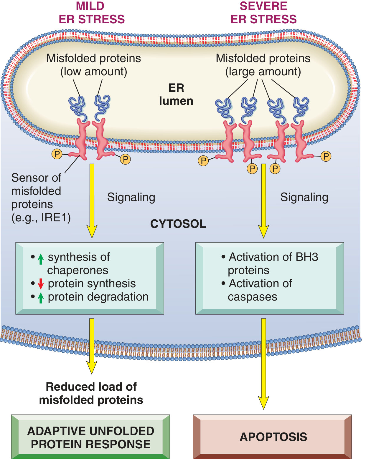
Diseases linked to misfolded proteins include cystic fibrosis (CFTR), familial hypercholesterolemia (LDL receptor), Tay-Sachs disease, and many neurodegenerative diseases (Alzheimer's, Parkinson's).
- Robbins & Kumar Basic Pathology, p. 13-14
3f. DNA Damage
DNA damage from radiation, chemotherapeutic drugs, ROS, or other mutagens activates the p53 tumor suppressor pathway:
- If damage is repairable → cell cycle arrest to allow time for repair
- If damage is too great → p53 activates pro-apoptotic genes (e.g., BAX) → apoptosis via the intrinsic pathway
This serves as a safeguard against propagation of cells with mutations that could lead to cancer.
- Robbins & Kumar Basic Pathology, p. 14
4. Reversible vs. Irreversible Injury
| Feature | Reversible Injury | Irreversible Injury |
|---|---|---|
| ATP depletion | Moderate | Severe |
| Cell swelling | Present | Severe |
| Membrane integrity | Intact (plasma) | Disrupted |
| Ribosomes | Detached | Degraded |
| Mitochondria | Swollen but intact | MPTP open, vacuolated |
| Lysosomes | Intact | Ruptured |
| Outcome | Full recovery if stimulus removed | Necrosis or apoptosis |
The point of no return from reversible to irreversible injury corresponds to: severe mitochondrial dysfunction with MPTP opening, profound membrane damage, and lysosomal rupture.
5. Clinicopathologic Examples
Hypoxia / Ischemia
The sequence: reduced oxidative phosphorylation → ATP depletion → Na+ pump failure → cell swelling → ribosome detachment → protein synthesis failure → fatty change. If ischemia continues: membrane damage → influx of Ca2+ → enzyme activation → irreversible injury and necrosis.
Ischemia-Reperfusion Injury
Paradoxically, restoring blood flow to ischemic tissue can worsen injury. On reperfusion:
-
Burst of ROS generation (from xanthine oxidase, neutrophils, mitochondria) damages membranes and proteins
-
Calcium overload into reversibly injured cells pushes them to irreversibility
-
Complement activation and neutrophil recruitment amplify tissue damage
-
This mechanism is relevant in myocardial infarction, stroke, and organ transplantation
-
Robbins & Kumar Basic Pathology, p. 15
Chemical/Toxic Injury
- Direct toxins (e.g., HgCl2): bind to membrane proteins and sulfhydryl groups → ↑ membrane permeability → loss of ion homeostasis
- Indirect toxins (e.g., CCl4): converted by cytochrome P-450 in the ER to CCl3• free radical → lipid peroxidation → membrane destruction → liver cell necrosis
6. Cell Death Pathways Summary
| Feature | Necrosis | Apoptosis |
|---|---|---|
| Trigger | Severe injury (ischemia, toxins) | DNA damage, loss of growth signals, immune attack |
| Morphology | Cell swelling, membrane rupture, cytoplasmic release | Cell shrinkage, chromatin condensation, apoptotic bodies |
| Inflammation | Yes (pro-inflammatory) | No (non-inflammatory; phagocytosed quickly) |
| Pathway | Uncontrolled, passive | Programmed (intrinsic or extrinsic caspase cascade) |
Other forms of cell death include necroptosis (programmed necrosis, RIPK3-mediated), pyroptosis (inflammatory caspase-1/11-mediated, with cytokine release), ferroptosis (iron-dependent lipid peroxidation), and autophagy-associated cell death (when the stress exceeds the recycling capacity).
Sources:
- Robbins & Kumar Basic Pathology (10th ed.), Chapter 1 - Cellular Responses to Stress and Toxic Insults
- Robbins, Cotran & Kumar Pathologic Basis of Disease (10th ed.), Chapter 2 - Cellular Responses to Stress and Toxic Insults
Tell me in short
Cell Injury Mechanisms - Quick Summary
Six core mechanisms cause most cell injury:
-
ATP Depletion (Mitochondrial dysfunction) - Loss of energy → Na+/K+ pump fails → cell swells → proteins stop synthesizing → eventually irreversible damage
-
Oxidative Stress (ROS) - Free radicals (O2•-, H2O2, •OH) damage membrane lipids, proteins, and DNA; major player in ischemia-reperfusion injury
-
Calcium Overload - Elevated cytosolic Ca2+ activates destructive enzymes: proteases, phospholipases, endonucleases → membrane breakdown + DNA fragmentation
-
Membrane Damage - From phospholipases, ROS, toxins, or complement → loss of ionic gradients → cell rupture (necrosis)
-
ER Stress / Misfolded Proteins - Overwhelms the unfolded protein response → apoptosis via the intrinsic (mitochondrial) pathway
-
DNA Damage - Activates p53 → either cell cycle arrest (for repair) or apoptosis if damage is irreparable
The outcome depends on severity:
- Mild/brief injury → Reversible (cell recovers)
- Severe/prolonged → Irreversible → Necrosis (inflammatory, passive) or Apoptosis (programmed, non-inflammatory)
inflammation
Reading File
Finding Sources
Finding Sources
Reading File
Finding Sources
Reading File
Reading File
Reading File
Loading Image
I have enough rich content for a comprehensive but well-organized answer. Here it is:
Inflammation
Inflammation is the response of vascularized tissues to infection and tissue damage. It brings cells and molecules of host defense from the circulation to the site of injury to eliminate the offending agents and start repair. Without it, infections would go unchecked - but excessive or misdirected inflammation underlies many diseases.
The cardinal signs are: rubor (redness), calor (heat), tumor (swelling), dolor (pain), and functio laesa (loss of function).
Acute Inflammation
Acute inflammation is rapid in onset (minutes to hours) and short in duration. It has three main components:
1. Vascular Changes
- Vasodilation (main mediator: histamine) → increased blood flow → redness and warmth
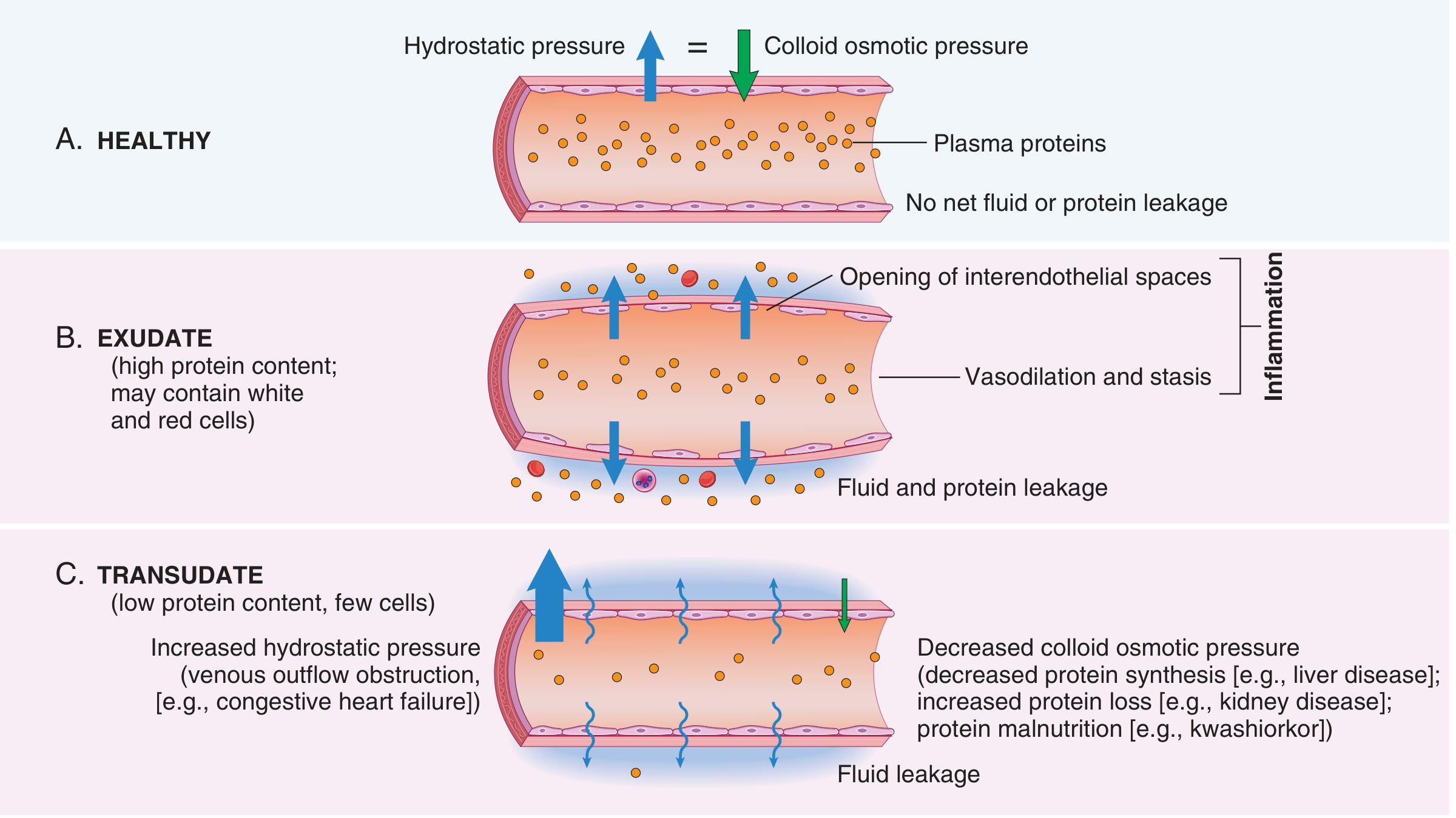
- Increased vascular permeability → protein-rich fluid leaks out of postcapillary venules into tissues → exudate forms → swelling (edema)
- Mechanism: endothelial cell contraction opens interendothelial gaps (triggered by histamine, bradykinin, leukotrienes)
- Slowed blood flow + increased viscosity → stasis
2. Leukocyte Recruitment (Key steps)
| Step | Process | Mediators |
|---|---|---|
| Margination | WBCs move to vessel periphery as flow slows | Stasis |
| Rolling | Loose adhesion to endothelium | Selectins (P- and E-selectin) |
| Firm adhesion | Tight binding to endothelium | Integrins (ICAM-1, VCAM-1) |
| Transmigration (diapedesis) | WBCs squeeze through gaps | CD31 (PECAM-1) |
| Chemotaxis | Migration toward injury | C5a, LTB4, IL-8, bacterial products |
Neutrophils arrive first (within hours); macrophages follow later and dominate in 24-48 hours.
3. Phagocytosis and Destruction
- Leukocytes recognize, engulf (phagocytosis), and destroy microbes/debris
- Killing occurs via:
- ROS (superoxide, H2O2, hypochlorous acid - from the respiratory burst via NADPH oxidase)
- Nitric oxide (NO)
- Lysosomal enzymes (elastase, cathepsins, myeloperoxidase)
Inflammatory Mediators
| Mediator | Source | Main Effects |
|---|---|---|
| Histamine | Mast cells, platelets | Vasodilation, ↑ permeability |
| Prostaglandins (PGE2, PGD2) | Mast cells, macrophages (via COX-1/COX-2) | Vasodilation, pain, fever |
| Leukotrienes (LTB4) | Neutrophils (via lipoxygenase) | Chemotaxis, ↑ permeability |
| LTC4/D4/E4 | Mast cells | Vasoconstriction, bronchospasm (asthma) |
| TNF & IL-1 | Macrophages | Endothelial activation, fever, acute phase response |
| Chemokines (IL-8/CXCL8) | Many cells | Leukocyte recruitment and activation |
| Complement (C3a, C5a) | Plasma (liver) | Vasodilation, chemotaxis, opsonization |
| Bradykinin | Plasma (kinin system) | ↑ Permeability, pain |
| Lipoxins | Neutrophils | Anti-inflammatory - suppress recruitment |
Key drug targets: Aspirin/NSAIDs block COX → reduce prostaglandins. Steroids block phospholipase A2 → reduce all arachidonic acid metabolites. Anti-TNF biologics (e.g., adalimumab) block TNF directly.
Outcomes of Acute Inflammation
- Complete resolution - injury removed, tissue regenerates fully (most common with mild, limited injury)
- Scarring/fibrosis - when tissue destruction is extensive or regeneration is impossible
- Progression to chronic inflammation - when the cause persists or healing is impaired
Chronic Inflammation
Prolonged (weeks to months), with simultaneous inflammation, tissue injury, and repair attempts. Characterized by:
- Mononuclear infiltrate: lymphocytes, macrophages, plasma cells (not neutrophils)
- Tissue destruction by activated macrophages and lymphocytes
- Fibrosis (scarring) as repair attempts continue
Causes
- Persistent infections (mycobacteria, fungi, viruses, parasites) - organisms resist eradication
- Autoimmune/hypersensitivity diseases (rheumatoid arthritis, multiple sclerosis, asthma)
- Prolonged toxic exposure (silica → silicosis; cholesterol → atherosclerosis)
Key Cells
- Macrophages - central orchestrators; activated by IFN-γ → secrete TNF, IL-1, ROS, proteases → cause tissue damage; also drive fibrosis via PDGF, TGF-β
- Lymphocytes (T and B cells) - amplify and sustain macrophage activation; B cells produce antibodies
- Plasma cells - antibody production
- Eosinophils - allergic reactions and parasitic infections
- Mast cells - IgE-mediated reactions
Granulomatous Inflammation
A specialized form of chronic inflammation - aggregates of activated macrophages (epithelioid cells), often surrounded by lymphocytes, sometimes with giant cells and central necrosis (caseation).
Causes: TB, leprosy, sarcoidosis, Crohn's disease, fungal infections, foreign bodies.
Systemic Effects (Acute Phase Response)
Triggered by TNF, IL-1, and IL-6:
- Fever (prostaglandins act on hypothalamus)
- Leukocytosis (WBC count rises)
- Acute phase proteins - CRP, fibrinogen, serum amyloid A rise (useful as clinical markers)
- Malaise, anorexia, weight loss in severe/chronic cases
Source: Robbins & Kumar Basic Pathology (10th ed.), Chapter 2 - Inflammation and Repair
/simplelangugage Inflammation
Inflammation — Simple Language
What is it?
When your body gets hurt or infected, it sends a rescue team to fix the problem. That response is inflammation. It's not the disease — it's your body fighting back.
Why does it happen?
Your body detects something bad — a cut, a germ, a toxin — and raises the alarm. Chemical signals tell blood vessels and immune cells: "Emergency here, come now."
What you see (and feel)
| Sign | What's actually happening |
|---|---|
| Redness | More blood rushing to the area |
| Heat | That extra blood is warm |
| Swelling | Fluid leaking out of blood vessels into tissue |
| Pain | Chemical signals irritating nerve endings |
| Can't use it | The body's way of saying "rest this area" |
Two types
🔴 Acute (Short-term)
- Starts fast (minutes to hours)
- Lasts days
- Main soldier: Neutrophil (arrives first, kills germs, then leaves)
- Example: a pimple, a sprained ankle, a sore throat
🟠 Chronic (Long-term)
- Lasts weeks to months
- Happens when the problem won't go away (e.g., TB, autoimmune disease)
- Main soldier: Macrophage + Lymphocytes
- Can quietly damage your own tissues over time
- Example: rheumatoid arthritis, asthma, atherosclerosis
How does it end?
- Full recovery - problem solved, tissue goes back to normal ✅
- Scarring - damage was too much to fully repair
- Becomes chronic - the cause sticks around
The key players (simplified)
| Player | Job |
|---|---|
| Histamine | Opens blood vessels (causes redness/swelling) |
| Prostaglandins | Cause pain and fever |
| Neutrophils | First responders — eat and kill bacteria |
| Macrophages | Clean up debris, direct long-term repair |
| TNF & IL-1 | Alarm signals — tell the whole body something is wrong |
| Complement | Chemical system that punches holes in bacteria |
When inflammation is the problem
Normally it helps you. But sometimes it misfires:
- Autoimmune disease - attacks your own body (e.g., lupus, rheumatoid arthritis)
- Allergy - overreacts to harmless things (pollen, food)
- Chronic disease - low-grade inflammation quietly damages organs (atherosclerosis, diabetes)
One line summary: Inflammation is your body's emergency response to harm — useful in the short term, damaging if it never turns off.
difference between acute and chronic inflammation
Based on the detailed content already retrieved from Robbins pathology in this session, here is a precise comparison:
Acute vs. Chronic Inflammation
| Feature | Acute Inflammation | Chronic Inflammation |
|---|---|---|
| Onset | Rapid (minutes to hours) | Slow (insidious or after acute) |
| Duration | Short (days to weeks) | Long (weeks to months to years) |
| Cause | Infection, trauma, burns, toxins | Persistent infection, autoimmunity, prolonged toxic exposure |
| Main cells | Neutrophils (early), macrophages (later) | Macrophages, lymphocytes, plasma cells |
| Vascular changes | Prominent - vasodilation, increased permeability | Less prominent |
| Exudate | Yes - protein-rich fluid leaks into tissue | Minimal |
| Tissue destruction | Usually limited | Often extensive and ongoing |
| Fibrosis | Absent | Common - scarring is a hallmark |
| Cardinal signs | Redness, heat, swelling, pain | Often absent or subtle |
| Systemic effects | Fever, leukocytosis, acute phase proteins | Weight loss, anemia, fatigue |
| Outcome | Resolution, scarring, or → chronic | Fibrosis, organ dysfunction, malignancy risk |
Key Cellular Difference
Acute → Neutrophils dominate
- Short-lived, arrive fast, kill microbes via ROS and enzymes
- Die after ~24-48 hours at the site
Chronic → Macrophages + Lymphocytes dominate
- Macrophages are activated by IFN-γ (from T cells)
- They release TNF, IL-1, proteases, ROS → prolonged tissue damage
- T cells and B cells sustain the immune response
- Plasma cells produce antibodies
Key Mediator Difference
| Acute | Chronic |
|---|---|
| Histamine, bradykinin | TNF, IL-1, IL-6 (sustained) |
| Prostaglandins, leukotrienes | TGF-β (drives fibrosis) |
| Complement (C3a, C5a) | IFN-γ (activates macrophages) |
| IL-8 (neutrophil chemotaxis) | PDGF (fibroblast activation) |
Examples
| Acute | Chronic |
|---|---|
| Acute appendicitis | Rheumatoid arthritis |
| Lobar pneumonia | Tuberculosis |
| Acute tonsillitis | Crohn's disease |
| Skin abscess | Silicosis |
| Sprained ankle | Atherosclerosis |
| Meningitis (bacterial) | Asthma |
Special Form of Chronic: Granulomatous Inflammation
A pattern unique to chronic inflammation - clusters of activated macrophages called epithelioid cells, surrounded by lymphocytes, sometimes with central caseous necrosis.
Seen in: TB, leprosy, sarcoidosis, Crohn's disease, fungal infections, foreign body reactions.
One-line summary:
Acute = fast, neutrophil-driven, usually self-limiting. Chronic = slow, macrophage/lymphocyte-driven, causes ongoing tissue damage and scarring.
events of acute inflammation
Finding Sources
Finding Sources
Finding Sources
Reading File
Reading File
Reading File
Loading Image
I have all the content needed. Here is the comprehensive answer:
Events of Acute Inflammation
Acute inflammation has three sequential events that always occur together:
Vascular Changes → Leukocyte Recruitment → Phagocytosis & Destruction
EVENT 1: Vascular Changes
Step 1 - Transient Vasoconstriction
Immediately after injury, arterioles briefly constrict (lasts only seconds).
Step 2 - Vasodilation
- Arterioles and then capillaries dilate → increased blood flow to the area
- Mediator: Histamine (primary), NO, prostaglandins
- Result: Redness (rubor) and Heat (calor)
Step 3 - Increased Vascular Permeability
- Inflammatory mediators cause endothelial cells to contract → interendothelial gaps open in postcapillary venules
- Protein-rich fluid leaks into tissue → Exudate forms
- Mediators: Histamine, bradykinin, leukotrienes, substance P
- Occurs within 15-30 minutes of stimulus
- Result: Swelling (tumor)
Exudate = high protein, cellular debris (inflammation) Transudate = low protein, no cells (heart failure, hypoalbuminemia)
Step 4 - Stasis
- Fluid loss + vasodilation → blood flow slows
- Red cells concentrate centrally; leukocytes shift to periphery (margination)
- Vessels become engorged → vascular congestion
Lymphatics also respond:
- Increased lymph flow drains excess fluid
- Lymphangitis (red streaks) and lymphadenopathy may develop
EVENT 2: Leukocyte Recruitment (Cellular Events)
This is a multistep, highly organized sequence — each step uses different molecules.
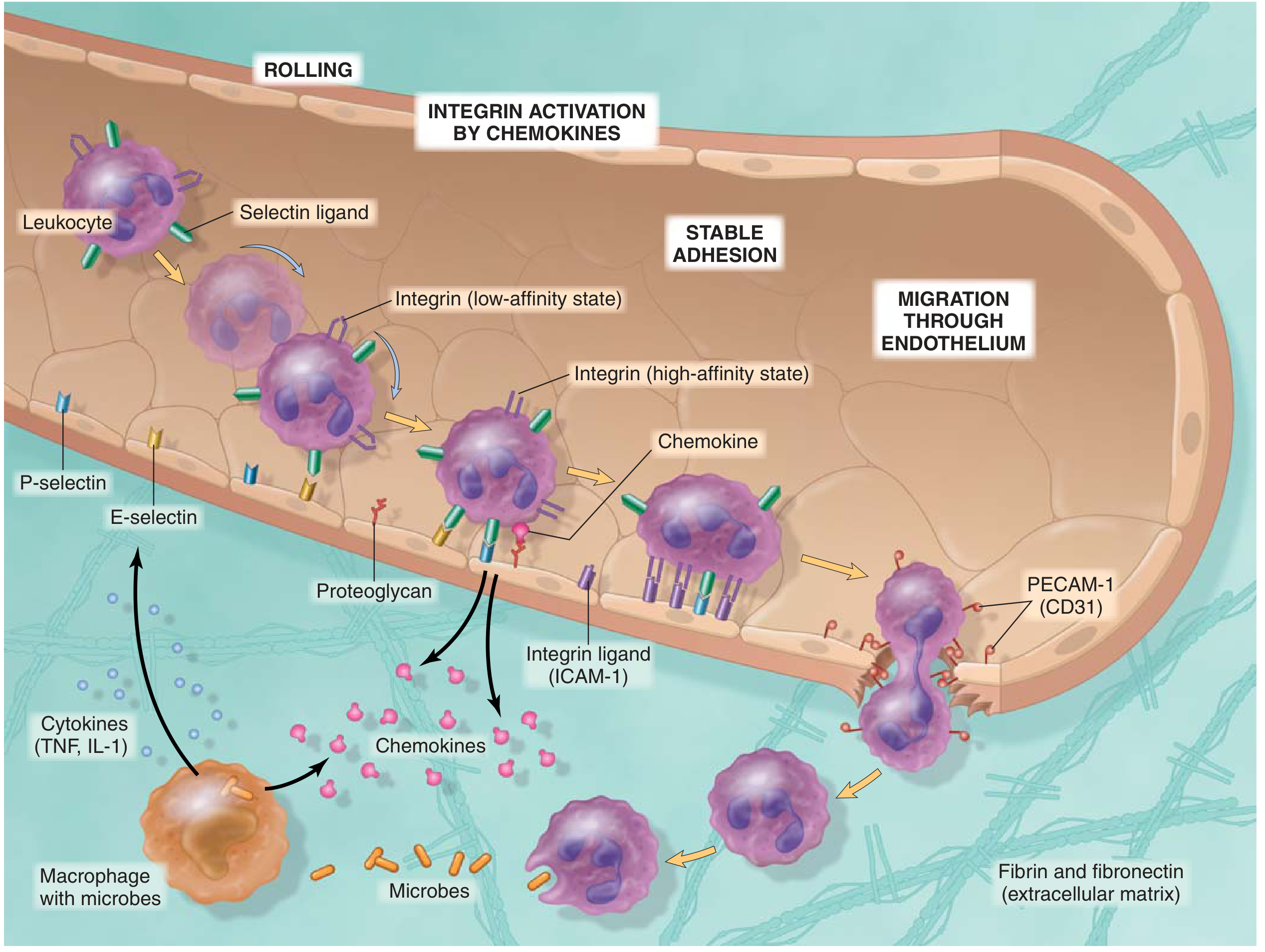
Step-by-step:
| Step | What Happens | Molecules Involved |
|---|---|---|
| 1. Margination | Slowed flow pushes WBCs to vessel wall | Stasis (physical) |
| 2. Rolling | Leukocytes loosely tumble along endothelium | Selectins (P-selectin, E-selectin on endothelium; L-selectin on leukocytes) bind sialic acid-containing ligands |
| 3. Activation | Chemokines displayed on endothelium activate leukocytes | IL-8/CXCL8, C5a, LTB4, fMLP |
| 4. Firm Adhesion | Leukocytes stick tightly and stop rolling | Integrins (LFA-1, Mac-1 on leukocytes) bind ICAM-1, VCAM-1 on endothelium |
| 5. Transmigration (Diapedesis) | Leukocytes squeeze through gaps between endothelial cells | CD31 (PECAM-1) at junctions, then pierce basement membrane (collagenases) |
| 6. Chemotaxis | Leukocytes migrate toward injury along a chemical gradient | C5a, LTB4, IL-8, bacterial products (fMLP) |
Key concept: Selectins handle rolling. Integrins handle firm adhesion. PECAM-1 handles transmigration. These can be blocked by drugs (e.g., anti-integrin therapies in IBD).
Which cells arrive when?
- Neutrophils - first (within 6-24 hours); rapid, short-lived, major killers
- Monocytes → Macrophages - follow (24-48 hours onward); slower but longer-lasting
EVENT 3: Phagocytosis and Destruction
Once at the site, leukocytes must recognize, ingest, and kill the offending agent.
Step 1 - Recognition and Opsonization
- Phagocytes recognize microbes via:
- Pattern recognition receptors (e.g., Toll-like receptors, mannose receptors)
- Opsonins - coat microbes and enhance phagocytosis: IgG antibodies (Fc receptor), C3b (complement receptor), plasma lectins
- Opsonization dramatically increases efficiency of phagocytosis
Step 2 - Engulfment
- Pseudopods extend around the particle → form a phagosome
- Phagosome fuses with lysosomes → phagolysosome
- Lysosomal contents discharged into phagolysosome
Step 3 - Killing and Degradation
Two main killing mechanisms:
A. Oxygen-dependent killing (Respiratory Burst)
- NADPH oxidase assembled on phagosome membrane → converts O2 to superoxide (O2•-)
- O2•- → H2O2 → then myeloperoxidase (MPO) converts H2O2 + Cl- → hypochlorous acid (HOCl) - the most potent microbicidal agent
- Defect in NADPH oxidase → Chronic Granulomatous Disease (CGD) - recurrent infections
B. Oxygen-independent killing
- Lysosomal enzymes: elastase, cathepsins, defensins
- Lysozyme: degrades bacterial cell walls
- Lactoferrin: chelates iron (needed for bacterial growth)
- Major basic protein (eosinophils): kills parasites
Leukocyte-mediated Tissue Damage
Leukocytes also release their killing agents extracellularly (especially when frustrated phagocytosis occurs - e.g., immune complexes on flat surfaces):
- ROS, proteases, elastase → damage host connective tissue
- This is the basis of tissue injury in many inflammatory diseases (e.g., glomerulonephritis, ARDS)
Summary Flow Chart
INJURY/INFECTION
      ↓
Mast cells/macrophages release histamine, TNF, IL-1
      ↓
VASODILATION + ↑ VASCULAR PERMEABILITY
      ↓
Blood flow slows → Stasis → Margination of WBCs
      ↓
Rolling (Selectins) → Activation (Chemokines) → Firm Adhesion (Integrins)
      ↓
Transmigration (PECAM-1) → Chemotaxis toward injury
      ↓
PHAGOCYTOSIS → Opsonization → Engulfment → Killing (ROS + enzymes)
      ↓
Clearance of agent → Resolution OR → Chronic inflammation
Source: Robbins & Kumar Basic Pathology (10th ed.), Chapter 2 - Inflammation and Repair
This is a shared conversation. Sign in to Orris to start your own chat.