
Hospital aqiured pnemonia
"hospital-acquired pneumonia" AND treatment
hospital acquired pneumonia chest X-ray consolidation
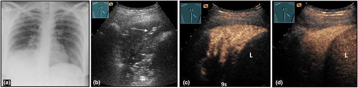
This composite figure illustrates the multimodal diagnostic imaging of community-acquired pneumonia in a 36-year-old female. (a) A posteroanterior chest X-ray reveals a dense, right-sided lower lobe consolidation. (b) B-mode lung ultrasound (LUS) demonstrates a corresponding hypoechoic peripheral consolidation featuring hyperechoic linear structures consistent with air bronchograms (indicated by an arrow). (c) Contrast-enhanced ultrasound (CEUS) at 9 seconds shows early pulmonary arterial perfusion within the lesion. (d) At 2 minutes, the lesion exhibits marked, homogeneous enhancement, reaching an intensity similar to the adjacent liver (L), which serves as a vascular reference. The progression from early arterial perfusion to late homogeneous enhancement is a hallmark of uncomplicated lobar pneumonia. This series demonstrates how CEUS adds functional perfusion data to the structural findings of B-mode LUS, assisting in the differentiation between pneumonic consolidation and other pulmonary pathologies like abscesses or infarcts.

Multi-modal diagnostic imaging of community-acquired pneumonia in a 37-year-old female. Panel (a) is a posterior-anterior chest X-ray showing right-sided basal consolidation. Panel (b) is a B-mode Lung Ultrasound (LUS) demonstrating an inhomogeneous, hypoechoic consolidation with visible air bronchograms (arrows). Panels (c), (d), and (e) present a Contrast-Enhanced Ultrasound (CEUS) time-series. At 3 seconds (c), the consolidation shows an inhomogeneous pulmonary arterial enhancement pattern with a significant non-perfused area (NPA), representing a primary absence of pulmonary arterial perfusion. At 9 seconds (d), the consolidation displays delayed secondary bronchial arterial perfusion within the previously non-perfused tissue. At 3 minutes (late phase, e), the area remains demarcated as a hypoechoic region (arrows). The sequence illustrates the hemodynamic differentiation between pulmonary and bronchial arterial blood supply in pneumonic lung tissue, highlighting the utility of CEUS in evaluating complex perfusion disturbances in pulmonary pathology.
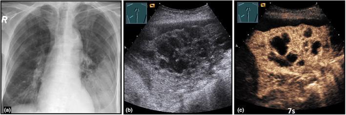
This composite figure illustrates a case of community-acquired abscessed pneumonia using three imaging modalities. Panel (a) is a posterior-anterior chest X-ray showing a low-field consolidation in the left lung. Panel (b) presents a B-mode Lung Ultrasound (LUS) of the same region, demonstrating a complex, hypoechoic consolidation. Within this consolidation, central anechoic areas are visible, representing confluent regions of necrosis or early abscess formation. Panel (c) displays a Contrast-Enhanced Ultrasound (CEUS) image at 7 seconds (pulmonary arterial phase). It reveals an inhomogeneous enhancement pattern throughout the consolidated lung tissue. Notably, there are multiple well-defined anechoic zones characterized by a total absence of enhancement, which confirms the presence of avascular abscess cavities. This multi-modal approach highlights the utility of CEUS in differentiating simple pneumonia from complicated, necrotizing, or abscessed pneumonia by visualizing microvascular perfusion and identifying non-perfused necrotic tissue.
Hospital-Acquired Pneumonia (HAP)
Definition
- Ventilator-Associated Pneumonia (VAP): occurs ≥3 days after the onset of mechanical ventilation
- Non-Ventilator HAP (NV-HAP): occurs in non-intubated hospitalized patients
Source: Goldman-Cecil Medicine, p. 1000; Current Surgical Therapy 14e, p. 1626
Epidemiology
- Affects approximately 1 in every 100 hospitalized patients
- Incidence: 5-10 cases per 1000 hospital admissions
- HAP/VAP is the 2nd most common cause of hospital-acquired infection
- ~3.5% of ventilated patients vs ~0.5% of non-ventilated patients develop pneumonia
- 30% of HAP cases occur in the ICU
- 16% of patients ventilated >1 day develop VAP, with 13% overall mortality
- Risk is highest on medical, surgical, oncology, and neurology services
Source: Goldman-Cecil Medicine, p. 1000; Current Surgical Therapy 14e, p. 1626
Pathogenesis
- Patients on mechanical ventilation are at particularly high risk
- Other risk factors: severe underlying disease, immunosuppression, prolonged antibiotic therapy, invasive devices, impaired consciousness, dysphagia, nasogastric tube feeding
Source: Goldman-Cecil Medicine, p. 1000; Robbins & Cotran, p. 665
Microbiology
Common Causative Organisms
| Organism | Frequency | Notes |
|---|---|---|
| Staphylococcus aureus | 30-40% | ~50% are MRSA |
| Pseudomonas aeruginosa | 15-20% | 28-35% resistant to cefepime |
| Enteric gram-negative bacilli (Klebsiella, E. coli, Enterobacter) | 20-40% | 19-29% resistant to pip-tazo |
| Acinetobacter baumannii | 5-10% | 56-61% carbapenem-resistant |
- MRSA rate: ~50% of S. aureus
- Pseudomonas resistance to antipseudomonal cephalosporins: ~26%
- Acinetobacter carbapenem resistance: 56-61%
Clinical Manifestations
- Fever (>38°C) or hypothermia (<36°C)
- Tachypnea and increased respiratory secretions / purulent sputum
- Leukocytosis (WBC ≥12,000) or leukopenia (WBC ≤4,000)
- Decline in oxygen saturation (SpO₂ drop)
- New or progressive radiographic infiltrates on chest X-ray
- Positive lower respiratory tract cultures
Diagnostic Accuracy of Clinical Signs (meta-analysis, 25 studies, 1639 patients)
| Sign | Sensitivity | Specificity |
|---|---|---|
| Fever | 66% | 54% |
| Purulent secretions | 77% | 39% |
| Leukocytosis | 64% | 59% |
| Infiltrate on chest X-ray | 89% | 26% |
| ETA culture (≥10⁴ CFU/mL) | 76% | 68% |
| BAL culture (≥10⁴ CFU/mL) | 71% | 80% |
Source: Goldman-Cecil Medicine, p. 1000
Diagnosis
- HAP (non-intubated): Expectorated sputum, induced sputum, nasotracheal suctioning
- VAP: Endotracheal aspirate (ETA) - preferred for semiquantitative analysis; BAL if needed
- ETA: ≥10⁵-10⁶ CFU/mL
- BAL: ≥10⁴ CFU/mL
- Protected specimen brush: ≥10³ CFU/mL
Source: Current Surgical Therapy 14e, p. 1626-1627; Goldman-Cecil Medicine, p. 1000
Treatment
Empiric Antibiotic Selection
Risk Factors for MDR Organisms
| MDR Risk | Key Risk Factors |
|---|---|
| MDR VAP | IV antibiotics within 90 days; septic shock at time of VAP; ARDS preceding VAP; ≥5 days in hospital before VAP; acute renal replacement therapy before VAP |
| MDR HAP | IV antibiotics within 90 days |
| MRSA VAP/HAP | IV antibiotics within 90 days; local MRSA rate >10-20% |
| MDR Pseudomonas | IV antibiotics within 90 days; local resistance rates |
Empiric Regimens
- Monotherapy with piperacillin-tazobactam, cefepime, imipenem, meropenem, or levofloxacin
- These agents cover MSSA, Pseudomonas, and gram-negative bacilli
- Dual coverage for gram-negatives (two agents with different mechanisms) PLUS
- MRSA coverage: Vancomycin OR Linezolid
| Pathogen | Preferred Agent |
|---|---|
| MSSA | Oxacillin, Nafcillin, or Cefazolin |
| MRSA | Vancomycin OR Linezolid |
| Pseudomonas | Based on susceptibility + local antibiogram (monotherapy if no septic shock) |
| Other GNB (Klebsiella, E. coli) | Monotherapy based on susceptibilities |
| MDR Acinetobacter | Carbapenem (if sensitive), colistin, or ampicillin-sulbactam |
Source: Current Surgical Therapy 14e, p. 1627-1628; Harrison's 22E, p. 1077; IDSA/ATS 2016 Guidelines
Ventilator-Associated Events (VAE) - CDC/NHSN Algorithm
| Category | Criteria |
|---|---|
| VAC (Ventilator-Associated Condition) | After ≥2 days stability: FiO₂ increase ≥0.20 OR PEEP increase ≥3 cmH₂O, sustained ≥2 days |
| IVAC (Infection-related VAC) | VAC + temp >38°C or <36°C or WBC ≥12 or ≤4 + new antibiotic ≥4 days (after ≥3 days ventilation) |
| Possible VAP | IVAC + purulent secretions (≥25 neutrophils, ≤10 squamous epithelial cells) |
| Probable VAP | Possible VAP + positive quantitative culture |
Source: Current Surgical Therapy 14e, p. 1626
Prevention
- Head-of-bed elevation to 30-45° (reduces aspiration risk)
- Oral decontamination / daily toothbrushing - A 2024 meta-analysis in JAMA Internal Medicine (PMID 38109100) found daily toothbrushing is associated with a reduced incidence of hospital-acquired pneumonia
- Ventilator bundles: daily sedation interruption, spontaneous breathing trials, early extubation
- Avoid unnecessary antibiotics (reduces colonization with resistant organisms)
- Strict hand hygiene
- Subglottic secretion drainage (for patients expected to be ventilated >48h)
- Minimize nasogastric tube use and feed enterally when possible
Prognosis
- HAP in non-ventilated patients: lower mortality than VAP due to better host immunity
- VAP: 13% overall mortality; higher in MDR organisms, septic shock, immunosuppression
- Blood culture positivity in HAP: <15% - limits culture-guided de-escalation
- Delays in empiric treatment and inadequate empiric coverage increase mortality
Key Differences: HAP vs VAP
| Feature | HAP (Non-ventilated) | VAP |
|---|---|---|
| Definition | Pneumonia ≥48h after admission, not intubated | Pneumonia ≥3 days after mechanical ventilation |
| MDR risk | Lower (IV abx within 90d only) | Higher (5 risk factors) |
| Sample collection | Sputum, induced sputum | Endotracheal aspirate, BAL |
| Anaerobic risk | Higher (macroaspiration) | Lower |
| Mortality | Lower | ~13% |
| Blood culture positivity | <15% | ~15% |
- Harrison's Principles of Internal Medicine 22E (2025), p. 1077
- Goldman-Cecil Medicine, p. 1000
- Current Surgical Therapy 14e, p. 1625-1631
- Robbins & Cotran Pathologic Basis of Disease, p. 665
- IDSA/ATS 2016 HAP/VAP Guidelines (Kalil AC et al., Clin Infect Dis 63:e61, 2016)
- Recent evidence: Toothbrushing & HAP prevention - PMID 38109100 (JAMA Intern Med, 2024)