
Lead poisoning fmt
"lead poisoning" treatment chelation
lead poisoning basophilic stippling blood smear
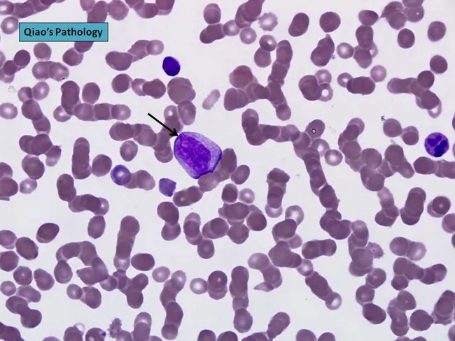
Comprehensive description: Brightfield light microscopy image of a peripheral blood smear stained with Wright-Giemsa, captured at high magnification to visualize circulating plasma cells. The central plasmablast shows an enlarged, irregular nucleus with a prominent nucleolus; the cytoplasm is moderately basophilic. This cell appears larger than surrounding erythrocytes and is consistent with malignant plasma cells typical of plasma cell leukemia. The background demonstrates rouleaux formation of red blood cells, a common feature in paraproteinemias due to increased serum proteins. The smear illustrates plasmacytosis that, when exceeding 20% of leukocytes, supports a diagnosis of plasma cell leukemia. In PCL, neoplastic plasma cells may be CD56 negative and often secrete monoclonal immunoglobulin with IgD or IgE isotypes or light-chain restriction; cytogenetic abnormalities are frequently observed. Morphology alone cannot definitively distinguish PCL from aggressive myeloma; immunophenotyping and cytogenetic studies are essential for precise classification. This image is valuable for hematology education, cytology review, and discussions of malignant plasma cell disorders, including primary versus secondary PCL, and their prognostic significance. Potential clinical utilities include morphologic confirmation in suspected PCL, training in recognition of plasmablasts, and correlation with CBC abnormalities and clinical features such as anemia, thrombocytopenia, lymphadenopathy, and organomegaly.
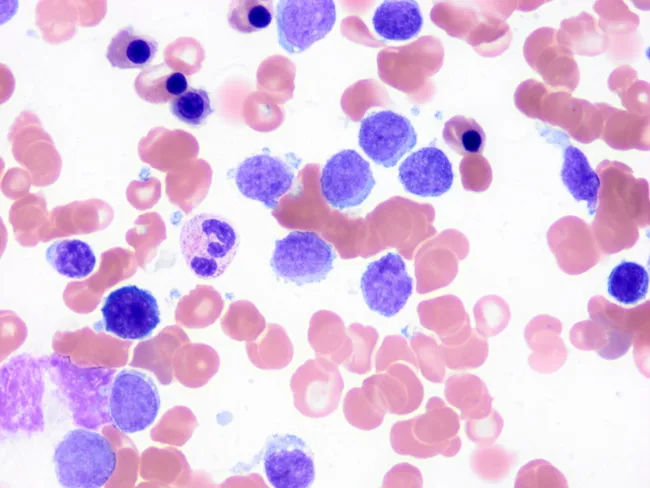
This image depicts a peripheral blood smear prepared with Wright-Giemsa stain and examined under light microscopy at high magnification. The predominant features are small, mature-appearing lymphocytes with scant cytoplasm and discrete basophilic nuclei interspersed among erythrocytes. A characteristic subset of circulating lymphocytes displays short, polarity-restricted cytoplasmic villi (finger-like projections) consistent with splenic marginal zone lymphoma (SMZL) involvement of peripheral blood. The villi are typically slender and localized to one pole of the cell, unlike the longer, circumferential villous extensions seen in hairy cell leukemia, which aids in differential diagnosis. Some lymphocytes appear slightly irregular or irregular nuclear contours; occasional larger atypical cells may be present but are less common. The background shows normocytic red cells with normal distribution; platelets are not prominent. This cytomorphology supports SMZL in the context of known splenomegaly or lymphoproliferative disease and is often corroborated by immunophenotyping and molecular studies. Clinically, peripheral blood involvement occurs in roughly half to two-thirds of SMZL cases and helps establish disease burden. The image illustrates the diagnostic utility of meticulous peripheral smear review for small-vessel lymphocytosis and villous lymphocytes, informing differential diagnoses, guiding flow cytometry panels, and contributing to monitoring and prognosis.
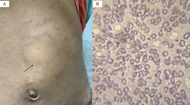
Educational medical image panel consisting of a clinical photograph and a diagnostic pathology image illustrating physical and hematological findings of a hemoglobinopathy such as Hb E/beta-thalassemia. Panel A is a clinical photograph of a patient's abdomen showing a prominent bulge in the left upper quadrant and mid-abdomen, with a black arrow indicating the anterior notch of a massively enlarged spleen (splenomegaly). The overlying skin shows subtle striae or discoloration. Panel B is a peripheral blood smear (Leishman stain, 200x) showing significant red blood cell (RBC) dysmorphology. Key findings include target cells (codocytes) marked by black arrows, teardrop cells (dacrocytes) marked by blue arrows, and basophilic stippling within microcytic, hypochromic RBCs marked by yellow arrows. These visual markers are classic indicators of disordered erythropoiesis and hemoglobin synthesis abnormalities, providing a diagnostic bridge between clinical examination (splenomegaly) and laboratory hematopathology.
Lead Poisoning (Plumbism)
Sources and Epidemiology
- Industrial: Storage battery manufacture, glass/rubber/ship-building/printing industries, smelting, lead oxide fumes
- Environmental (non-occupational): Leaded paint (pre-1960 housing), lead pipes in water supply, contaminated soil, lead in toys/jewelry
- Children: Pica - chewing leaded paint chips from windowsills/walls is the most common pediatric mechanism
- Organic lead: Tetraethyl lead (gasoline antiknock additive, now largely phased out); absorbed through skin - only organic compounds have significant dermal absorption; inorganic lead does NOT penetrate intact skin
Routes of Absorption
| Route | Notes |
|---|---|
| Inhalation | Most common in occupational poisoning (fumes and dust) |
| Ingestion | Lead paint, contaminated food/water; 90% of ingested lead is excreted in feces |
| Skin | Only organic lead compounds (e.g., tetraethyl lead) |
Toxicokinetics
- 95% of absorbed lead enters erythrocytes, then transported to liver, kidneys, and finally bones (main storage depot - ~95% of total body burden in adults)
- Body store in average adult: 150-400 mg; blood level averages ~25 mcg/100 mL
- Lead exerts toxicity by binding SH-groups of enzymes involved in porphyrin synthesis and carbohydrate metabolism
- Bone lead is theoretically "metabolically inactive" but can be remobilized during bone resorption (e.g., pregnancy, osteoporosis)
- USA geometric mean blood lead concentration (2017-2018): 0.753 mcg/dL - reflecting the major decline after removal of leaded gasoline
Mechanism of Toxicity
- ALA dehydratase (delta-aminolevulinic acid dehydratase) - blocks conversion of ALA to porphobilinogen
- Ferrochelatase - blocks incorporation of iron into protoporphyrin IX
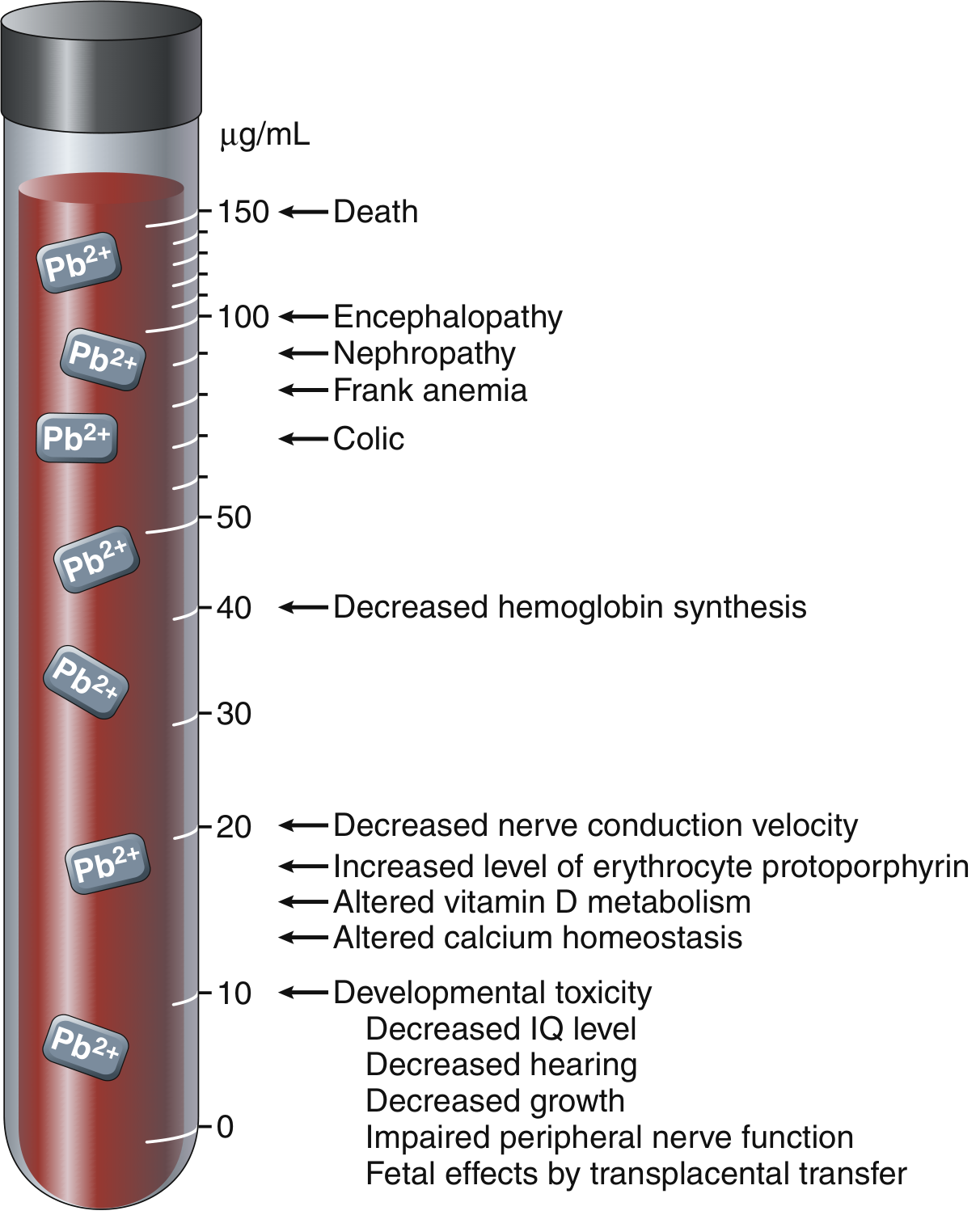
Clinical Features by Blood Lead Level (BLL)
By Organ System
- Microcytic, hypochromic anemia with characteristic basophilic stippling of RBCs (due to aggregated ribosomes)
- Caused by inhibition of heme synthesis at multiple steps
- BLL >40 mcg/dL: decreased hemoglobin synthesis; BLL >70-80: frank anemia
- Children (CNS primarily affected): irritability, anorexia, decreased attention, learning disabilities, decreased IQ, growth retardation, developmental delay, seizures; severe: lead encephalopathy with cerebral edema, demyelination, cortical necrosis - mortality 5-20%, permanent deficits in >25% survivors
- Adults (peripheral nervous system primarily): segmental demyelinating peripheral neuropathy affecting most-used motor neurons - classic presentation is wrist-drop (extensor muscles of wrist/fingers) and foot-drop (peroneal muscles)
- BLL >20: decreased nerve conduction velocity
- Lead "colic" - severe, poorly localized abdominal pain mimicking an acute abdomen
- Anorexia, nausea, vomiting, constipation; mechanism unclear
- Proximal tubular damage with intranuclear lead inclusions (pathognomonic on biopsy)
- Chronic: interstitial fibrosis, renal failure
- Saturnine (lead) gout - due to impaired renal urate excretion
- Hypertension (chronic exposure)
- Altered calcium homeostasis and vitamin D metabolism
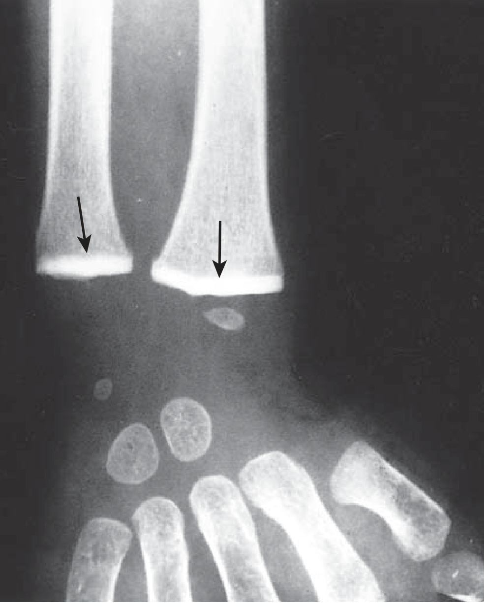
- Lead lines at metaphyses of long bones (especially distal radius, distal tibia/fibula, proximal tibia) on X-ray - represent failure of bone remodeling, not actual lead deposition; seen in children with chronic exposure
- Burton's line - bluish-black line along the gingival margin due to lead sulfide deposition (from reaction with H₂S from oral bacteria)
Acute vs. Chronic Presentation
Acute Inorganic Lead Poisoning
- Uncommon; results from industrial inhalation of large lead oxide fumes or large acute ingestion
- Onset of severe symptoms requires days to weeks of recurrent exposure
- Presents with encephalopathy or lead colic, hemolytic anemia, elevated liver transaminases
- Misdiagnosed as: appendicitis, peptic ulcer, biliary colic, pancreatitis, infectious meningitis
Chronic Lead Poisoning
- Typical multisystemic picture: headache + abdominal pain + anemia (classic triad to suspect lead)
- Fatigue, anorexia, malaise; neurologic complaints; weakness, arthralgias
- Less commonly: motor neuropathy, gout, renal insufficiency
- Children: neurocognitive deficits, growth retardation, developmental delay
Organic Lead Poisoning
- Now very rare after phase-out of tetraethyl lead
- Neurological symptoms dominate: behavioral changes, irritability, insomnia, restlessness → tremor, chorea, convulsions, mania
Diagnosis
Key Test: Venous Blood Lead Level (BLL)
- Best confirmatory test; reflects current circulating lead in blood and soft tissues
- CDC reference value (2021): elevated if BLL ≥ 3.5 mcg/dL in children
- Clinical chelation threshold: ≥45 mcg/dL in children (CDC recommendation)
- Capillary screens may be falsely elevated - always confirm with venous sample
Other Tests
| Test | Finding | Significance |
|---|---|---|
| Zinc protoporphyrin (ZPP) | Elevated (BLL >20) | Reflects chronic/subacute exposure; elevated also in iron deficiency |
| Urinary ALA (delta-ALA) | Elevated | Reflects heme pathway inhibition |
| Urinary coproporphyrin (UCP) | Reddish fluorescence under Wood lamp when ether-extracted urine is acidified | Strongly positive when BLL >80 mcg/dL |
| CBC + peripheral smear | Basophilic stippling | Characteristic but not always present |
| Abdominal X-ray | Radiopaque flecks (lead paint chips, objects) | Confirms recent ingestion; guides decontamination |
| Long bone X-ray (wrist/knee) | Dense metaphyseal lead lines | Chronic pediatric exposure |
| CT head | Cerebral edema | Rule out other causes of altered mental status |
| Urine after CaNa₂EDTA challenge | >500 mcg/24h lead = positive | Rarely used; primarily reflects soft-tissue lead |
Management
Step 1: Remove from Source
Step 2: Decontamination (if acute ingestion)
- Activated charcoal does NOT bind lead
- If abdominal X-ray shows lead in GI tract: whole bowel irrigation (WBI) with polyethylene glycol
- Radiopaque lead objects in stomach/small intestine may need GI decontamination
Step 3: Chelation Therapy
| Situation | BLL Threshold | Agent | Regimen |
|---|---|---|---|
| Asymptomatic children | <44 mcg/dL | None (source removal only) | Environmental investigation + follow-up |
| Symptomatic or BLL 45-69 mcg/dL (no CNS signs) | ≥45 mcg/dL | Succimer (DMSA) oral | 10 mg/kg q8h × 5 days, then 10 mg/kg q12h × 14 days (max 500 mg/dose) |
| BLL ≥70 mcg/dL OR any encephalopathy | Any | BAL + CaNa₂EDTA parenteral | Admit; BAL 4 mg/kg IM q4h; start CaNa₂EDTA IV with 2nd BAL dose |
| Severe encephalopathy | Any | BAL → CaNa₂EDTA | CaNa₂EDTA 1500 mg/m²/day continuous IV infusion × 5 days |
- Oral; FDA-approved for children with BLL ≥45 mcg/dL; also effective in adults
- Adverse effects: nausea, vomiting, diarrhea, transient LFT elevation
- Discharge to lead-free environment if treated outpatient
- IV infusion at 1000-1500 mg/m²/day (30-75 mg/kg/day) × up to 5 days
- Do NOT start alone in encephalopathy - may paradoxically increase lead transport across blood-brain barrier
- Adverse effects: renal tubular injury, chelation of iron and zinc; requires adequate urine output
- Give with hemodialysis if renal failure present
- Deep IM injection (diluted in peanut oil - contraindicated in peanut allergy)
- Initiates chelation before CaNa₂EDTA in encephalopathy
- Dose: 4 mg/kg q4h
- Adverse effects: nausea, vomiting, urticaria, fever, hypertension, hemolysis in G6PD deficiency
- Parenteral chelation limited to ≤5 days; may continue with oral succimer
- After cessation, BLL may rebound as lead re-equilibrates from bone stores (watch for this in chronic exposure)
- Chelation does NOT reverse established neurologic damage
- Prophylactic chelation in the workplace is never a substitute for exposure reduction - Katzung's Basic and Clinical Pharmacology, 16th Ed.
Supportive Care for Lead Encephalopathy
- Intensive care monitoring
- Cerebral edema: corticosteroids, mannitol, or hypertonic saline
- Seizures: anticonvulsants
- Maintain adequate urine output; avoid overhydration
Occupational Exposure Limits (Adults)
- OSHA mandates worker removal if BLL >50-60 mcg/dL (outdated standard)
- Expert panel (2007) recommended removal if single BLL >30 mcg/dL or two successive levels ≥20 mcg/dL (over 4 weeks)
- Long-term goal: BLL <10 mcg/dL for occupational workers - Katzung's Basic and Clinical Pharmacology, 16th Ed.
Differential Diagnosis
- Acute abdomen / colic (appendicitis, biliary colic, pancreatitis)
- Peripheral neuropathy (Guillain-Barré, carpal tunnel)
- CNS: viral meningoencephalitis, tuberculous meningitis
- Children: ADHD, behavioral disorder, iron deficiency anemia, thalassemia
- Gout (in adults with renal involvement)
Key Facts Summary for Exams
| Feature | Detail |
|---|---|
| Classic triad | Headache + abdominal pain + anemia |
| Blood finding | Basophilic stippling (pathognomonic but not always present) |
| Bone finding | Metaphyseal dense "lead lines" (children) |
| Adult neuropathy | Wrist-drop + foot-drop (motor > sensory) |
| Biochemical marker | Elevated ZPP, elevated urine ALA and coproporphyrin |
| Gum sign | Burton's bluish-black gingival line |
| Chelation threshold (child) | BLL ≥45 mcg/dL |
| Oral chelator | Succimer (DMSA) |
| IV chelator | CaNa₂EDTA |
| Encephalopathy: sequence | BAL first → CaNa₂EDTA 4 hours later |
| BAL contraindication | G6PD deficiency, peanut allergy |
| EDTA contraindication | Give alone in encephalopathy (↑ BBB penetration of lead) |
| CDC reference value (2021) | ≥3.5 mcg/dL = elevated in children |
- Katzung's Basic and Clinical Pharmacology, 16th Edition (Inorganic Lead Poisoning chapter)
- Rosen's Emergency Medicine - Concepts and Clinical Practice
- Robbins & Kumar Basic Pathology (Environmental/Nutritional Disease)
- Adams and Victor's Principles of Neurology, 12th Edition (Lead Poisoning in Children)
- Park's Textbook of Preventive and Social Medicine
- P.C. Dikshit Textbook of Forensic Medicine and Toxicology (Acute/Chronic Lead Poisoning)