
vaginitis cervicitis
Reading File
Now let me search the medical library for comprehensive coverage of vaginitis and cervicitis in parallel.
Finding Sources
Finding Sources
Finding Sources
Good results. Now let me read the most comprehensive sources in parallel - Harrison's (most up to date, 2025), Goldman-Cecil, Tintinalli's, and Berek & Novak's.
Reading File
Reading File
Reading File
Reading File
Excellent content. Let me get the cervicitis treatment sections and the Berek & Novak detail, plus search for relevant images.
I now have comprehensive material from multiple authoritative sources. Let me also get the Harrison's MPC treatment and the Tintinalli trichomonas section.
Reading File
Reading File
I have very comprehensive data from Harrison's 22E (2025), Goldman-Cecil, Tintinalli's, and Robbins Pathology. Now let me compose the full, well-organized answer.
Vaginitis and Cervicitis
VAGINITIS
Vaginitis is inflammation of the vaginal mucosa, almost always presenting with vaginal discharge (leukorrhea), odor, pruritus, or irritation. The three most common infectious causes are bacterial vaginosis (BV), vulvovaginal candidiasis (VVC), and trichomoniasis. Normal vaginal flora is dominated by lactobacilli, which maintain a low pH (~3.8-4.5) that protects against pathogens.
1. Bacterial Vaginosis (BV)
Pathogenesis: BV is not a true infection but a polymicrobial disruption of normal flora. The lactobacillus-dominant flora is replaced by anaerobes - primarily Gardnerella vaginalis, Bacteroides spp., Mobiluncus, and Mycoplasma hominis. The loss of lactobacilli raises vaginal pH above 4.5.
Clinical features:
- Thin, homogeneous, gray-white, malodorous ("fishy") discharge
- Odor worse after unprotected intercourse (alkaline semen volatilizes amines)
- Usually no significant itching or burning
- May be asymptomatic
Amsel's criteria (3 of 4 required):
- Thin, homogeneous, gray-white discharge
- Vaginal pH > 4.5
- Positive whiff (amine) test - fishy odor with 10% KOH
- Clue cells on wet mount (squamous epithelial cells coated with bacteria, obscuring cell margins)
Clue cells are the hallmark finding. The Nugent score (Gram stain) is the gold standard for research.
Treatment (Harrison's 22E):
- First-line: Oral metronidazole 500 mg twice daily × 7 days
- Alternatives: Intravaginal 2% clindamycin cream (5 g nightly × 7 nights), intravaginal 0.75% metronidazole gel (5 g twice daily × 5 days)
- Single-dose oral: Secnidazole 2 g once; or tinidazole 2 g daily × 3 days
- Other oral: Clindamycin 300 mg twice daily × 7 days
- Recurrence is common; suppressive twice-weekly intravaginal metronidazole gel reduces recurrence
- Lactobacillus crispatus CTV-05 (LACTIN-V) weekly vaginal administration reduces recurrent BV by ~one-third (RCT evidence)
- BV in pregnancy increases risk of preterm delivery and spontaneous abortion; however, treatment has not consistently reduced adverse outcomes
2. Vulvovaginal Candidiasis (VVC)
Pathogenesis: Candida albicans accounts for 85-92% of cases. It is part of normal vaginal flora in ~20% of women; symptomatic infection requires a disruption of balance. Predisposing factors:
- Systemic antibiotics (suppress competing lactobacilli)
- Diabetes mellitus, pregnancy, oral contraceptive use (alter epithelial glycogen)
- Immunosuppression, HIV
- Tight synthetic undergarments
C. glabrata and C. tropicalis are non-albicans strains, often more resistant.
Classification:
- Uncomplicated: Sporadic, mild-moderate, C. albicans, immunocompetent non-pregnant host
- Complicated: Recurrent (≥4 episodes/year), severe, non-albicans species, or abnormal host (DM, pregnancy, immunosuppression)
Clinical features:
- Vulvar pruritus - the most common and specific symptom
- White, cottage cheese-like or thick curdy discharge adhering to vaginal walls (sometimes thin/watery)
- Vulvar erythema, edema, fissures
- External dysuria, dyspareunia
- No odor (odor favors BV)
- Vaginal pH is normal (3.8-4.5) - distinguishes from BV and trichomoniasis
Diagnosis:
- Wet mount (KOH prep) shows yeast, pseudohyphae, or hyphae - sensitivity ~50%
- A classic clinical presentation warrants treatment even if fungal elements are not seen
- Culture for non-albicans species in refractory/recurrent cases
Treatment:
- Uncomplicated: Single oral dose fluconazole 150 mg (preferred by many patients), or short-course intravaginal azole (clotrimazole 100 mg vaginal tablets twice daily × 3 days; miconazole 1200 mg vaginal suppository once)
- OTC topicals: Clotrimazole 2% cream 5 g intravaginally × 3 days; miconazole 4% cream 5 g × 3 days
- Rx topicals: Butoconazole 2% cream 5 g once; terconazole 0.8% cream 5 g × 3 days
- Complicated/recurrent: Prolonged or maintenance oral fluconazole; otesoconazole 150 mg daily × 7 days then weekly × 11 weeks (newer option per Goldman-Cecil)
- Treatment of sexual partners is NOT indicated - Goldman-Cecil Medicine
- VVC is not classified as an STI, though it can be sexually transmitted
3. Trichomoniasis
Pathogenesis: Trichomonas vaginalis is a flagellated protozoan - the most common nonviral STI worldwide (estimated annual incidence exceeds gonorrhea and chlamydia combined - Harrison's 22E). It attaches to vaginal mucosa, causing local inflammation.
Clinical features:
- Frothy, yellow-green, malodorous vaginal discharge (classic but not universal)
- Vulvar pruritus and irritation
- "Strawberry cervix" - punctate hemorrhages on the cervix (pathognomonic when present, but seen in only ~2% clinically and ~45% by colposcopy)
- Symptoms within 5-28 days of exposure
- Many infections are asymptomatic (especially in men)
Diagnosis:
- Wet mount: motile trichomonads (pear-shaped flagellates) - only ~50-70% sensitive
- NAAT (vaginal, endocervical, or urine specimens): highly sensitive and specific; preferred when available
- Culture: 95% sensitive, gold standard but takes 2-5 days
Treatment:
- First-line: Metronidazole 2 g orally as a single dose (preferred: lower cost, better adherence, fewer side effects)
- Alternative: Tinidazole 2 g orally single dose
- If single-dose fails: Metronidazole 500 mg twice daily × 7 days (resistance is quantitative, not qualitative)
- Pregnancy: Metronidazole 500 mg twice daily × 7 days preferred in women; 2 g single dose in men
- Partners must be treated simultaneously; abstain from sex until both are treated and asymptomatic
- Avoid alcohol during treatment and 24 h (metronidazole) / 72 h (tinidazole) after - disulfiram-like reaction
- Metronidazole gel is NOT effective for trichomoniasis
- CDC resistance hotline: 414-718-4141
Complications of T. vaginalis: Preterm birth, low birth weight, PID, increased HIV transmission risk
4. Other/Less Common Causes
| Type | Key Features | Treatment |
|---|---|---|
| Atrophic vaginitis | Postmenopausal or prolonged breastfeeding; decreased estrogen → thin, pale, dry epithelium; pH elevated | Topical or systemic estrogen replacement |
| Desquamative inflammatory vaginitis (DIV) | Neutrophils, parabasal cells, gram-positive cocci on smear | 2% clindamycin cream ± topical steroid × several weeks |
| Toxic shock syndrome vaginitis | Staphylococcus aureus - Gram stain + culture of vaginal fluid | Antistaphylococcal antibiotics, supportive care |
| Allergic/contact vaginitis | Latex, spermicides, douches, antiseptics | Remove offending agent |
| Foreign body | Retained tampon, cervical cap - usually malodorous | Removal |
Comparative Summary Table
| Feature | Bacterial Vaginosis | VVC | Trichomoniasis |
|---|---|---|---|
| Discharge | Thin, gray-white, homogeneous | White, thick/curdy | Yellow-green, frothy |
| Odor | Fishy (amine) | None | Offensive |
| pH | >4.5 | Normal (3.8-4.5) | >4.5 |
| Pruritus | Minimal | Marked (hallmark) | Present |
| Wet mount | Clue cells | Hyphae/pseudohyphae (KOH) | Motile trichomonads |
| Whiff test | Positive | Negative | Variable |
| STI | No | No | Yes |
| Treatment | Metronidazole/clindamycin | Fluconazole/azole | Metronidazole/tinidazole |
- Jawetz Medical Microbiology 28E (Table 48-5)
- Tintinalli's Emergency Medicine
- Harrison's Principles of Internal Medicine 22E
CERVICITIS
Definition and Overview
Cervicitis is inflammation of the cervix - the most common manifestation of serious bacterial STIs in women. It can be:
- Infectious (most clinically important)
- Noninfectious (chemical, traumatic, physical)
Differentiation is difficult due to the presence of normal vaginal flora.
Etiology
Major pathogens (Robbins Pathology / Harrison's 22E):
- Chlamydia trachomatis - most common (up to 40% of STI-clinic cases)
- Neisseria gonorrhoeae
- Mycoplasma genitalium (increasingly recognized)
- Trichomonas vaginalis
- HSV-2 - less common but important because of neonatal transmission risk
- Ureaplasma urealyticum
- HPV (certain types)
- BV-associated bacteria may also cause a cervical inflammatory reaction
Nearly half of MPC cases have no identifiable microbial etiology even with NAAT testing.
Mucopurulent Cervicitis (MPC)
MPC is cervicitis of the columnar epithelium and subepithelium of the endocervix - the "silent partner" of urethritis in men.
Clinical features:
- Often asymptomatic - discovered on routine pelvic examination
- Abnormal vaginal discharge (mucopurulent or purulent)
- Endocervical tenderness on bimanual examination
- Yellow or green mucopus visible at the endocervical os
- Easily induced endocervical bleeding (friable cervix)
- ≥10 PMNs per high-power field on endocervical Gram stain
Key clinical point: MPC can be a harbinger of PID. In pregnant women, MPC can cause obstetric complications.
Cervical ectopy ("cervical erosion") is commonly confused with infectious cervicitis - it represents columnar epithelium extending onto the ectocervix. Ectopy shows intact epithelium on colposcopy and contains clear or cloudy (not yellow) mucus. It is more susceptible to STI acquisition.
Diagnosis
- NAAT of cervical or vaginal swab: gold standard for C. trachomatis, N. gonorrhoeae, M. genitalium, and T. vaginalis
- Gram stain of endocervical discharge: ≥10 PMNs/HPF supports diagnosis
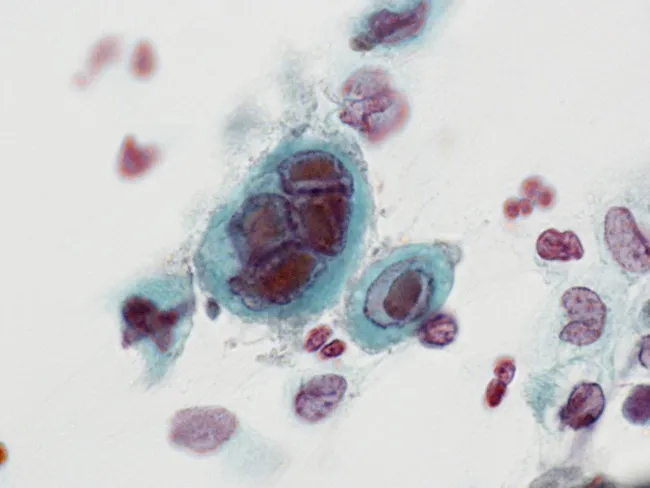
- Pap smear may show inflammation; HSV cytopathic changes (multinucleation, ground-glass nuclei, intranuclear inclusions) are characteristic
- Culture for gonorrhea when antibiotic sensitivities are needed
Treatment
Based on Goldman-Cecil Medicine and Harrison's 22E (2025):
Empiric treatment (when follow-up cannot be ensured, or high-risk population):
- For Chlamydia: Doxycycline 100 mg orally twice daily × 7 days (preferred), or azithromycin 1 g orally single dose
- For Gonorrhea (if prevalence is high or risk factors present): Ceftriaxone 500 mg IM once
- For M. genitalium: Doxycycline 100 mg twice daily × 7 days followed by moxifloxacin 400 mg once daily × 7 days (M. genitalium often persists after doxycycline alone; azithromycin resistance is now recognized)
Targeted treatment (when NAAT results available):
- Treat based on identified pathogen(s)
- Treat partners with a similar regimen unless results warrant different therapy
Risk factors for empiric treatment:
- Age < 25 years
- New sexual partner
- Sexual partner with an STI
- Partners who have other partners
- Follow-up cannot be assured
Complications of Untreated Cervicitis
- Pelvic inflammatory disease (PID) - infection ascends to cause endometritis, salpingitis, tubo-ovarian abscess
- Infertility due to tubal damage
- Ectopic pregnancy
- Chronic pelvic pain
- Neonatal infection - HSV (potentially fatal), gonococcal ophthalmia, neonatal chlamydia (pneumonia, conjunctivitis)
- HIV acquisition - cervical ectopy and inflammation increase HIV susceptibility
Pathology (Robbins & Kumar)
Cervicitis is extremely common and affects the cervical columnar epithelium. Histologically:
- Acute cervicitis: neutrophilic infiltrate, edema, vascular congestion
- Chronic cervicitis: lymphocytic/plasma cell infiltrate, may show squamous metaplasia
- Herpetic cervicitis: intranuclear Cowdry type A inclusions, multinucleated giant cells
- Treatment is usually empiric with antibiotics active against Chlamydia and Neisseria gonorrhoeae; NAATs on vaginal fluid can confirm organisms
Key Distinguishing Points: Vaginitis vs Cervicitis
| Feature | Vaginitis | Cervicitis |
|---|---|---|
| Source of discharge | Vaginal walls | Endocervical glands |
| Main symptoms | Discharge, odor, pruritus | Often asymptomatic, purulent discharge |
| pH changes | Yes (BV, Trichomonas) | Not primary diagnostic tool |
| Main organisms | G. vaginalis, Candida, T. vaginalis | C. trachomatis, N. gonorrhoeae, M. genitalium |
| Clue cells | Yes (BV) | No |
| Risk of PID | Low directly | High - gateway to upper tract infection |
| STI classification | Only Trichomonas is STI | Almost always STI-associated |
Sources:
- Harrison's Principles of Internal Medicine 22E (2025), Chapter 141
- Goldman-Cecil Medicine, Chapter 264
- Robbins & Kumar Basic Pathology
- Tintinalli's Emergency Medicine, Chapter 102
This is a shared conversation. Sign in to Orris to start your own chat.