
Layers of scrotum
Reading File
The layers of the scrotum (from outside in) are: skin, dartos muscle/fascia, external spermatic fascia, cremasteric fascia and muscle, internal spermatic fascia, and tunica vaginalis (parietal then visceral layer). A common mnemonic is "Some Damn Englishmen Called It Testis."
Searching sources...
Finding Sources
Finding Sources
Reading File
Reading File
Reading File
Reading File
Loading Image
Here is a complete, textbook-based breakdown of the layers of the scrotum, from superficial to deep:
Layers of the Scrotum (Superficial to Deep)
The wall of the scrotum is a direct continuation of the anterior abdominal wall layers, acquired during the descent of the testes in fetal life. Each layer corresponds to an abdominal wall layer.
1. Skin
- Thin, corrugated (wrinkled), and more darkly pigmented than surrounding skin due to a high concentration of melanocytes in the basal layer of the epidermis.
- Consists of keratinized stratified squamous epithelium.
- The dermis contains hair follicles, apocrine and eccrine sweat glands, and sebaceous glands.
- No subcutaneous adipose tissue - this is replaced by the dartos layer below.
- Corresponds to the skin of the abdominal wall.
2. Dartos Layer (Tunica Dartos)
- Replaces the subcutaneous fat (hypodermis) of the abdominal wall.
- Contains smooth muscle fibers arranged in two sublayers: a superficial dartos muscle layer (DMSL) and a deep dartos muscle layer (DMDL).
- Also contains myofibroblasts embedded in elastic-fiber-rich extracellular matrix.
- Contraction of the dartos wrinkles the scrotal skin, reducing surface area and heat loss to maintain optimal temperature for spermatogenesis (about 2-3°C below body temperature).
- Apocrine and eccrine sweat glands and hair follicle roots are found between its two layers.
- Corresponds to Camper's and Scarpa's fascia (superficial abdominal fascia / Colles' fascia in the perineum).
3. External Spermatic Fascia
- An extension of the external abdominal oblique aponeurosis.
- Thin but organized layer with thick collagen bundles, nerve fibers, and blood vessels.
- A few smooth muscle fibers are detectable on its outer surface.
4. Cremaster Muscle and Cremasteric Fascia
- An elongation of the internal oblique muscle into the scrotal wall.
- Contains loosely arranged striated (skeletal) muscle fascicles surrounded by cremasteric fascia.
- Predominantly type I (slow oxidative) fibers - fatigue resistant.
- Innervated by the genital branch of the genitofemoral nerve (L1-L2).
- The cremasteric reflex (stroking the inner thigh causes ipsilateral testicular elevation) tests this at spinal levels L1-L2; it is absent on the side of testicular torsion.
5. Internal Spermatic Fascia
- Derived from the transversalis fascia (deep to the transversus abdominis muscle).
- Positioned between the cremaster muscle and the parietal layer of the tunica vaginalis.
- Mostly dense irregular connective tissue containing nerves, blood vessels, and lymphatics.
6. Parietal Layer of Tunica Vaginalis
- The innermost layer; derived from the parietal peritoneum of the abdominal cavity (processus vaginalis).
- A serous membrane lined by simple squamous (mesothelial) epithelium supported by a thin submesothelial connective tissue layer.
- Together with the visceral layer (adherent to the testis), it forms the tunica vaginalis cavity, which contains a small amount of lubricating fluid.
- Abnormal fluid accumulation here = hydrocele (congenital if processus vaginalis remains patent; acquired from infection, injury, or malignancy).
Histological Section of the Scrotal Wall
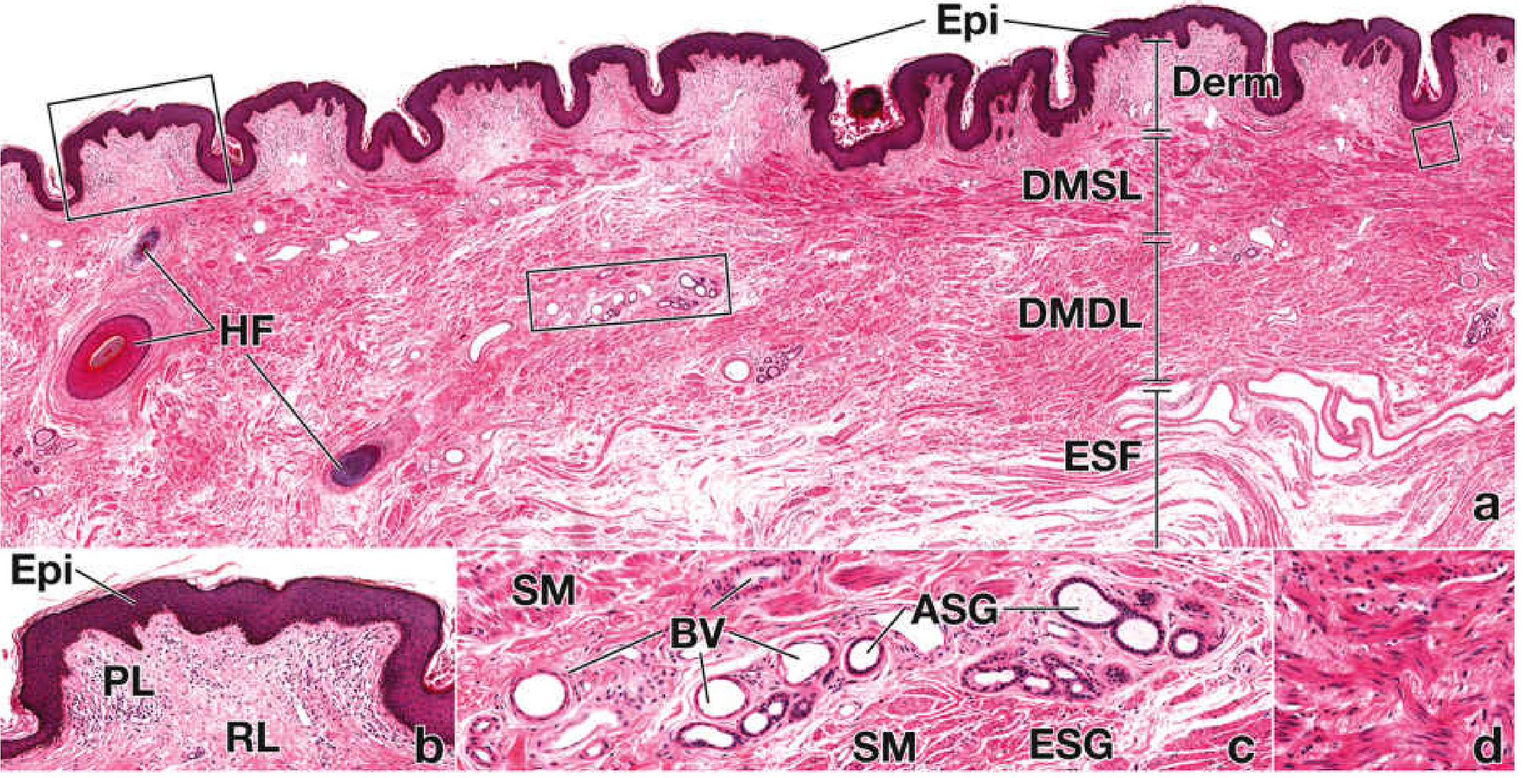
H&E section of the scrotal wall. Epi = epidermis; Derm = dermis; DMSL = dartos muscle superficial layer; DMDL = dartos muscle deep layer; ESF = external spermatic fascia; HF = hair follicle; ASG = apocrine sweat gland; ESG = eccrine sweat gland; BV = blood vessel.
Summary Table
| Layer | Abdominal Wall Equivalent |
|---|---|
| Skin | Skin |
| Dartos muscle/fascia | Camper's + Scarpa's fascia (Colles' fascia) |
| External spermatic fascia | External oblique aponeurosis |
| Cremaster muscle + cremasteric fascia | Internal oblique muscle |
| Internal spermatic fascia | Transversalis fascia |
| Parietal tunica vaginalis | Parietal peritoneum |
Mnemonic
"Some Damn Englishmen Called It Testis"
- Skin
- Dartos
- External spermatic fascia
- Cremaster muscle and cremasteric fascia
- Internal spermatic fascia
- Tunica vaginalis (parietal layer)
- Histology: A Text and Atlas with Correlated Cell and Molecular Biology, pp. 2158-2161
- Smith and Tanagho's General Urology, 19th Edition, p. 526
This is a shared conversation. Sign in to Orris to start your own chat.