
what is congenital cholesteatoma
congenital cholesteatoma ear middle ear
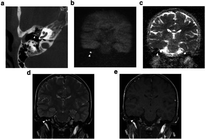
This multi-modal diagnostic image panel demonstrates a congenital middle ear cholesteatoma (CMEC) in an 11-year-old patient. Image (a) is an axial Cone Beam CT (CBCT) showing a well-defined, rounded soft tissue mass (asterisk) in the anterior-superior quadrant of the middle ear, extending medially to the malleus handle (arrowhead). Images (b-e) provide Magnetic Resonance Imaging (MRI) correlations of the same lesion (arrowheads). The lesion exhibits pathognomonic signal characteristics: high signal intensity on non-echo planar diffusion-weighted imaging (DWI b1000, image b) and low signal on the corresponding Apparent Diffusion Coefficient (ADC) map (image c), confirming true restricted diffusion. Coronal T2-weighted (d) and T1-weighted (e) images show intermediate and low signal intensities, respectively. This combination of CT morphology and MRI diffusion restriction is essential for differentiating cholesteatoma from other middle ear pathologies like cholesterol granuloma or inflammatory tissue. The case illustrates the standard radiological protocol for evaluating conductive hearing loss and localizing intratympanic masses in pediatric patients.
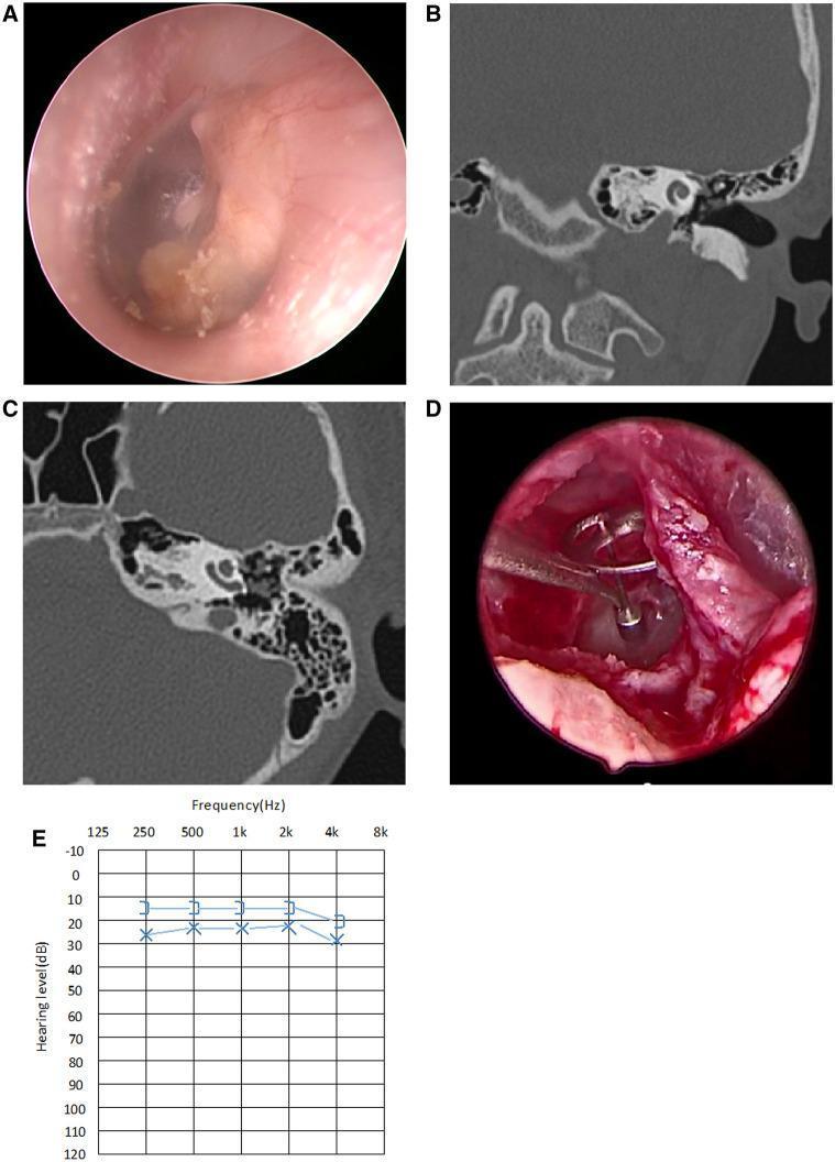
A multi-panel clinical figure documenting a case of Stage III Congenital Middle Ear Cholesteatoma (CMEC). Panel A presents an endoscopic view of the left tympanic membrane, showing a prominent, whitish, opalescent mass localized behind the eardrum, involving multiple quadrants. Panels B and C are high-resolution CT scans of the temporal bone in coronal and axial planes, respectively, demonstrating a soft tissue density filling the middle ear cavity with associated erosion or impairment of the ossicular chain, while the mastoid air cells remain well-pneumatized and uninvolved. Panel D shows an intraoperative view following cholesteatoma excision, highlighting a Total Ossicular Replacement Prosthesis (TORP) placed for hearing reconstruction due to an absent stapes superstructure. Panel E displays a postoperative audiogram showing mild conductive hearing loss with a 10 dB air-bone gap, indicating successful functional reconstruction. This composite serves as an educational tool for the diagnosis, surgical management via endoscopic ear surgery (EES), and functional outcomes of pediatric middle ear cholesteatoma.
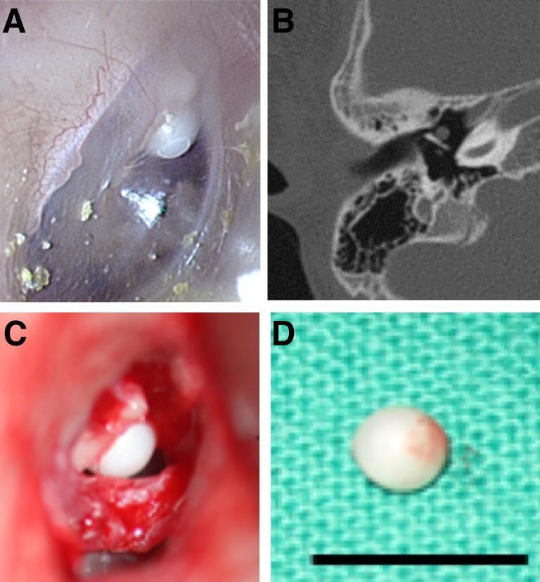
This composite clinical figure illustrates the diagnostic and surgical characteristics of a congenital cholesteatoma in a pediatric patient. Panel A shows an otoscopic view of a pearly, white, spherical mass situated behind an intact, translucent tympanic membrane in the anterior-superior quadrant. Panel B provides an axial non-contrast computed tomography (CT) scan of the temporal bone, identifying a localized soft tissue density within the middle ear cavity without evidence of significant ossicular erosion. Panel C displays the intraoperative finding via a retroauricular approach, revealing the intact spherical keratinous cyst before excision. Panel D shows the gross pathology of the excised specimen, a small pearl-like mass approximately 5mm in diameter (scale bar provided). The figure highlights the classic presentation of congenital cholesteatoma as a 'keratin pearl' in a child with no prior history of ear discharge, surgery, or tympanic membrane perforation. This content is relevant for otolaryngology education regarding middle ear pathologies and surgical management.
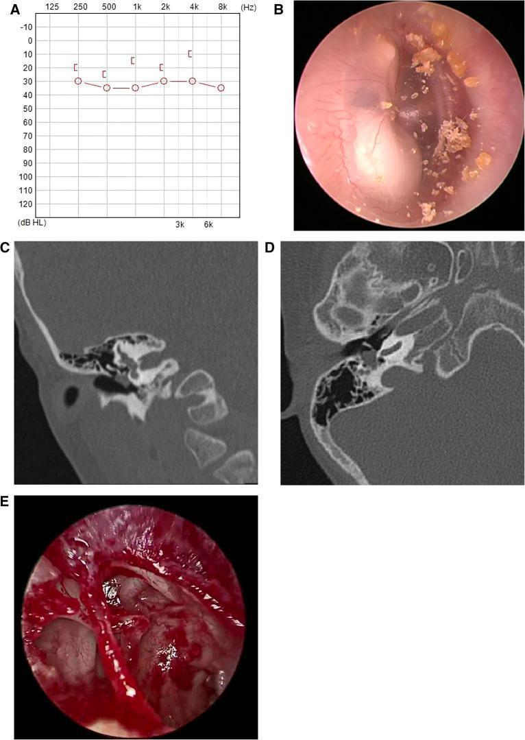
A multi-panel clinical figure documenting a case of Stage II Congenital Middle Ear Cholesteatoma (CMEC). Panel A presents a preoperative audiogram showing mild conductive hearing loss. Panel B is an endoscopic view of the right tympanic membrane, revealing a characteristic opaque, white mass located behind the posterior-superior and inferior quadrants, accompanied by surface keratinous debris. Panels C and D are high-resolution computed tomography (CT) scans of the temporal bone in coronal and axial views, respectively. These demonstrate a soft-tissue density localized within the middle ear cavity without evidence of mastoid air cell involvement or significant bony erosion. Panel E shows an intraoperative endoscopic view after complete surgical excision, confirming an intact ossicular chain. This compilation illustrates the diagnostic triad of pediatric CMEC: audiometric assessment, characteristic otoscopic white reflex (leukocoria of the ear), and radiological localization for surgical planning.
"congenital cholesteatoma"[MeSH Terms]
Congenital Cholesteatoma
Definition
- Scott-Brown's Otorhinolaryngology, Vol 2, p. 4511
- Cummings Otolaryngology, p. 728
Pathogenesis / Embryology
- The Developing Human - Clinically Oriented Embryology, p. 2580
- Scott-Brown's Otorhinolaryngology, Vol 2, p. 4517
Anatomic Classification
| Group | Location |
|---|---|
| 1 | Middle ear (most common) |
| 2 | Perigeniculate area (around facial nerve genu) |
| 3 | Petrous apex |
| 4 | Cerebellopontine angle |
- Cummings Otolaryngology, p. 728
Clinical Features
- White, opalescent mass behind an intact tympanic membrane (the classic "keratin pearl")
- Conductive hearing loss (usually the presenting complaint)
- ~50% of patients present with recurrent otitis media
- May fistulize through the tympanic membrane if untreated
- Facial nerve paralysis (insidious, progressive)
- Sensorineural hearing loss or vestibular dysfunction from labyrinthine/IAC erosion
- May expand dramatically before producing symptoms
- Cummings Otolaryngology, p. 733-735
Staging (Potsic System)
| Stage | Description |
|---|---|
| I | Single quadrant; no ossicular involvement or mastoid extension |
| II | Multiple quadrants; no ossicular involvement or mastoid extension |
| III | Ossicular involvement (erosion/removal); no mastoid extension |
| IV | Mastoid extension (regardless of other findings) |
- Scott-Brown's Otorhinolaryngology, Vol 2, p. 4542-4557
Diagnostic Evaluation
-
Otoscopy: White, pearly mass behind an intact tympanic membrane
-
High-resolution CT (temporal bone): Demonstrates soft tissue density in the middle ear; identifies ossicular erosion and bony destruction
-
MRI: CC shows slightly higher signal than CSF on T1, and moderately high signal on T2-weighted images. Diffusion-weighted imaging (DWI) shows restricted diffusion (bright on DWI, low on ADC map) -- key for differentiating from cholesterol granuloma (which is bright on both T1 and T2) or arachnoid cysts
-
Cummings Otolaryngology, p. 740
Surgical Management
-
Goal: Complete removal of the cholesteatoma matrix or permanent exteriorization
-
Isolated middle ear CC: Can be removed transtympanically with full visualization of the extent of the lesion, especially on the medial aspect of the eardrum
-
Ossicular erosion: Standard middle ear reconstructive techniques (ossiculoplasty, TORP/PORP)
-
Perigeniculate / Petrous apex CC: Approaches include middle cranial fossa, transotic, or transsphenoidal, depending on hearing status and location
-
Cummings Otolaryngology, p. 745
Key Distinguishing Features (vs. Acquired Cholesteatoma)
| Feature | Congenital | Acquired |
|---|---|---|
| Tympanic membrane | Intact | Perforated or retraction pocket |
| History of otorrhea | Absent | Usually present |
| Prior ear surgery/AOM | Absent (by definition) | Often present |
| Age at presentation | Infancy/early childhood | Any age |
| Location | Anterior middle ear most common | Posterior/superior most common |
Can you tell me why there is a labyrinthitis or sensorineural hearing loss in cases of Com?
Why Labyrinthitis and SNHL Occur in COM
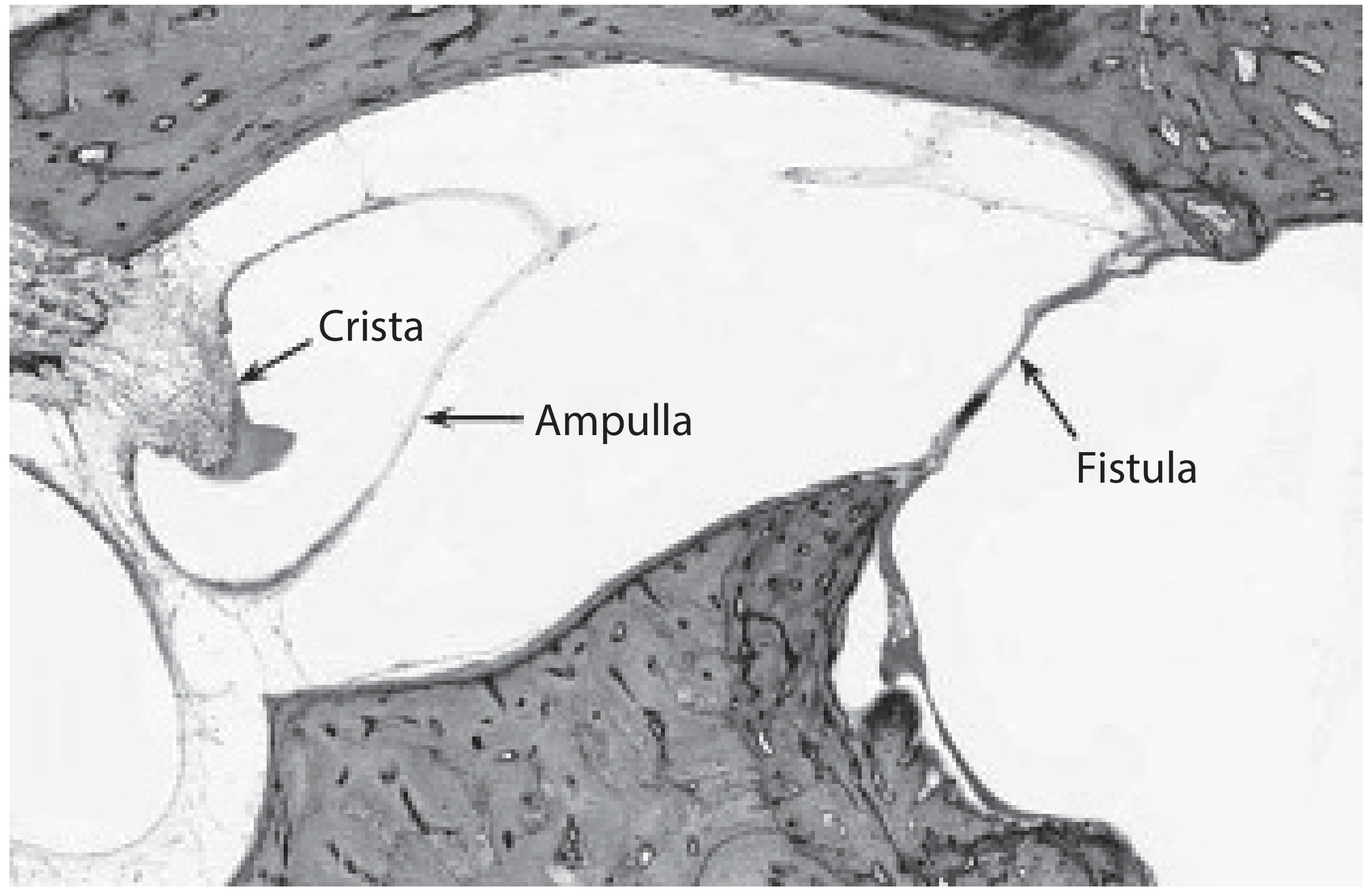
1. Labyrinthine Fistula (Most Common Mechanism)
- It is the most prominent structure in the aditus ad antrum
- It lies directly in the path of an enlarging posterior-superior cholesteatoma
- The cholesteatoma matrix produces pro-inflammatory cytokines that activate osteoclasts, causing progressive demineralization and bone resorption
- Cholesteatoma matrix contacts the endochondral bone of the labyrinth
- Osteoclast-mediated bone erosion -- the bone first demineralizes, then disappears
- Intraoperatively, a thin residual bone layer appears as a "blue line" parallel to the canal lumen
- Eventually, only the endosteum (and sometimes just perilymph) separates the middle ear from the inner ear
"Labyrinthine fistula represents an erosive loss of the endochondral bone that overlies the semicircular canals without loss of perilymph... This loss of the overlying protective bone allows pressure or mass-induced motion of the underlying endosteum, perilymph, and, by contiguity, the endolymphatic compartment, which evokes vestibular and sometimes auditory symptoms."
- Cummings Otolaryngology, p. 2307
2. Spread of Infection: Serous vs. Suppurative Labyrinthitis
| Type | Mechanism | Effect on Inner Ear |
|---|---|---|
| Serous labyrinthitis | Bacterial toxins and inflammatory mediators diffuse across the intact endosteum or round window membrane into the perilymph, without actual bacterial invasion | Reversible or partial SNHL; vertigo |
| Suppurative labyrinthitis | Frank bacterial invasion of the labyrinth, usually via the fistula or round window | Irreversible, profound SNHL; total vestibular loss (dead labyrinth) |
- Shambaugh Surgery of the Ear, p. 5672-5675
- Scott-Brown's Otorhinolaryngology, Vol 2, p. 915-940
3. Diffusion of Ototoxic Substances Through the Round Window Membrane
- Chole and Chiu demonstrated loss of cochlear hair cell stereocilia in animals with experimental cholesteatomas, with or without infection
- McGinn and Chole showed hair cell loss in areas of the cochlea subjacent to areas of bone erosion, suggesting ototoxic substances may traverse the bony cochlear wall directly
- Meyerhoff et al. found that 17.9% of temporal bones with COM showed histologic evidence of labyrinthitis
- Vartiainen and Karjalainen compared 874 chronically infected ears to 609 controls and found significantly worse bone conduction (cochlear function surrogate) in infected ears -- ears with cholesteatoma were worse than those without
"The destructive effects of an expanding cholesteatoma within the middle ear or mastoid and concomitant chronic infection are not limited to the bony structures of the temporal bone."
- Cummings Otolaryngology, p. 1191
-
Cochlear hair cells (outer > inner)
-
Spiral ganglion cells
-
Stria vascularis -- type I fibrocytes in the spiral ligament express NF-κB, which alters ion channel function and impairs endocochlear potential
-
Scott-Brown's Otorhinolaryngology, Vol 2, p. 918-923
Summary of Mechanisms
COM (cholesteatoma/infection)
        |
        ├── Bone erosion → Labyrinthine fistula (lateral SCC most common)
        │           ├── Pressure on endosteum → Vestibular symptoms
        │           └── Bacterial invasion → Suppurative labyrinthitis → Dead labyrinth
        │
        ├── Toxin/mediator diffusion → Round window membrane
        │           └── Serous labyrinthitis → Cochlear hair cell damage → SNHL
        │
        └── Direct bony cochlear wall traversal by ototoxic substances
                    └── Hair cell loss → SNHL
Clinical Note on Surgical Management of Labyrinthine Fistula
-
Removing the matrix risks perilymph loss and SNHL (~10% risk of SNHL in reported series)
-
Leaving the matrix intact at first surgery (canal wall up or canal wall down) and removing it at a second stage -- when the matrix has walled off into a small cyst -- results in SNHL avoidance in >96% of cases
-
Shambaugh Surgery of the Ear, p. 5651-5663
Can you please explain me how cholestatoma spreads and what is the physiology of destruction?
cholesteatoma spread pathway middle ear mastoid ossicular erosion diagram
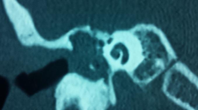
This diagnostic image is a coronal Computed Tomography (CT) scan of the temporal bone, specifically the middle ear and surrounding mastoid structures. The scan reveals a large, non-enhancing soft tissue density within the middle ear cavity and epitympanum, characteristic of a cholesteatoma. Key pathological findings include the significant erosion and partial lysis of the ossicular chain, involving the malleus and incus. There is also evident blunting and erosion of the scutum (the lateral wall of the attic), a classic radiographic sign of an acquired cholesteatoma. The bony labyrinth, including the cochlea and semicircular canals, appears relatively spared. The tegmen tympani remains intact without obvious intracranial extension. This image demonstrates the destructive nature of middle ear cholesteatoma on the conductive hearing apparatus and its impact on the local bony architecture of the temporal bone, serving as a primary tool for preoperative planning in otolaryngology.
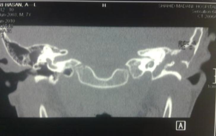
This diagnostic image is a coronal Computed Tomography (CT) scan of the temporal bones focusing on the middle ear and mastoid air cell systems. The scan demonstrates significant bilateral pathology characterized by extensive bony erosion and the loss of normal mastoid air cell septation, a condition described as automastoidectomy. The middle ear cavities and enlarged mastoid regions are opacified by heterogeneous soft tissue density material, consistent with congenital cholesteatoma. Key visible landmarks include the ossicular chain, which appears indistinct or eroded, and the tegmen tympani. On the right side, measurements are visible indicating clinical assessment of the bony structures. The anatomical context relates to a rare case of a first branchial cleft fistula opening into the middle ear through the zygomatic root. This visual is significant for otolaryngology and pediatric radiology, illustrating the advanced bony remodeling and middle ear obliteration associated with congenital ear malformations and chronic cholesteatoma.
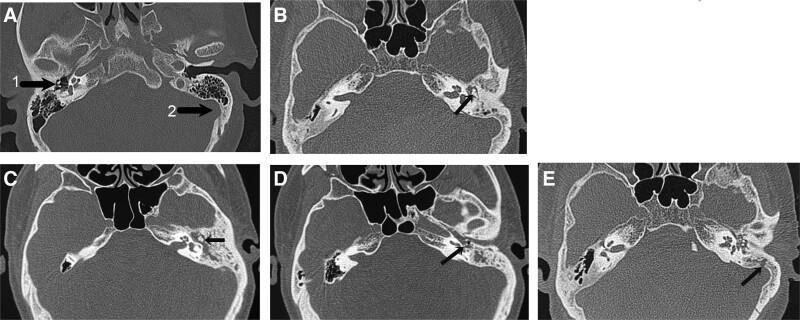
This composite of high-resolution computerized tomography (HRCT) axial scans of the temporal bone demonstrates a progressive classification of middle ear cholesteatoma based on bone destruction. (A) Baseline normal anatomy showing an intact ossicular chain (malleus, incus, and stapes) and a well-defined sigmoid sinus bone plate. (B) Grade (+) showing early soft tissue opacification without definitive ossicular erosion. (C) Grade (++) demonstrating clear destruction of the malleus and/or incus (indicated by arrow). (D) Grade (+++) showing advanced erosion involving the incus and stapes. (E) Grade (++++) exhibiting the most extensive destruction, involving the complete ossicular chain, mastoid tegmen, and/or the sigmoid sinus bone plate (indicated by arrow). Key visual indicators of pathology across the series include increased radiolucency, irregular bone margins, and loss of structural integrity of the auditory ossicles and mastoid air cells. This visual scale serves as a diagnostic tool for assessing the severity of cholesteatoma invasion and its correlation with clinical outcomes such as hearing loss.
How Cholesteatoma Spreads and Destroys - The Full Picture
Part 1: Structure of the Lesion (What Is Spreading)
| Layer | Description |
|---|---|
| Matrix | Keratinizing squamous epithelium - the "skin" lining the sac; this is the actively destructive layer |
| Perimatrix | Subepithelial connective tissue richly infiltrated with inflammatory cells (fibroblasts, macrophages, T-cells, osteoclast precursors) |
| Keratin debris | Dead, desquamated cells that accumulate inside the sac, expanding it under pressure |
- Cummings Otolaryngology, p. 1186
Part 2: Routes and Direction of Spread
Pars Flaccida (Attic/Epitympanic) Cholesteatoma - Most Common
- Retraction pocket forms in the pars flaccida → invaginates into the Prussak space (lateral epitympanic recess)
- Keratin accumulates → sac expands medially
- Scutum (lateral attic wall) is eroded first
- Ossicles are displaced medially and eroded - the long process of the incus is the most frequently destroyed ossicle
- Spreads posteriorly into the aditus ad antrum → mastoid antrum → mastoid air cells (automastoidectomy)
- Can track medially to the tegmen (roof), causing tegmen dehiscence and dural exposure
- Can erode the lateral semicircular canal (labyrinthine fistula)
- Can track along the fallopian canal → facial nerve involvement
Pars Tensa (Posterosuperior) Cholesteatoma
- Arises from posterior or posterosuperior marginal perforation
- Grows medial to the ossicular chain (unlike pars flaccida type which is lateral to ossicles)
- Erodes the posterior lateral attic wall and posterosuperior EAC wall
- Tends to erode the stapes superstructure and long process of incus early
- Higher tendency for inner ear erosion compared to pars flaccida type
- Shambaugh Surgery of the Ear, p. 2069-2083
- Scott-Brown's Otorhinolaryngology, Vol 2, p. 767-793
Part 3: The Physiology of Bone Destruction
Step 1: The Old (Wrong) Theory - Pressure Necrosis
- Cummings Otolaryngology, p. 1133
Step 2: Osteoclast-Mediated Bone Resorption (The Core Mechanism)
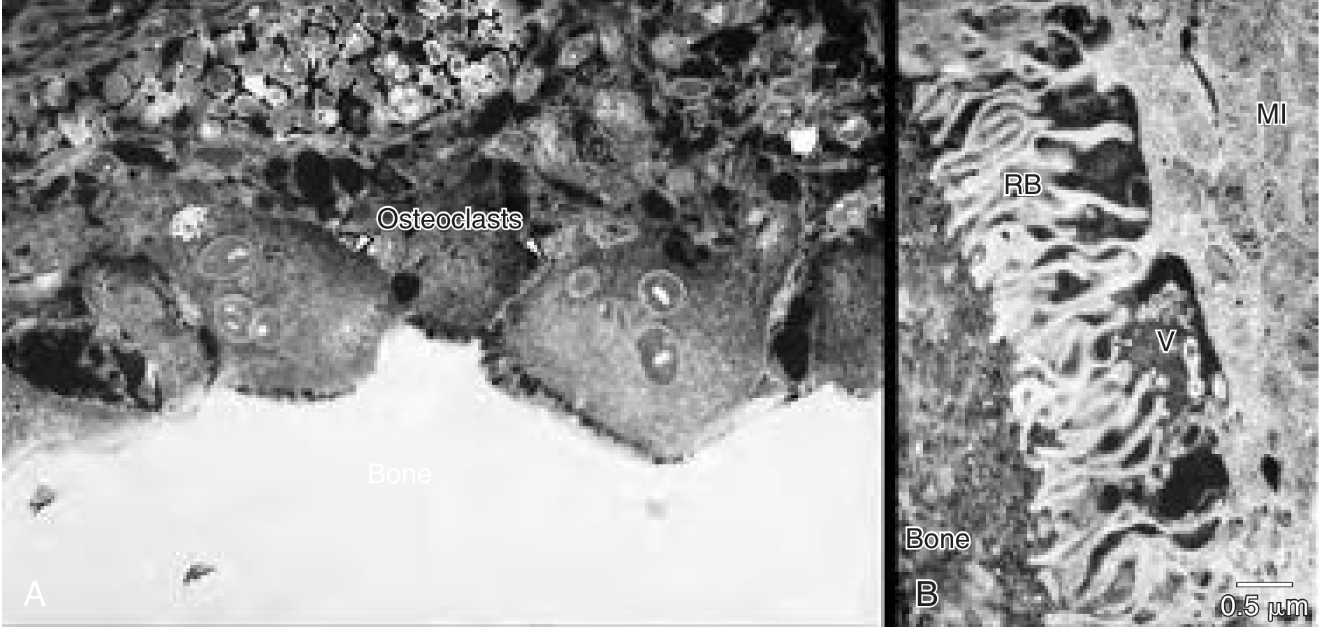
- Osteoclasts form a tight sealing zone against bone
- They create a ruffled border -- a highly folded membrane that dramatically increases surface area
- The space beneath the ruffled border is acidified (pH 4.0)
- Acid phosphatase dissolves the mineral (hydroxyapatite) phase of bone
- Acid proteases (cathepsin-like enzymes) and collagenase dissolve the organic collagen matrix
- The resulting resorption lacunae progressively deepen, eating through bone
Step 3: The RANK/RANKL Signaling Cascade
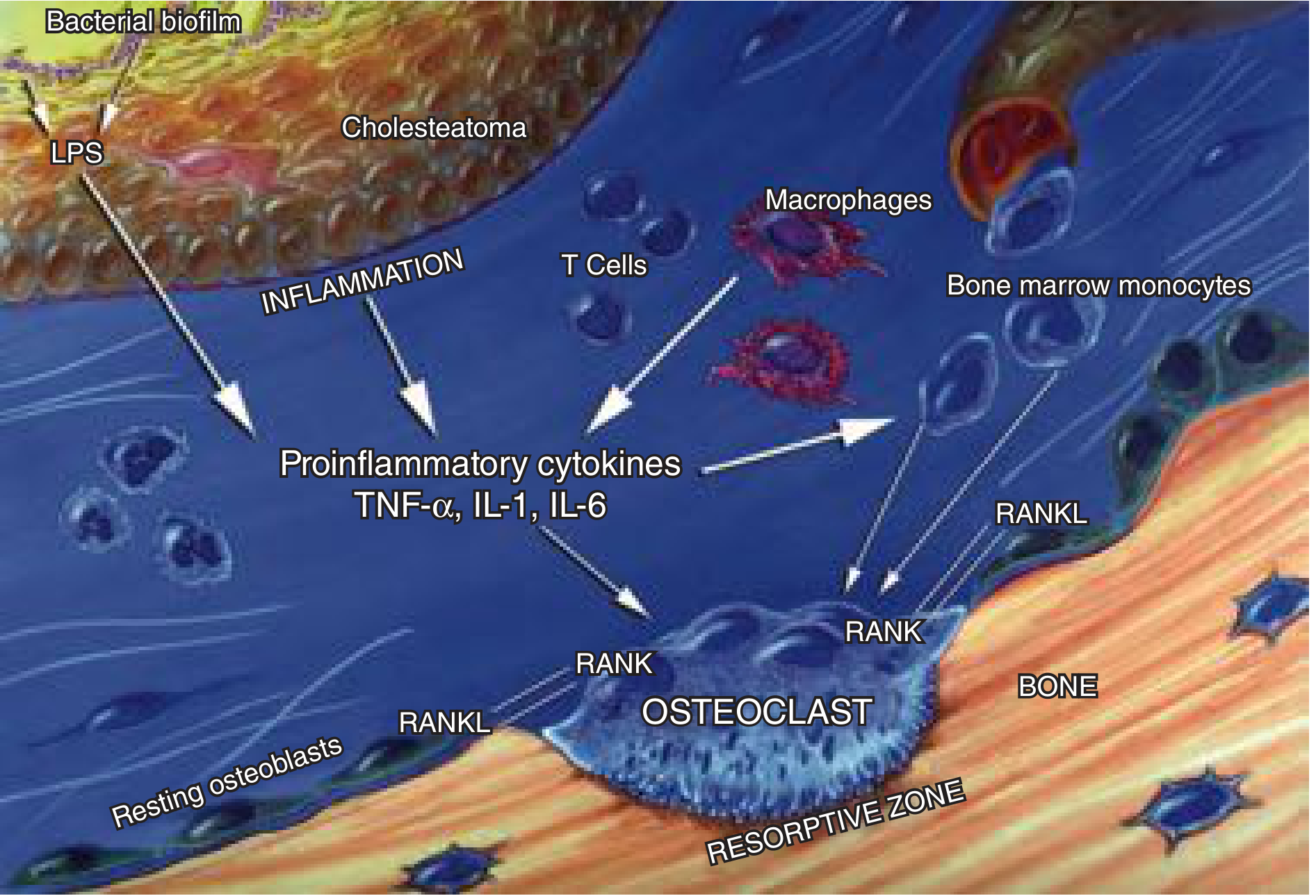
- The cholesteatoma matrix and surrounding bacterial products (LPS from biofilm) trigger inflammation
- This activates T cells, macrophages, and stromal cells in the perimatrix
- These cells express RANKL (Receptor Activator of NF-κB Ligand) on their surface
- RANKL binds to RANK receptors on osteoclast precursors (bone marrow-derived monocytes)
- This drives osteoclastogenesis -- differentiation of precursors into active multinucleated osteoclasts
- M-CSF (macrophage colony-stimulating factor) potentiates this process
- Osteoprotegerin (OPG) is the natural decoy receptor that would normally inhibit RANKL -- in cholesteatoma, OPG is overwhelmed by the pro-inflammatory state
- Pro-inflammatory cytokines TNF-α, IL-1, IL-6 further amplify osteoclast activation
Step 4: Matrix Metalloproteinases (MMPs)
- MMPs 1, 2, 3, and 9 -- expressed in basal and suprabasal cell layers of the matrix
- Neutrophil collagenase -- found throughout the epithelium and granulation tissue
- TIMP-1 (tissue inhibitor of MMPs) is present only in tiny amounts in granulation tissue -- meaning the inhibitory check is essentially absent
-
MMPs degrade the extracellular matrix around bone (collagen, fibronectin, gelatin)
-
Neutral collagenase specifically degrades the osteoid (unmineralized collagen) surface of bone, stripping away the organic layer and directly exposing mineralized bone to osteoclastic attack
-
This "primes" the bone surface for osteoclast binding and resorption
-
Cummings Otolaryngology, p. 1147-1150
Step 5: Keratin Debris as a Direct Inflammatory Trigger
- Keratin itself triggers a foreign body granuloma response
- This elicits further macrophage and inflammatory cell infiltration
- Which amplifies the osteoclastic response locally
Part 4: Why Endochondral Bone Is More Resistant
-
The mastoid air cells and middle ear walls are destroyed early
-
The otic capsule resists erosion longer, but once breached causes labyrinthine fistula
-
Cummings Otolaryngology, p. 1143
Summary Flowchart
Cholesteatoma matrix expansion
          │
          ├── Physical: Sac grows along paths of least resistance
          │     (Prussak space → aditus → antrum → mastoid → tegmen/labyrinth/skull base)
          │
          └── Biochemical bone destruction:
                │
                ├── Bacteria (LPS) + cholesteatoma products
                │       ↓
                ├── Inflammation → TNF-α, IL-1, IL-6
                │       ↓
                ├── RANKL on T-cells/stromal cells → RANK on osteoclast precursors
                │       ↓
                ├── Osteoclast activation → acid phosphatase + cathepsins
                │       → demineralize + digest bone collagen
                │
                ├── MMPs from epithelium → degrade extracellular matrix
                │   Neutral collagenase → strips osteoid → exposes bone
                │
                └── Keratin extrusion → foreign body granuloma → amplifies cycle
- Cummings Otolaryngology, p. 1131-1165
- Scott-Brown's Otorhinolaryngology, Vol 2, p. 767-793
- Shambaugh Surgery of the Ear, p. 2051-2097
when does we reconstruct the attic in cholesteatoma disease when it is medaial or lateral to ossicles?
attic reconstruction cholesteatoma lateral medial ossicles scutum when to reconstruct surgery
https://sydneyearendoscopy.com/wp-content/uploads/2015/07/ee…
Attic Reconstruction in Cholesteatoma: When and Why
First, Understand the Anatomy: What Is the "Attic"?
- A lateral wall = the scutum (the bony lateral attic wall, part of the EAC superior wall)
- A medial wall = the tegmen, facial nerve, and otic capsule structures
- Contents = the head of malleus, body and short process of incus
The Two Types and Their Position Relative to Ossicles
| Type | Origin | Position | Structures Destroyed First |
|---|---|---|---|
| Pars flaccida (attic) cholesteatoma | Pars flaccida retraction into Prussak space | Lateral to ossicles | Scutum eroded; ossicles displaced medially |
| Pars tensa (posterosuperior) cholesteatoma | Posterior/posterosuperior marginal perforation | Medial to ossicles | Long process of incus + stapes eroded; ossicles displaced laterally |
Pars Flaccida (Lateral to Ossicles): Attic/Scutum Reconstruction
- After complete removal of the cholesteatoma, the scutum defect must be reconstructed in almost all canal-wall-up (CWU) approaches
- The goal is to restore the lateral bony buttress so the tympanic membrane/pars flaccida cannot re-retract
- This is done at the primary surgery or at second-look surgery depending on disease extent
- Tragal cartilage with perichondrium -- most popular; rigid, resistant to resorption, good for defects up to ~3 mm
- Cortical bone pate (crushed mastoid bone mixed with blood)
- Temporalis fascia alone -- less rigid, higher retraction/recurrence risk
- Calcium phosphate ceramics / hydroxyapatite -- synthetic option
- Marchioni et al., Otolaryngol Clin N Am 2013: In 146 patients with attic cholesteatoma managed endoscopically, 52.7% had cartilage scutum reconstruction, 14.4% bone pate, and 32.9% temporalis fascia.
Pars Tensa (Medial to Ossicles): Ossiculoplasty, Not Scutum Repair
- Incus interposition -- reshaped removed incus placed between malleus and stapes head (if stapes suprastructure intact = Type III tympanoplasty equivalent)
- PORP (Partial Ossicular Replacement Prosthesis) -- from stapes head to tympanic membrane/malleus, when incus is gone but stapes suprastructure present
- TORP (Total Ossicular Replacement Prosthesis) -- from footplate to tympanic membrane, when stapes suprastructure also eroded
- Staged reconstruction -- if disease was extensive or infected, leave the middle ear with a Silastic sheet and reconstruct at a second-look operation (6-12 months later)
The Decision Framework: Lateral vs. Medial
Cholesteatoma position relative to ossicles
         │
         ├── LATERAL to ossicles (Pars flaccida / Prussak space)
         │      │
         │      ├── Scutum eroded → ATTIC (SCUTUM) RECONSTRUCTION needed
         │      │      Material: cartilage > bone pate > fascia
         │      │
         │      └── If ossicles also eroded → ossiculoplasty ALSO needed
         │
         └── MEDIAL to ossicles (Pars tensa / posterosuperior)
                │
                ├── Incus long process / stapes eroded → OSSICULOPLASTY needed
                │      (PORP or TORP depending on stapes status)
                │
                └── Scutum less commonly affected → attic repair less critical
                    (posterior attic wall may need repair if eroded posteriorly)
The Bigger Picture: Why Attic Reconstruction Matters for Recurrence
- Eustachian tube dysfunction → negative middle ear pressure
- Thinned, unsupported pars flaccida is sucked medially → retraction pocket
- Keratin accumulates → cholesteatoma
"The recurrence consists in a new dangerous tympanic retraction pocket caused by inadequate reconstruction of the scutum and tympanic loss of substance, inducing persistence of the physiopathologic process of middle ear depression."
- Marchioni et al., Otolaryngol Clin N Am 2013
Summary Table
| Situation | Position | What to Reconstruct | Timing |
|---|---|---|---|
| Pars flaccida, early (scutum eroded, ossicles intact) | Lateral | Scutum only (cartilage/bone pate) | Primary surgery |
| Pars flaccida, advanced (ossicles also eroded) | Lateral | Scutum + ossiculoplasty (PORP/TORP) | Scutum at primary; ossiculoplasty at primary or second-look |
| Pars tensa (ossicles eroded, scutum intact) | Medial | Ossiculoplasty (PORP/TORP) | Primary if safe; otherwise staged |
| Matrix on stapes footplate (any type) | Medial | Leave matrix; second-look ossiculoplasty | Staged (to avoid perilymph leak) |
| Canal wall down (open cavity) | Any | No scutum repair needed; cavity obliteration optional | Primary surgery |
why is there 4k notch or boilers notch seen in pta in noise induced hearing loss
Search for clinical images of 4kHz notch in PTA audiograms