


Draw fundus colour diagram
fundus photography
clinical fundus colour diagram recording conventions ophthalmology
Create a clinical colour fundus diagram on standard ophthalmology Amsler-Dubois charts, with two separately labelled eyes: Right eye (OD) and Left eye (OS). In each diagram draw normal optic disc and retinal vessels. At the macula show a well-circumscribed round central yellow-orange elevated lesion surrounded by a dense dark grey-black pigmented ring or halo, with adjacent small pale yellow hard exudate-like deposits and subtle surrounding grey retinal change. In OD, place the disc nasal-left and lesion temporal-right; in OS, place the disc nasal-right and lesion temporal-left. Use conventional clinical fundus-diagram colour coding, clear and legible, and include a brief key stating that the drawing is based on images and lesion identity requires clinical confirmation.
Colour fundus diagram
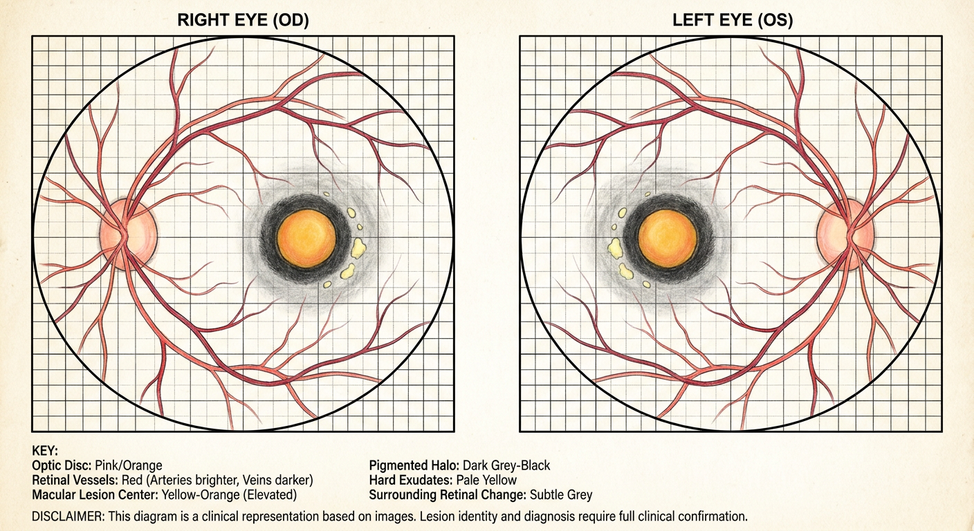
- Optic discs and major retinal vessels appear grossly normal in the visible field.
- A round, well-demarcated yellow-orange macular lesion is present.
- The lesion is bordered by a dark grey-black pigmented halo/ring.
- There are adjacent pale yellow deposits/exudate-like changes and subtle surrounding retinal disturbance.
Diagnosis stargard, re with central corneal choreoretinal atrophy and in le copper beater appearance without chorioretinal atrophy
Stargardt disease fundus atrophy
Stargardt disease beaten bronze macular appearance chorioretinal atrophy fundus
Create a neat hand-drawn clinical colour fundus diagram on standard ophthalmology Amsler-Dubois charts, clearly labelled Right eye (OD) and Left eye (OS), for Stargardt disease. OD: show the optic disc nasal to the macula, a well-defined central macular chorioretinal atrophy area with a central pale grey-white atrophic zone, surrounding retinal pigment epithelial mottling and pigment clumps, and a few small yellow-white pisciform flecks. OS: show a central macular beaten-bronze or coppery granular retinal pigment epithelial appearance, with subtle pigment mottling and a few yellow-white pisciform flecks but no central chorioretinal atrophy. Draw vessels and discs normally. Add below the diagrams: OD - Stargardt disease with central chorioretinal atrophy. OS - Stargardt disease with beaten-bronze appearance, no chorioretinal atrophy. Use only conventional clinical fundus-diagram colours, with a clean labelled legend.
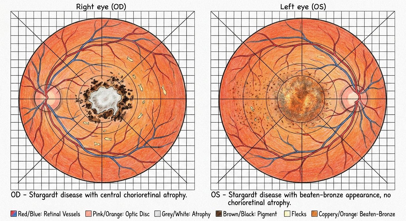
- OD (RE): Stargardt disease with central macular chorioretinal atrophy, RPE mottling/pigmentary change, and flecks.
- OS (LE): Stargardt disease with beaten-bronze (coppery granular) macular appearance, RPE mottling and flecks, without chorioretinal atrophy.