
Beningn and malignant
I'll clarify what aspect you need before diving in.
Asking for Preferences
What topic would you like to cover regarding benign and malignant? · What level of detail do you need?
Benign tumors grow slowly and stay localized, while malignant tumors invade surrounding tissue and can spread (metastasize) to distant sites. This distinction drives diagnosis, prognosis, and treatment decisions.
Searching sources...
Reading File
Finding Sources
Finding Sources
Finding Sources
The most relevant source is Robbins Pathology - "CHARACTERISTICS OF BENIGN AND MALIGNANT" and "Benign and Malignant Tumors". Let me read those sections.
Reading File
Reading File
Let me read more of the characteristics section (local invasion and metastasis) and also grab the relevant histology images.
Reading File
I now have rich content. Let me also fetch the key histology images showing benign vs malignant differences.
Loading Image
Loading Image
I have all the content needed. Here is a comprehensive overview:
Benign vs. Malignant Tumors (Neoplasms)
Definition
A neoplasm (tumor) is an abnormal mass of tissue resulting from uncontrolled cell proliferation that persists even after the stimulus that initiated it is removed. All neoplasms have two basic components: the proliferating tumor cells (parenchyma) and the supporting stromal framework (blood vessels, connective tissue).
Key Distinguishing Characteristics
Three features reliably separate benign from malignant tumors, according to Robbins & Kumar Basic Pathology: differentiation/anaplasia, local invasion, and metastasis.
| Feature | Benign | Malignant |
|---|---|---|
| Differentiation | Well differentiated; closely resembles tissue of origin | Poorly/undifferentiated (anaplastic); abnormal morphology |
| Anaplasia | Absent | Often present (pleomorphism, large nuclei, prominent nucleoli) |
| Capsule | Usually encapsulated | Non-encapsulated (exception: renal cell carcinoma has pseudocapsule) |
| Border | Well-circumscribed, smooth | Irregular, poorly defined |
| Growth rate | Slow | Usually rapid (but not always - not a reliable discriminator alone) |
| Mitoses | Rare; normal configuration | Frequent; atypical/abnormal mitotic figures |
| Local invasion | Absent - expands without invading | Present - infiltrates and destroys surrounding tissue |
| Metastasis | Never | Hallmark of malignancy |
| Desmoplasia | Absent | Can induce dense fibrous stroma (scirrhous tumors) |
| Necrosis | Rare | Common (due to outgrowing blood supply) |
| Recurrence after excision | Rare | More common |
1. Differentiation and Anaplasia
Benign tumors are composed of well-differentiated cells that closely resemble their normal counterparts. A lipoma contains mature fat cells with cytoplasmic lipid vacuoles; a chondroma contains mature cartilage cells synthesizing cartilaginous matrix. Mitoses are rare and of normal configuration.
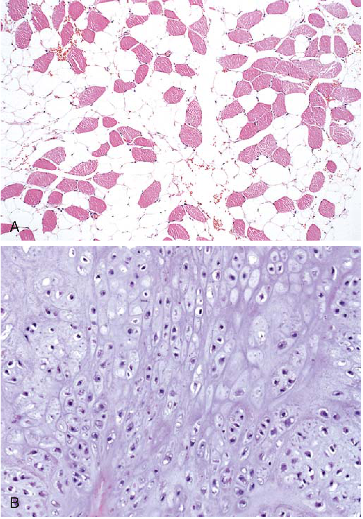
Malignant tumors show morphologic alterations - cytologic features of malignancy include:
- Pleomorphism - variation in size and shape of cells and nuclei
- Abnormal nuclear morphology - hyperchromatic nuclei, increased nucleus:cytoplasm ratio (normally 1:4-6, may approach 1:1)
- Prominent nucleoli ("owl-eye" nucleoli)
- Atypical mitoses - tripolar, quadripolar, or other bizarre spindle forms
- Loss of polarity - cells grow in disorganized patterns
Anaplasia (literally "backward formation") - total loss of differentiation - is a reliable indicator of malignancy.
2. Local Invasion
-
Benign tumors grow as cohesive, expansile masses. They do not penetrate the capsule or invade the surrounding normal tissue. This well-defined plane of cleavage makes surgical excision straightforward and complete.
-
Malignant tumors lack a well-defined capsule. They infiltrate, invade, and destroy surrounding normal tissue. Even seemingly encapsulated cancers (e.g., renal cell carcinoma) show microscopic invasion. Fibrous stroma induced by the tumor (desmoplasia) makes some cancers (e.g., breast carcinoma) rock-hard and "scirrhous."
Well-differentiated adenocarcinoma of the thyroid may look almost normal histologically - its malignant nature revealed only by invasion into adjacent tissues. - Robbins & Kumar Basic Pathology
3. Metastasis
Metastasis is the defining hallmark of malignancy. Benign tumors never metastasize.
Routes of metastatic spread:
- Lymphatic spread - most common for carcinomas (e.g., breast cancer to axillary lymph nodes)
- Hematogenous spread - most common for sarcomas; also used by carcinomas (liver, lung are common sites)
- Seeding of body cavities (transcoelomic) - e.g., ovarian carcinoma seeding the peritoneal cavity
Nomenclature
The name of a tumor encodes its behavior and tissue of origin:
| Tissue of Origin | Benign | Malignant |
|---|---|---|
| Fibrous tissue | Fibroma | Fibrosarcoma |
| Fat | Lipoma | Liposarcoma |
| Cartilage | Chondroma | Chondrosarcoma |
| Bone | Osteoma | Osteosarcoma |
| Blood vessels | Hemangioma | Angiosarcoma |
| Smooth muscle | Leiomyoma | Leiomyosarcoma |
| Striated muscle | Rhabdomyoma | Rhabdomyosarcoma |
| Melanocytes | Nevus (mole) | Melanoma |
| Glandular epithelium | Adenoma | Adenocarcinoma |
| Squamous epithelium | Squamous papilloma | Squamous cell carcinoma |
| Urothelium (bladder) | Urothelial papilloma | Urothelial carcinoma |
| Liver | Hepatic adenoma | Hepatocellular carcinoma |
| Kidney | Renal tubular adenoma | Renal cell carcinoma |
| Placenta | Hydatidiform mole | Choriocarcinoma |
| Uterus | Leiomyoma (fibroid) | Leiomyosarcoma |
Key naming rules:
- "-oma" = benign (exception: lymphoma, melanoma, hepatoma - all malignant)
- "Carcinoma" = malignant tumor of epithelial origin
- "Sarcoma" = malignant tumor of mesenchymal origin
Benign Cystic Teratoma (Example)
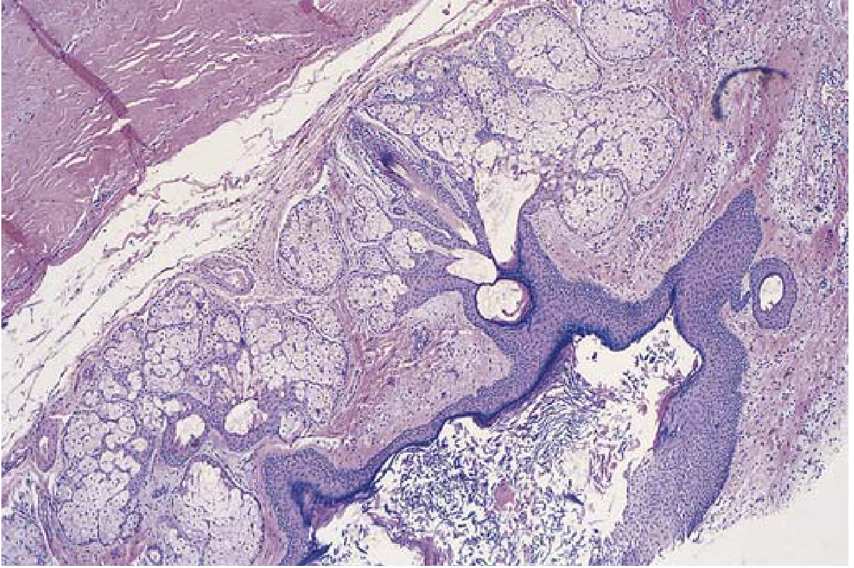
This is an example of a benign tumor arising from all three germ cell layers - note the well-organized, mature tissue elements with no atypia.
Clinical Significance
| Benign | Malignant | |
|---|---|---|
| Prognosis | Usually excellent after excision | Variable; depends on stage, grade |
| Recurrence | Rare | More likely |
| Effect on host | Usually local compression only | Local destruction + systemic effects (cachexia, paraneoplastic syndromes) |
| Treatment | Surgery (usually curative) | Surgery + radiation + chemo + targeted therapy |
Source: Robbins & Kumar Basic Pathology; Robbins, Cotran & Kumar Pathologic Basis of Disease
This is a shared conversation. Sign in to Orris to start your own chat.