give clear detials about dengue fever with all possible things for pediatric patients

Reading File
Finding Sources
Finding Sources
Finding Sources
Searching the Web

WHO dengue classification warning signs pediatric management guidelines 2024

Searching PubMed

"dengue" AND "children" AND management

Reading File
Reading File
Reading File
Reading File
Reading File
Reading File
Reading File
Reading File
Reading File
Reading File
Searching Images

dengue fever petechial rash tourniquet test child

Searching Images

dengue hemorrhagic fever rash skin

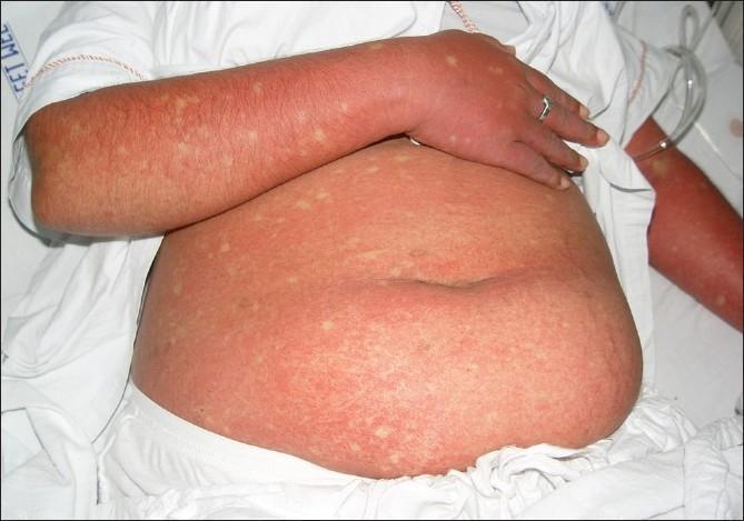
This clinical photograph displays a patient's abdomen and arm presenting a generalized, confluent erythematous rash, a characteristic cutaneous manifestation of Dengue Fever. The exanthem is marked by widespread, intense redness resulting from capillary dilatation and coalesced maculopapular lesions. A diagnostic hallmark visible here is the presence of 'islands of sparing'—small, rounded, or oval-shaped patches of normal-colored skin scattered throughout the erythematous background, often described as 'white islands in a sea of red'. These spared areas are randomly distributed across the affected trunk and limbs. The image serves as a key educational reference for recognizing the secondary rash phase of Dengue viral infection, typically occurring 3 to 6 days after the onset of fever. The visual presentation is essential for clinical diagnosis and differentiation from other viral exanthems, highlighting the specific immunologic response patterns seen in Dengue Fever (DF) and Dengue Hemorrhagic Fever (DHF).
This clinical photograph displays a patient's abdomen and arm presenting a generalized, confluent erythematous rash, a characteristic cutaneous manifestation of Dengue Fever. The exanthem is marked by widespread, intense redness resulting from capillary dilatation and coalesced maculopapular lesions. A diagnostic hallmark visible here is the presence of 'islands of sparing'—small, rounded, or oval-shaped patches of normal-colored skin scattered throughout the erythematous background, often described as 'white islands in a sea of red'. These spared areas are randomly distributed across the affected trunk and limbs. The image serves as a key educational reference for recognizing the secondary rash phase of Dengue viral infection, typically occurring 3 to 6 days after the onset of fever. The visual presentation is essential for clinical diagnosis and differentiation from other viral exanthems, highlighting the specific immunologic response patterns seen in Dengue Fever (DF) and Dengue Hemorrhagic Fever (DHF).

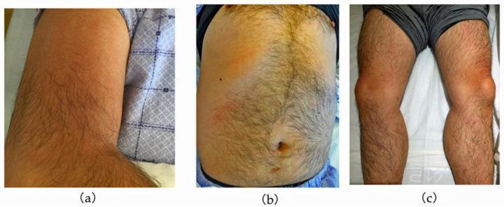
A series of three clinical photographs demonstrating cutaneous manifestations in a patient with dengue fever. (a) Right upper arm showing confluent, blanchable erythema with a diffuse distribution. (b) Anterior trunk (abdomen and chest) displaying a patchy, mottled erythematous rash interspersed with areas of normal skin tone, characteristic of the 'islands of white in a sea of red' pattern often seen in viral exanthems. (c) Lower limbs showing localized, more intense erythema focused around the knees and pretibial regions. The visual findings illustrate the secondary maculopapular rash typical of the recovery or late febrile phase of dengue virus infection. These images serve as an educational resource for identifying the clinical presentation of viral hemorrhagic fever rashes and their variable morphology across different anatomical regions, including the trunk, extremities, and periarticular areas.
A series of three clinical photographs demonstrating cutaneous manifestations in a patient with dengue fever. (a) Right upper arm showing confluent, blanchable erythema with a diffuse distribution. (b) Anterior trunk (abdomen and chest) displaying a patchy, mottled erythematous rash interspersed with areas of normal skin tone, characteristic of the 'islands of white in a sea of red' pattern often seen in viral exanthems. (c) Lower limbs showing localized, more intense erythema focused around the knees and pretibial regions. The visual findings illustrate the secondary maculopapular rash typical of the recovery or late febrile phase of dengue virus infection. These images serve as an educational resource for identifying the clinical presentation of viral hemorrhagic fever rashes and their variable morphology across different anatomical regions, including the trunk, extremities, and periarticular areas.

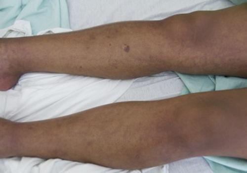
This clinical photograph displays the lower extremities of a patient exhibiting a diffuse, non-blanching purpuric rash. The lesions are characterized by a wide distribution of erythematous to violaceous macules and patches across both legs, including the shins and thighs. The morphology consists primarily of small petechiae and larger ecchymotic areas, with some regions showing confluent purpura. A notable, isolated round purpuric lesion is visible on the anterior surface of the upper lower leg. The skin texture appears generally intact without evidence of edema, ulceration, or necrosis, though some areas exhibit mild xerosis. Clinically, this presentation is consistent with a systemic hemorrhagic process or vasculopathy, often associated with infectious diseases such as Dengue fever (Dengue Hemorrhagic Fever), meningococcemia, or hematological conditions like thrombocytopenia. This image serves as an educational tool for identifying purpura in diverse skin tones and recognizing cutaneous manifestations of systemic viral or bacterial infections.
This clinical photograph displays the lower extremities of a patient exhibiting a diffuse, non-blanching purpuric rash. The lesions are characterized by a wide distribution of erythematous to violaceous macules and patches across both legs, including the shins and thighs. The morphology consists primarily of small petechiae and larger ecchymotic areas, with some regions showing confluent purpura. A notable, isolated round purpuric lesion is visible on the anterior surface of the upper lower leg. The skin texture appears generally intact without evidence of edema, ulceration, or necrosis, though some areas exhibit mild xerosis. Clinically, this presentation is consistent with a systemic hemorrhagic process or vasculopathy, often associated with infectious diseases such as Dengue fever (Dengue Hemorrhagic Fever), meningococcemia, or hematological conditions like thrombocytopenia. This image serves as an educational tool for identifying purpura in diverse skin tones and recognizing cutaneous manifestations of systemic viral or bacterial infections.

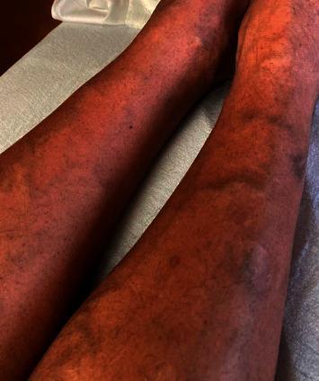
This clinical photograph shows the bilateral lower extremities of a 47-year-old male presenting with a diffuse dermatologic manifestation of thrombocytopenia. The skin demonstrates widespread, non-blanching petechiae, appearing as numerous pinpoint-sized (1-2 mm), reddish-purple macules. These lesions are densely scattered across the anterior and lateral aspects of the shins and calves. In addition to the petechial rash, several larger, coalescent purpuric lesions and subtle ecchymoses are visible, indicating small-vessel bleeding into the dermis. The background skin exhibits generalized erythema. This visual finding is characteristic of severe thrombocytopenia, in this clinical context associated with a viral hemorrhagic fever such as dengue. The image serves as an educational example of the cutaneous signs of low platelet counts and the systemic progression of infectious diseases like dengue fever in a hospital setting.

This clinical photograph shows the bilateral lower extremities of a 47-year-old male presenting with a diffuse dermatologic manifestation of thrombocytopenia. The skin demonstrates widespread, non-blanching petechiae, appearing as numerous pinpoint-sized (1-2 mm), reddish-purple macules. These lesions are densely scattered across the anterior and lateral aspects of the shins and calves. In addition to the petechial rash, several larger, coalescent purpuric lesions and subtle ecchymoses are visible, indicating small-vessel bleeding into the dermis. The background skin exhibits generalized erythema. This visual finding is characteristic of severe thrombocytopenia, in this clinical context associated with a viral hemorrhagic fever such as dengue. The image serves as an educational example of the cutaneous signs of low platelet counts and the systemic progression of infectious diseases like dengue fever in a hospital setting.

Dengue Fever in Pediatric Patients

1. Overview & Etiology

Dengue is caused by one of four related RNA flaviviruses (DENV-1 to DENV-4), transmitted mainly by the day-biting Aedes aegypti mosquito (less commonly A. albopictus). Infection with one serotype gives lifelong immunity to that serotype and 1-3 years of cross-protection against the others - after which a second, heterologous infection carries a much higher risk of severe disease, which is why severe dengue disproportionately affects children with secondary infections (Red Book 2021, p. 520-522; Jawetz, Melnick & Adelberg's Medical Microbiology).
  • Incubation: 3-14 days (commonly 4-7 days) after the mosquito bite
  • Transmission: mosquito bite is the main route; rare routes include vertical (maternal-fetal, ~20% rate, higher if maternal infection is near delivery), breastfeeding, blood/organ donation, and needlestick injury
  • Roughly 75% of infections are asymptomatic; about 25% are symptomatic

2. Clinical Course - Three Phases

Febrile phase (days 1-7): Abrupt high fever (39-40°C), often biphasic ("saddleback" curve), severe headache, retro-orbital pain, myalgia/arthralgia ("breakbone fever"), facial flushing, injected oropharynx, and a macular/maculopapular rash. Small children may present with only a mild upper respiratory illness, which can be mistaken for influenza, measles, or rubella (Tintinalli's Emergency Medicine, p. 1122-1123).
Critical phase (24-48 hours, around defervescence, day 3-7): This is when plasma leakage occurs - the vascular permeability rises in parallel with hemoconcentration (rising hematocrit). This is the window in which children can silently progress to shock. Warning signs typically appear here.
Recovery phase (48-72 hours after critical phase): Gradual reabsorption of extravascular fluid, return of appetite, hemodynamic stabilization, diuresis. A convalescent rash described as "isles of white in a sea of red" and generalized pruritus can occur. Bradycardia is common. Caution: giving excess IV fluid during this phase can precipitate pulmonary edema/heart failure because fluid is now returning to the vasculature (Park's Textbook of PSM, p. 291-292).
Dengue rash showing islands of white in a sea of red

3. WHO 2009 Classification (used for pediatric triage)

CategoryDefinition
Dengue without warning signsFever + 2 of: nausea/vomiting, rash, aches/pains, leukopenia, positive tourniquet test
Dengue with warning signsAbove, plus any of: abdominal pain/tenderness, persistent vomiting, clinical fluid accumulation (ascites/pleural effusion), mucosal bleeding, lethargy/restlessness, liver enlargement >2 cm, rising hematocrit with rapidly falling platelets
Severe dengueSevere plasma leakage causing shock or respiratory distress from fluid accumulation, and/or severe bleeding, and/or severe organ involvement (AST/ALT ≥1000 IU/L, impaired consciousness, cardiac/organ failure)
Dengue shock syndrome (DSS) - pediatric-specific criteria (Park's Textbook of PSM, p. 293): shock is defined in a child as pulse pressure ≤20 mmHg, OR signs of poor perfusion (cold extremities, delayed capillary refill, weak/rapid pulse), OR age-based hypotension: systolic BP <80 mmHg if <5 years old, or 80-90 mmHg in older children. A dangerous feature is that children in early/compensated shock often remain alert and lucid with a "normal" systolic pressure - an inexperienced clinician can miss the narrowing pulse pressure and underestimate severity until sudden decompensation occurs.
Less common severe manifestations in children include myocarditis, pancreatitis, hepatitis, hemophagocytic lymphohistiocytosis, meningoencephalitis, and post-dengue acute disseminated encephalomyelitis (ADEM).

4. Pediatric-Specific Risk Factors for Severe Disease

  • Infants of dengue-immune mothers (maternal antibody-enhanced infection)
  • Second/subsequent (heterologous serotype) infections
  • Chronic comorbidities: asthma, sickle cell disease, diabetes mellitus
  • Higher body mass index
  • Concomitant NSAID/steroid use

5. Diagnosis

  • Baseline CBC at first visit - establishes the child's own hematocrit baseline; falling platelets with rising hematocrit signals progression to the critical/leakage phase
  • Virologic (days 1-7 of fever): RT-PCR for viral RNA, or NS1 antigen immunoassay (rapid, minutes to 1 day)
  • Serologic (from day 3-5 onward): anti-dengue IgM (detectable by day 3-5, ~99% positive by day 10); IgG rise (4-fold between acute and convalescent sample) confirms recent infection
  • Combined NS1 + IgM testing in the first 10 days identifies ≥90% of cases
  • Note: IgM can cross-react with Zika, West Nile, and other flaviviruses, so serology needs cautious interpretation
  • Serial hematocrit and platelet monitoring is the single most important bedside tool for guiding admission/fluid decisions during the critical window (Park's Textbook of PSM, p. 293-296)

6. Management (WHO tiered approach, adapted for children)

No specific antiviral exists - treatment is entirely supportive. Avoid aspirin, NSAIDs (ibuprofen), and other salicylates because of bleeding risk and Reye syndrome association; use paracetamol (acetaminophen) only, dosed no more frequently than every 6 hours, plus tepid sponging for fever.
Group A - Dengue without warning signs (outpatient/home care):
  • Encourage oral rehydration solution, fruit juice, and electrolyte fluids (caution with sugary fluids in diabetic children)
  • Paracetamol for fever/discomfort
  • Daily follow-up: temperature, oral intake/output, urine output (should void at least every 6 hours), watch for warning signs
  • Educate caregivers to return immediately for: no improvement, worsening around defervescence, severe abdominal pain, persistent vomiting, cold/clammy extremities, lethargy or irritability, any bleeding (black stools, coffee-ground emesis), or no urination for 4-6 hours
Group B - Dengue with warning signs (hospital admission required):
  • IV fluids started early (isotonic crystalloids) - early rehydration at this stage often prevents progression to shock
  • Serial hematocrit (daily from day 3 until afebrile for 1-2 days), platelet counts, and close vital sign monitoring
  • Fluid administered cautiously and titrated to clinical response, avoiding both under- and over-resuscitation
Group C - Severe dengue / dengue shock syndrome (intensive/critical care):
  • Aggressive but carefully titrated IV crystalloid resuscitation as first-line
  • If shock is refractory after an initial crystalloid trial, IV colloids and/or blood/blood products are used
  • Continuous monitoring for: early shock signs, occult bleeding, plasma leak (pleural effusion, ascites), and, critically, fluid overload as capillary permeability normalizes in the recovery phase
  • Early recognition and aggressive supportive therapy reduces mortality from severe dengue from roughly 5-10% down to under 1% (Red Book 2021, p. 523)

7. Complications to Watch For in Children

  • Hypovolemic shock, multi-organ failure
  • Pleural effusion, ascites, respiratory distress
  • Hemorrhage (mucosal bleeding, GI bleeding, petechiae/purpura - can mimic meningococcemia)
  • Hepatitis, myocarditis, pancreatitis
  • Neurologic: encephalopathy, encephalitis, ADEM
  • Fluid overload/pulmonary edema (iatrogenic, during recovery phase)
  • Overlap/mimicry with Kawasaki disease has been reported in children with near-identical presentations, complicating diagnosis (Andrews' Diseases of the Skin, p. 2461-2474)

8. Prevention

  • Mosquito bite avoidance: EPA-registered repellents, protective clothing, screened/air-conditioned housing, bed nets for daytime sleep (Aedes bites mainly during the day)
  • Vector control programs
  • Dengvaxia (CYD-TDV): a live-attenuated tetravalent vaccine, 3-dose schedule at 0/6/12 months, FDA-approved only for children aged 9-16 years with laboratory-confirmed prior dengue infection who live in an endemic area. It is NOT approved for seronegative children - vaccinating a dengue-naive child raises their risk of severe dengue upon subsequent natural infection (Red Book 2021, p. 524).

Supplementary Evidence Check (PubMed)

A 2024 systematic review/meta-analysis on risk factors for severe dengue in Latin America (PMID: 38263345) and a meta-analysis of routine blood parameters in dengue-infected children vs. adults (PMID: 36593636) are consistent with the textbook guidance above (secondary infection, comorbidities, and hematologic trends as key severity predictors) - no contradictory findings were found. A separate 2024 study specifically validated the WHO 2009 warning-signs framework's utility for detecting severe dengue in children, supporting its continued clinical use.
Bottom line for pediatric care: classify every febrile child using the WHO framework, watch hematocrit/platelet trends (not just absolute fever), remember that children in early shock can look deceptively well, avoid NSAIDs/aspirin, and match the fluid strategy to the disease phase - aggressive early rehydration for warning signs, careful titrated resuscitation for shock, and vigilance for fluid overload once the recovery phase begins.

why plasma Leakage.

Finding Sources
Finding Sources
Finding Sources
Searching PubMed

dengue plasma leakage endothelial glycocalyx mechanism

Reading File
Reading File

Why Plasma Leakage Happens in Dengue

Plasma leakage is the central event that turns "just a fever" into dengue hemorrhagic fever/dengue shock syndrome (DHF/DSS). It is an immune-mediated phenomenon, not a direct destructive effect of the virus itself. Here is the chain of events:

1. It starts with a second, different-serotype infection (or maternal antibody in infants)

There are four dengue serotypes (DENV-1 to DENV-4). A first infection produces antibodies that neutralize that serotype well, but only weakly and non-protectively cross-react with the other three. When a child is later infected with a different serotype - or when an infant carries maternal anti-dengue antibody from a previous maternal infection - these old antibodies bind the new virus but fail to neutralize it (Robbins, Cotran & Kumar Pathologic Basis of Disease, p. 336).

2. Antibody-Dependent Enhancement (ADE)

Instead of protecting, these non-neutralizing "enhancing" antibodies actually help the virus. The antibody-virus immune complexes bind Fc receptors on monocytes/macrophages, which increases viral uptake into these cells and boosts viral replication - effectively handing the virus a bigger foothold in the body. This is why severe dengue occurs almost exclusively on secondary heterotypic infection (or in antibody-carrying infants), and is more frequent with DENV-2 (Robbins Pathology, p. 336; Sherris & Ryan's Medical Microbiology, p. 287).

3. Cytokine storm

The infected macrophages, along with dengue-specific CD4+ and CD8+ T lymphocytes that are activated against the virus, secrete large amounts of pro-inflammatory cytokines - interferon-γ, TNF-α, IL-6, IL-8, and others. This surge is often described as a "cytokine storm." These cytokines act directly on the vascular endothelium, increasing its permeability (Sherris & Ryan's Medical Microbiology, p. 287).

4. Immune complex and complement activation

The circulating antigen-antibody complexes also activate the complement cascade, adding a further layer of vascular injury and immunopathology on top of the cytokine effect (Sherris & Ryan's Medical Microbiology, p. 287).

5. Endothelial dysfunction

The net result of the cytokine storm plus complement activation plus direct effects of peak viremia is transient endothelial dysfunction - the tight junctions between endothelial cells loosen and the vessel wall becomes "leaky." This is typically evident 3-6 days after fever onset, which is exactly the window in which defervescence and the critical phase occur (Fitzpatrick's Dermatology, p. 826). Some of the vascular injury also directly damages capillary endothelium in organs like the kidney, contributing to organ-specific complications such as dengue-associated AKI (Comprehensive Clinical Nephrology, p. 1867).

6. Why "plasma" leaks and not whole blood

The leaky endothelial junctions are large enough to let plasma water and smaller proteins escape into extravascular spaces (pleural cavity, peritoneal cavity, interstitial tissue) but generally too small to let red blood cells through in bulk. Blood cells are left behind in a shrinking plasma volume, which is why the hematocrit rises (hemoconcentration) even as the child is objectively losing intravascular volume. This is the paradox that makes dengue dangerous: a rising hematocrit with a falling platelet count is a laboratory signature of ongoing plasma leak, often before clinical shock is obvious - which is exactly why serial CBCs are the cornerstone of monitoring.

7. Why it is reversible

Because this is a functional cytokine/complement-mediated increase in permeability rather than structural vessel destruction, it is self-limited - lasting only 24-48 hours (the critical phase). Once the cytokine surge subsides, the endothelium reseals, and the leaked plasma is reabsorbed back into the vasculature during the recovery phase (which is why over-aggressive IV fluids given during recovery can cause dangerous fluid overload/pulmonary edema - the vessels are no longer leaky, so extra fluid has nowhere to go but into the circulation and lungs).

In short

Secondary/heterotypic infection → non-neutralizing antibodies enhance viral uptake into macrophages (ADE) → massive cytokine release from infected macrophages and activated T cells + complement activation from immune complexes → transient endothelial permeability increase → plasma (not cells) leaks into pleural/peritoneal/interstitial spaces → hemoconcentration, effusions, ascites, and, if uncorrected, hypovolemic shock (dengue shock syndrome).
This is precisely why the WHO warning signs (rising hematocrit, falling platelets, ascites, pleural effusion, abdominal pain) are used to catch this process early - because it is short-lived and reversible with timely fluid support, but rapidly fatal if it progresses unrecognized to decompensated shock.

How the. plasma Leakage. occurs.

Finding Sources
Finding Sources
Searching the Web

dengue NS1 protein endothelial glycocalyx disruption mechanism plasma leakage

Searching PubMed

dengue NS1 endothelial glycocalyx vascular leakage

How Plasma Leakage Actually Happens (Mechanistic Detail)

My previous answer explained why the immune system creates the problem (ADE, cytokine storm). Here is how that translates into fluid physically leaving the blood vessels.

1. The vessel wall has two barriers, and both get breached

Normally, capillaries retain plasma water and proteins inside the vessel because of two structures:
  • The endothelial glycocalyx layer (EGL) - a thin, gel-like coat of proteoglycans (heparan sulfate, syndecan-1) and glycoproteins lining the inner surface of every blood vessel, acting like a molecular sieve
  • Endothelial cell-cell junctions - tight/adherens junctions (proteins like VE-cadherin, claudins) that seal endothelial cells together side by side
Plasma leakage in dengue happens because both of these barriers are chemically disrupted, not because vessels are torn or endothelial cells die en masse.

2. The viral protein NS1 is the direct trigger

During peak viremia, the dengue virus secretes large amounts of a soluble protein called NS1 into the bloodstream (this is the same NS1 used for rapid diagnostic testing). Circulating NS1:
  • Binds directly to endothelial cells and activates enzymes (heparanase, sialidase, cathepsin L) that strip away the heparan sulfate and sialic acid components of the glycocalyx - essentially shaving off the protective coat
  • Cooperates with matrix metalloproteinase-9 (MMP-9) to disassemble the endothelial tight junctions and disrupt cell-cell adhesion
The combined effect of "stripped glycocalyx + loosened junctions" opens up paracellular gaps between endothelial cells wide enough for plasma water and small proteins to escape, while red blood cells largely stay behind (hence hemoconcentration rather than gross bleeding into tissue).

3. The cytokine storm amplifies it

Layered on top of the direct NS1 effect, the cytokines described earlier (TNF-α, IFN-γ, IL-6, IL-8) further activate the endothelium, pushing the same permeability pathways (junction protein internalization, cytoskeletal contraction of endothelial cells) even further open. This is why the leak is worst exactly when the immune/cytokine response peaks - around defervescence - rather than during the initial viral invasion.

4. What this looks like at the tissue level

Histologically, affected small vessels (particularly in the dermis and serosal surfaces) show endothelial cell swelling and perivascular edema with mononuclear cell infiltration - a functional, inflammatory change rather than destructive necrosis (Sherris & Ryan's Medical Microbiology, p. 718-731). This is consistent with why the process is reversible: the endothelium is "stunned and leaky," not destroyed, so it reseals once the trigger subsides.

5. Where the fluid goes

Because the leak occurs preferentially across capillaries in serosal-lined cavities, plasma accumulates in the:
  • Pleural space (pleural effusion)
  • Peritoneal cavity (ascites)
  • Gallbladder wall (edema, seen on ultrasound)
  • Interstitial soft tissue
This is why Red Book 2021 explicitly labels the shock mechanism in dengue a "capillary leak syndrome" (p. 1555-1556) - the shock is driven by plasma volume loss out of the vasculature, not by blood loss.

6. Why fluid therapy works

Since the vessel wall integrity issue is temporary and reversible, replacing the lost intravascular volume with IV crystalloids (e.g., lactated Ringer's) during the 24-48 hour critical window is usually enough to support the circulation until the leak seals itself in the recovery phase (Brenner and Rector's The Kidney, p. 803-812). Overshooting fluid replacement after the leak has resolved is what causes iatrogenic pulmonary edema, since the vessels can no longer "absorb" excess administered fluid the way they could during the leaky phase.

Summary chain

Secondary infection → ADE increases viral load in macrophages → NS1 secreted into blood + cytokine storm → NS1/MMP-9 strip the endothelial glycocalyx and disrupt tight junctions → cytokines further loosen endothelial junctions → paracellular gaps open → plasma water/protein leaks into pleural/peritoneal/interstitial spaces → hemoconcentration, effusions, and (if severe) hypovolemic shock → self-limited over 24-48 hours as the trigger resolves and the endothelium reseals.
Note on sources: The glycocalyx/NS1/MMP-9 mechanistic detail above comes from recent primary molecular research (not yet consolidated into a systematic review) - key papers include Pan et al., PLoS Pathogens 2021 (PMID: 34310658, NS1-MMP9 cooperation disrupting tight junctions) and Buijsers et al., Frontiers in Immunology 2021 (PMID: 34987504, heparanase-mediated glycocalyx degradation correlating with plasma leakage in dengue patients). These supplement, and are consistent with, the general immunopathology (ADE, cytokine storm, capillary leak) described in the standard textbooks cited above.
This is a shared conversation. Sign in to Orris to start your own chat.