
How to differentiate with real mri image in acute vs traumatic spine injury on basis of mri
Reading File
Finding Sources
Searching Images
MRI acute traumatic spinal injury STIR ligament cord edema compression fracture
Searching PubMed
spinal trauma AND MRI
Searching the Web
ACR Appropriateness Criteria Acute Spinal Trauma MRI ligament spinal cord injury
Finding Sources
query_images_db
Searching the Web
site:radsource.us spine trauma MRI STIR spinal cord edema epidural hematoma images
Loading Image
“Acute” and “traumatic” are not opposite diagnoses.
- Traumatic spinal injury means it resulted from trauma, such as a fall, road-traffic collision, sports injury, or violence.
- Acute spinal injury means it is recent, generally hours to days old.
So an injury can be an acute traumatic spine injury.
On MRI, the usual practical distinction is acute traumatic injury versus old/chronic injury or degenerative disease.
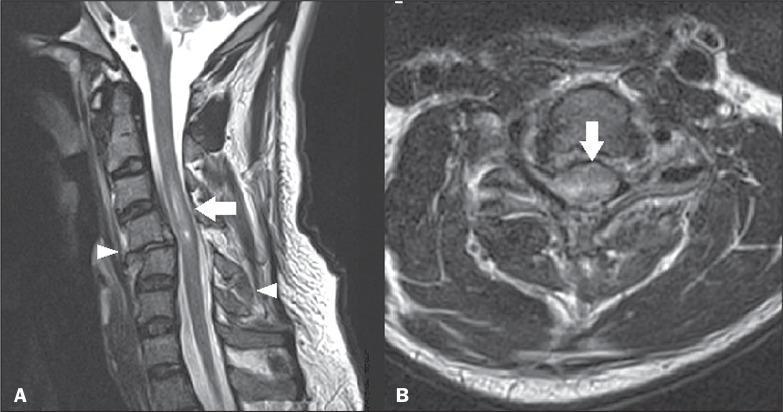
MRI differentiation
| MRI feature | Acute traumatic injury | Chronic/old traumatic injury or degeneration |
|---|---|---|
| Bone marrow | Marrow edema: low signal on T1, high signal on T2/STIR, often adjacent to an endplate, vertebral body, pedicle, or posterior element | No edema in healed fracture. Marrow may show fatty replacement, sclerosis, or stable deformity |
| Fracture | Fracture line may be low signal on all sequences, with surrounding STIR edema; acute compression/burst fracture may have retropulsion | Healed wedge deformity with no STIR edema; corticated/sclerotic margins, stable height loss |
| Spinal cord | Cord swelling and T2/STIR hyperintensity from edema/contusion; intramedullary low signal on T2 or GRE/SWI suggests hemorrhage | Cord atrophy and focal T2 hyperintensity from myelomalacia/gliosis, without acute swelling |
| Ligaments | Discontinuity or high STIR/T2 signal in anterior longitudinal ligament, posterior longitudinal ligament, ligamentum flavum, interspinous/supraspinous ligaments | Thickening, scarring, or no active high-signal edema |
| Paraspinal soft tissue | Prevertebral edema, posterior paraspinal muscle edema, soft-tissue hematoma | Usually absent, or chronic fatty muscle atrophy |
| Disc | Acute traumatic disc disruption, annular tear, disc-space widening, traumatic herniation, or edema | Disc desiccation, height loss, osteophytes, broad chronic bulge |
| Epidural space | Acute epidural hematoma: extradural collection causing thecal-sac/cord compression. Signal changes with age of blood | Resolved hematoma or chronic organizing collection; chronic stenosis is more often due to disc-osteophyte/facet disease |
| Alignment | New translation, perched/locked facets, widened interspinous distance, abnormal facet spacing, kyphosis | Fixed chronic malalignment with degenerative remodeling, but no edema |
Sequence-based approach
-
Sagittal T1
- Look for fracture anatomy, marrow replacement, epidural fat, and subacute blood.
- Acute marrow injury is typically T1 dark.
-
Sagittal T2 and STIR/fat-suppressed T2
- Most useful for acuity.
- Look for bright edema in bone marrow, cord, ligaments, disc, and paraspinal tissues.
- A fracture/deformity without STIR edema is usually old, although timing can vary.
-
Axial T2
- Defines canal compromise, cord compression, root injury, epidural hematoma, and facet injury.
-
GRE/SWI
- Detects susceptibility from hemorrhage in the spinal cord or epidural space.
-
Post-contrast T1 fat-suppressed images
- Not routinely needed for straightforward acute trauma.
- Useful if infection, tumor, inflammatory disease, or uncertain epidural collection is a consideration.
Key acute traumatic MRI patterns
- Cord edema/contusion: T2/STIR bright intramedullary signal with cord expansion.
- Cord hemorrhage: T2/GRE/SWI low-signal focus within the cord, often with surrounding edema. This is generally a more severe injury pattern.
- Posterior ligamentous complex injury: high STIR signal or disruption in interspinous/supraspinous ligaments, ligamentum flavum, facet capsules, and posterior paraspinal muscles. This can indicate instability even when CT has no major fracture.
- Acute compression or burst fracture: vertebral marrow edema, endplate disruption, posterior-wall retropulsion, and possible canal narrowing.
- Fracture-dislocation: translation/rotation with facet disruption and associated disc, ligament, cord, and prevertebral injury.
- Epidural hematoma: extra-axial epidural collection compressing the thecal sac or cord. Signal depends on blood age, so assess morphology and clinical timing rather than one sequence alone.
Important pitfalls
- Degenerative Modic type 1 endplate change can mimic acute fracture edema. It is centered at a degenerative disc/endplate, often with disc height loss and osteophytes, rather than a trauma-related fracture line or posterior soft-tissue injury.
- Cord T2 hyperintensity is not always acute trauma. Chronic cervical spondylotic myelopathy, demyelination, ischemia, infection, and tumor can also cause it. Acute trauma is supported by swelling, hemorrhage, new compression, ligament injury, and matching clinical mechanism.
- MRI does not replace CT for bone detail. CT is better for subtle cortical fractures and posterior-element fractures; MRI adds cord, disc, ligament, hematoma, and marrow assessment. This aligns with the ACR acute spine-trauma guidance, which uses CT first for many high-risk blunt-trauma patients and MRI when cord, nerve-root, or ligament injury is suspected.
High-yield reporting phrase
“Acute traumatic injury is favored when there is marrow edema on STIR, associated paraspinal/prevertebral edema, ligamentous disruption, acute disc injury, epidural hematoma, or cord edema/hemorrhage. Absence of edema with fixed deformity, fatty marrow change, cortication, or cord atrophy favors chronic injury.”
MRI is particularly sensitive for ligament injury, disc disruption, cord compression, and hematoma, although CT remains superior for detailed osseous assessment, as described in Rockwood and Green’s Fractures in Adults, 10th ed., MRI section, lines 2955-2959. A 2026 systematic review also evaluated MRI’s role in acute spinal-cord injury (PMID 42406832).
This is a shared conversation. Sign in to Orris to start your own chat.