
Cyst like in bladder wall in usg
cyst like lesion bladder wall ultrasound
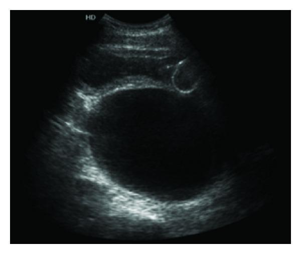
An abdominal ultrasound image in the transverse plane demonstrating a large, predominantly anechoic (dark), well-circumscribed cystic lesion located in the retrovesical space. The cyst exhibits a thin, regular wall without visible internal septations, solid components, or debris, suggesting a simple fluid-filled nature. Anteriorly, the urinary bladder is visible and appears compressed by the extrinsic mass effect of the cyst. The lesion is documented to have an approximate volume of 780 cc. The surrounding pelvic tissues show heterogeneous echogenicity, and the presence of acoustic enhancement posterior to the cyst is characteristic of a fluid-filled structure. This clinical finding is associated with bilateral obstructive uropathy and bladder outlet obstruction symptoms. The image is an essential diagnostic example for identifying retroperitoneal masses and pelvic cystic lesions, such as a periureteral diverticulum, within a urological context.
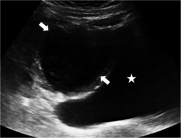
This diagnostic ultrasound image presents a sagittal view of the pelvic region in a pediatric patient. The primary finding is a round-shaped cystic lesion, demarcated by white arrows, situated immediately superior to the bladder. The bladder is identified by a white star and appears as a standard anechoic, fluid-filled structure with well-defined margins. In contrast, the superiorly located cystic lesion exhibits mixed internal echogenicity, characterized by low-level internal echoes and a mildly thickened wall, which differentiates it from a simple, clear cyst. This appearance suggests the presence of debris or proteinaceous material within the fluid. The anatomical position along the midline between the bladder and the umbilicus, combined with the signs of inflammation or infection (mixed echogenicity), is highly characteristic of an infected urachal cyst. The surrounding pelvic soft tissues show a heterogeneous echotexture within normal limits. This visual evidence supports the clinical evaluation of suprapubic pain in a pediatric context and illustrates the diagnostic features of urachal remnants.
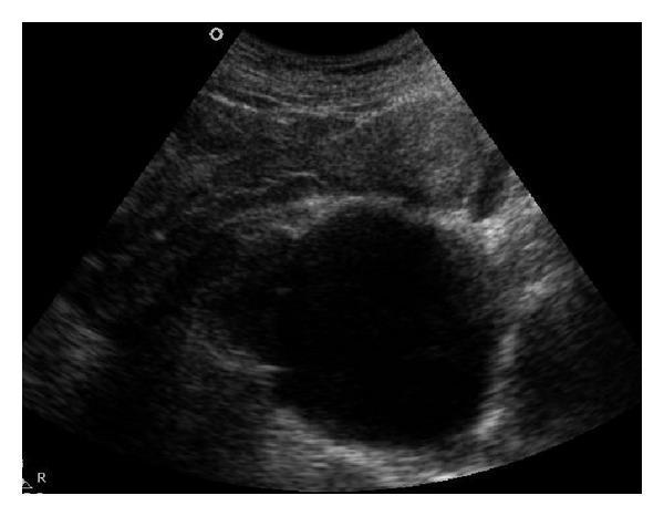
A sagittal pelvic ultrasound image demonstrating a large, well-defined cystic lesion in the midline of the pelvic region. The lesion is characterized by a smooth, thin wall and anechoic internal echogenicity, consistent with a fluid-filled structure. Measuring approximately 7.5 x 8.5 cm, the cyst is located posterior to the uterus and is exerting significant mass effect, resulting in the anterior compression and displacement of the uterus, rectum, and bladder. The visual findings are characteristic of an anterior sacral meningocele, a clinical entity associated with sacral agenesis where the spinal subarachnoid space herniates through a defect in the anterior sacrum. The image highlights key diagnostic markers including morphology, lesion relationship to pelvic viscera, and the lack of internal solid components or septations, which is critical for differentiating congenital pelvic cysts from adnexal or ovarian pathologies.
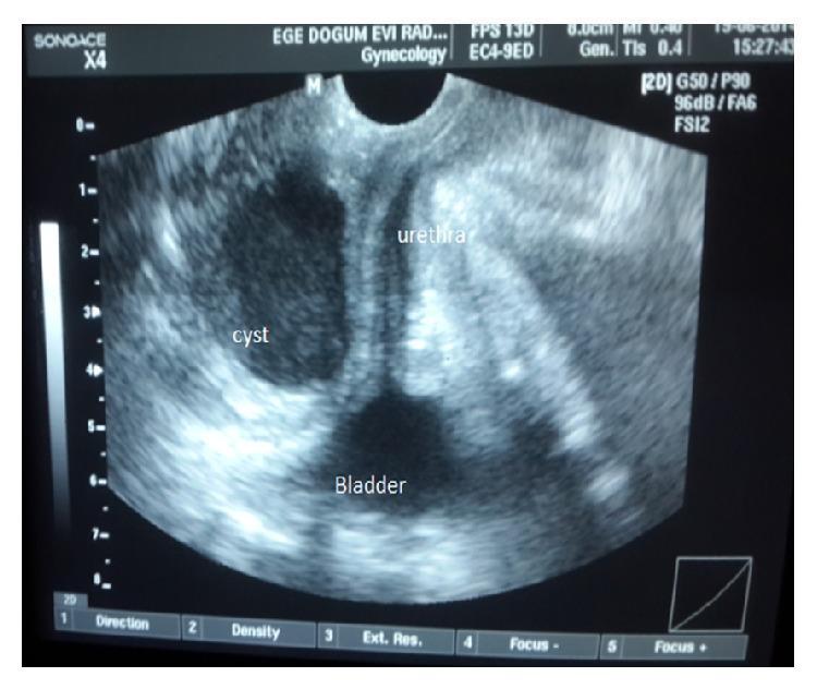
This diagnostic image is a gray-scale perineal ultrasonography of the female lower urinary tract. The ultrasound demonstrates the anatomical relationship between the bladder, urethra, and a vaginal wall cyst. The bladder is visible as a hypoechoic structure at the bottom of the field (inferior). Emerging from the bladder neck, the urethra is shown in a longitudinal orientation, appearing centrally. A well-defined, hypoechoic, oval-shaped cyst is positioned lateral to the urethra and superior to the bladder. The cyst displays distinct borders and lack of internal echoes, suggesting a fluid-filled nature, such as a Müllerian cyst. Notably, the image highlights the lack of communication or continuity between the cystic lesion and the urinary tract structures (bladder and urethra), which is a key diagnostic feature in differentiating vaginal cysts from urethral diverticula or cystoceles. This clinical imaging is relevant for gynecology and urology education regarding pelvic floor masses and differential diagnosis of anterior vaginal wall lesions.
Cyst-Like Lesion in Bladder Wall on USG
Differential Diagnosis
1. Urachal Cyst (Most Common Cause at Bladder Dome)
- Midline cystic structure between the anterior abdominal wall and peritoneum, at or near the bladder dome
- Well-defined, anechoic/hypoechoic, thin-walled
- If infected: mixed internal echoes, debris, thickened wall
- More common in adults than children
- Infected urachal cysts: lower abdominal pain, voiding symptoms, palpable mass
- Most common infecting organism: Staphylococcus aureus
- Can perforate into bladder or peritoneal cavity if untreated
- Diagnosis confirmed by USG; CT/MRI for complex cases
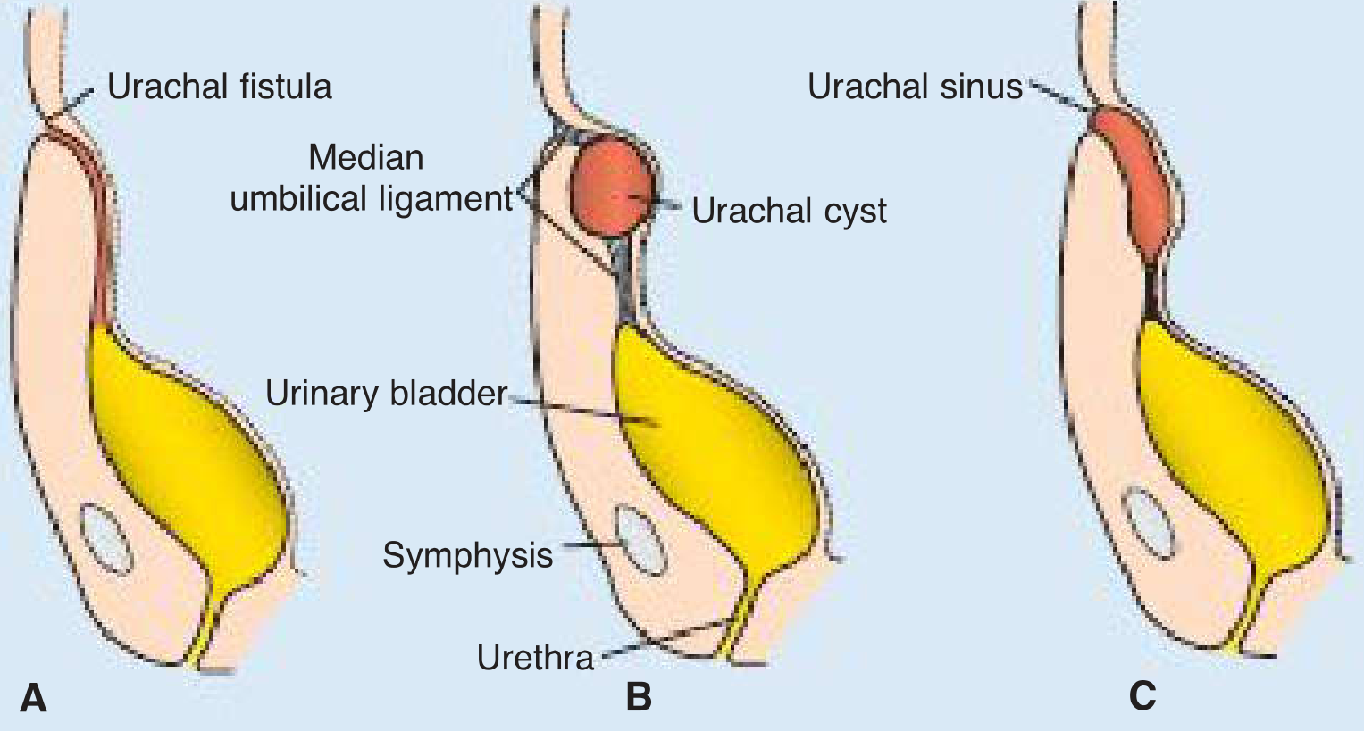
2. Urachal Diverticulum (Vesico-Urachal Diverticulum)
- Urachus obliterates except at the bladder apex, forming a diverticulum
- Appears as a cyst-like outpouching at the bladder dome on USG
- Usually asymptomatic, found incidentally
- May communicate with bladder lumen (fills during cystography)
3. Bladder Diverticulum
- True/false outpouching of bladder mucosa through the bladder wall
- USG: cystic lesion contiguous with bladder, may appear separate when collapsed
- Associated with bladder outlet obstruction (PUV in children) or connective tissue disorders (multiple diverticula)
- Key USG sign: increases in size during voiding, fills from bladder
- Risk of stasis, infection, stone formation, and rarely carcinoma within the diverticulum
4. Bladder Endometriosis
- Cyst-like hypoechoic nodule in the posterior bladder wall or dome
- More common in women of reproductive age
- May show cyclic changes in size
- TVUS can detect bladder detrusor nodules - appearance is a hypoechoic irregular lesion, not a simple cyst
- Associated with symptoms: cyclical hematuria, urgency
5. Cystic Tumors of the Bladder
- Rare; includes cystic transitional cell carcinoma, cystitis cystica, or cystic glandular lesions
- Cystitis cystica/glandularis: multiple small submucosal cyst-like nodules, often at the trigone
- Malignant cystic lesions: irregular walls, internal septations, vascularity on Doppler
- Require cystoscopy and biopsy for definitive diagnosis
6. Müllerian/Vaginal Wall Cyst (in Women)
- Cyst adjacent to the bladder neck or urethra, separate from the bladder lumen
- No communication with the urinary tract
- Appears as a smooth, well-defined anechoic lesion on perineal or transvaginal USG
7. Periureteral/Paraureteral Diverticulum
- Located adjacent to the ureteric orifice ("Hutch diverticulum")
- Appears as a cyst-like structure at the lateral bladder wall near the ureterovesical junction
- Can cause vesicoureteric reflux
USG Approach: Key Differentiating Features
| Feature | Urachal Cyst | Bladder Diverticulum | Endometriosis | Cystic Tumor |
|---|---|---|---|---|
| Location | Midline, dome to umbilicus | Any wall, common lateral | Posterior wall/dome | Any, often trigone |
| Wall | Thin, smooth | Thin (or absent) | Thickened, irregular | Irregular, nodular |
| Internal echoes | Anechoic (simple) / debris if infected | Anechoic, may have debris | Heterogeneous | Septations, solid areas |
| Bladder communication | None (usually) | Yes | No | No |
| Doppler | No flow | No flow | Variable | Vascularity in solid parts |
| Change with voiding | No | Enlarges during voiding | No | No |
Next Steps When a Bladder Wall Cyst is Found
- Note exact location - dome (urachal), lateral wall (diverticulum, paraureteral), trigone (cystitis cystica)
- Assess wall thickness and internal complexity - simple vs. complex
- Doppler to check for vascularity (suggests neoplasm)
- Fill status - compare full and post-void scans (diverticulum changes size)
- CT Urogram or MRI pelvis for complex lesions or if malignancy is suspected
- Cystoscopy + biopsy for intraluminal or submucosal lesions
Urachal diverticulum
urachal diverticulum bladder dome ultrasound imaging
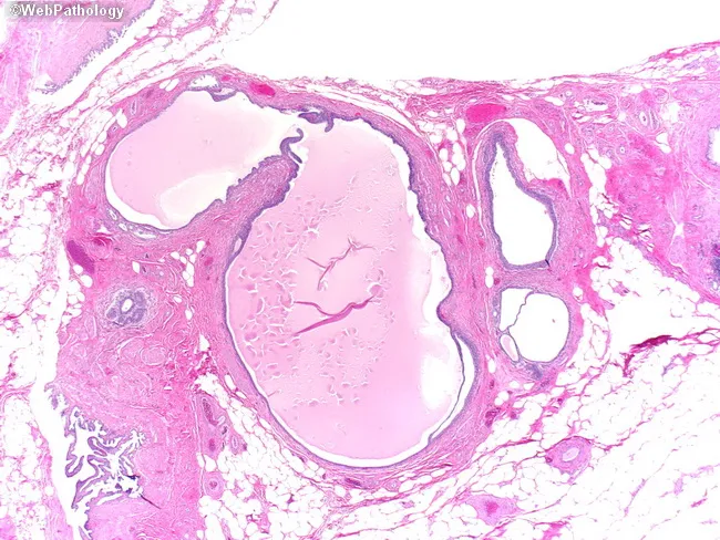
Imaging modality: Histopathology (light microscopy) of a bladder dome/anterior wall biopsy with urachal remnant. In this section, a cystically dilated luminal space is seen embedded in a thin fibromuscular stroma. The cyst is lined by epithelium that may resemble urothelium, be flattened and atrophic, or display glandular (enteric-type) differentiation. The mucosa is variably thinned with focal cuboidal-to-columnar lining, and the surrounding wall consists of a delicate fibromuscular capsule without invasion by neoplastic tissue. The midline, extravesical location supports urachal origin, and there is no overt inflammatory destructive process evident. Normal bladder mucosa above and around the lesion is unremarkable. The architecture, epithelium, and stromal configuration are characteristic of a urachal remnant or urachal cyst rather than a malignant neoplasm. Clinically, this finding is important to distinguish benign urachal remnants from urachal carcinoma and other cystic bladder lesions, since management and prognosis differ markedly. Differential diagnoses include bladder diverticulum, cystitis cystica/glandularis, and enteric metaplasia. Pathologic recognition informs clinical correlation, imaging review, and surveillance strategies. This entity, commonly asymptomatic or presenting with recurrent infections, carries a favorable prognosis when confined to the urachal tract and without invasive features on histology. Correlative imaging and clinical data reinforce benign interpretation and help guide follow-up.
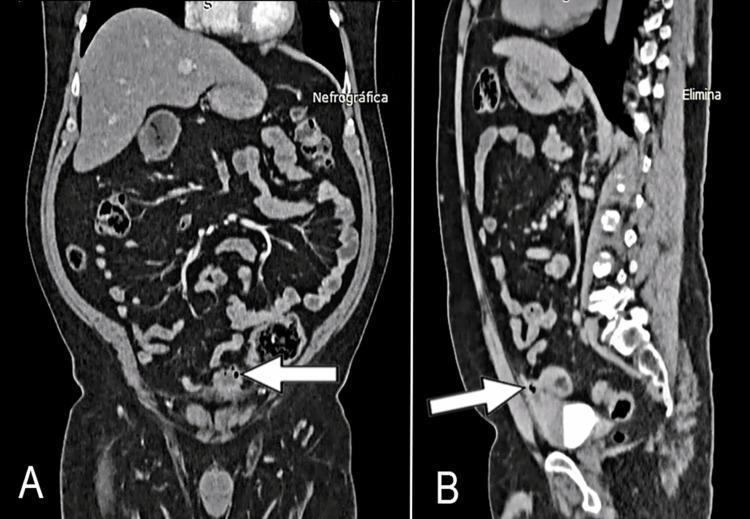
This diagnostic image displays a contrast-enhanced abdominal CT scan in coronal (A) and sagittal (B) views, illustrating a urachal remnant pathology. Image A (coronal) reveals a heterogeneous mass located at the anterosuperior aspect of the bladder dome (indicated by a white arrow). The mass displays irregular morphology and internal foci of gas (hypodense pockets). Image B (sagittal) demonstrates the anatomic continuity of this mass with a patent urachus, appearing as a tubular structure extending superiorly towards the umbilicus within the space of Retzius. Key clinical features include the well-defined margins of the lesion and its relationship with the embryological remnant. These findings are highly characteristic of a urachal cyst or urachal diverticulum, potentially complicated by infection (urachitis) or a urachal neoplasm. The imaging is critical for surgical planning of a partial cystectomy and urachal resection.
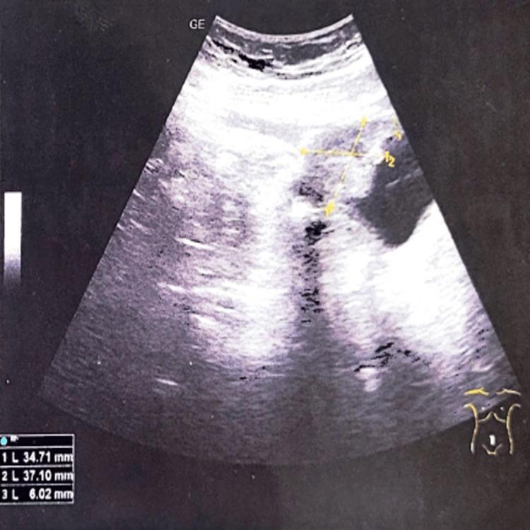
This diagnostic ultrasound image in sagittal view demonstrates the pelvic region, specifically focusing on the urinary bladder. A significant, solid, heterogeneous mass is visible originating from the bladder dome (anterosuperior wall). The lesion displays mixed echogenicity with irregular borders compared to the adjacent bladder wall. Electronic calipers measure the tumor dimensions as approximately 34.71 mm by 37.10 mm. The bladder lumen appears relatively clear of internal debris. This imaging finding is characteristic of a bladder dome tumor, which in this clinical context is associated with a urachal remnant or urachal adenocarcinoma. The image serves as an educational example of identifying bladder wall thickening and localized masses using transabdominal sonography, emphasizing the importance of evaluating the dome for urachal pathology.
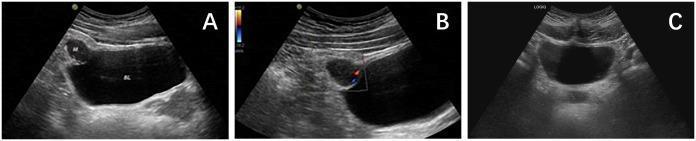
This set of diagnostic images consists of three transabdominal ultrasound panels (A, B, and C) focused on the urinary bladder. Panel A is a grayscale B-mode image showing a well-demarcated, hypoechoic mass measuring approximately 2.6 x 1.8 cm located at the anterior superior aspect (bladder dome), consistent with an urachal lesion. The mass displays an irregular shape and heterogeneous internal echogenicity. Panel B utilizes Color Doppler Flow Imaging (CDFI), demonstrating sparse, dot-linear blood flow signals primarily localized at the peripheral margins of the hypoechoic mass. Panel C represents a follow-up grayscale ultrasound showing a normal bladder configuration with no evidence of the previously seen mass, indicating successful surgical resection or resolution. These images are clinically significant for evaluating urachal pathologies, such as urachal PEComas or abscesses, and documenting treatment outcomes in urological oncology.
Urachal Diverticulum
What Is It?
All Four Urachal Anomalies (for context)
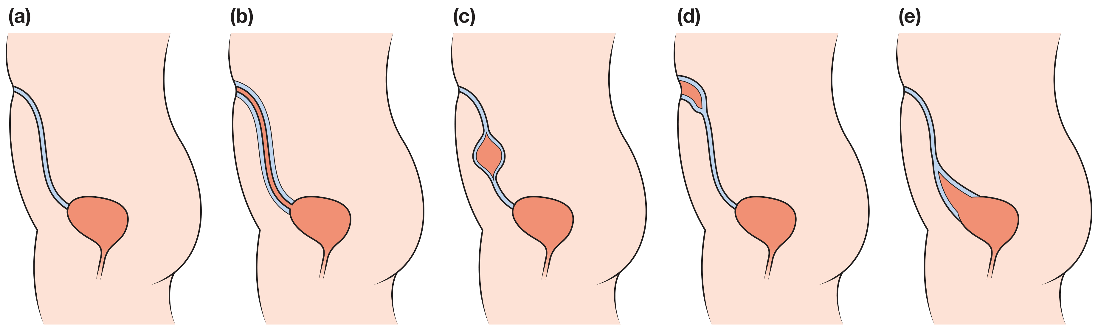
| Anomaly | Patent segment | Communication |
|---|---|---|
| (a) Normal | Fully obliterated | None |
| (b) Patent urachus | Entire urachus | Bladder ↔ Umbilicus (urinary fistula) |
| (c) Urachal cyst | Mid-segment only | None (isolated cyst) |
| (d) Urachal sinus | Umbilical end | Open at umbilicus only |
| (e) Urachal diverticulum | Vesical end only | Open into bladder dome |
Key Anatomical Features
- Located at the bladder dome / apex, in the midline
- Forms a blind-ending sac that opens freely into the bladder lumen
- No connection to the umbilicus (unlike patent urachus)
- Size varies - can be small (a few mm) or large enough to cause urinary stasis
- Because the neck is typically wide and drains freely into the bladder, it rarely causes obstruction
Clinical Features
- Recurrent UTIs - from urinary stasis within the sac
- Stone formation - calcium deposits in the diverticulum (from stagnant urine)
- Lower abdominal/suprapubic discomfort
- Haematuria - from infection or stone
Imaging
Ultrasound
- Cyst-like anechoic structure at the bladder dome, midline
- Communicates with the bladder (may be harder to confirm on USG alone)
- Look for it on a full bladder scan - fills with urine from the bladder
- Post-void: may partially empty (if neck is wide) or retain fluid (if narrow-necked with stasis)
VCUG (Voiding Cystourethrogram)
- Best for demonstrating communication with bladder
- Contrast fills the diverticulum from the bladder dome
CT Urogram / MRI
- Shows midline cystic structure at bladder apex
- Useful to assess size, wall, and any complications
Complications
- Urinary stasis - predisposes to infection and stone formation
- Recurrent UTIs
- Calculi within the diverticulum
- Malignant transformation - rare but important; urachal adenocarcinoma can arise from remnant epithelium (more relevant in adults with persistent remnants)
Management
- Asymptomatic, incidentally found: Observe; discuss excision risk vs. benefit with the patient/family given long-term malignant transformation risk (low but non-zero)
- Symptomatic (infection, stones, recurrent UTIs): Surgical excision of the urachal remnant including a bladder cuff
- Prune-belly or PUV context: Address the underlying obstruction first; the diverticulum may resolve or require concurrent excision