
gastrulation.list out derivatives of 3 germ layers
Gastrulation and Derivatives of the Three Germ Layers
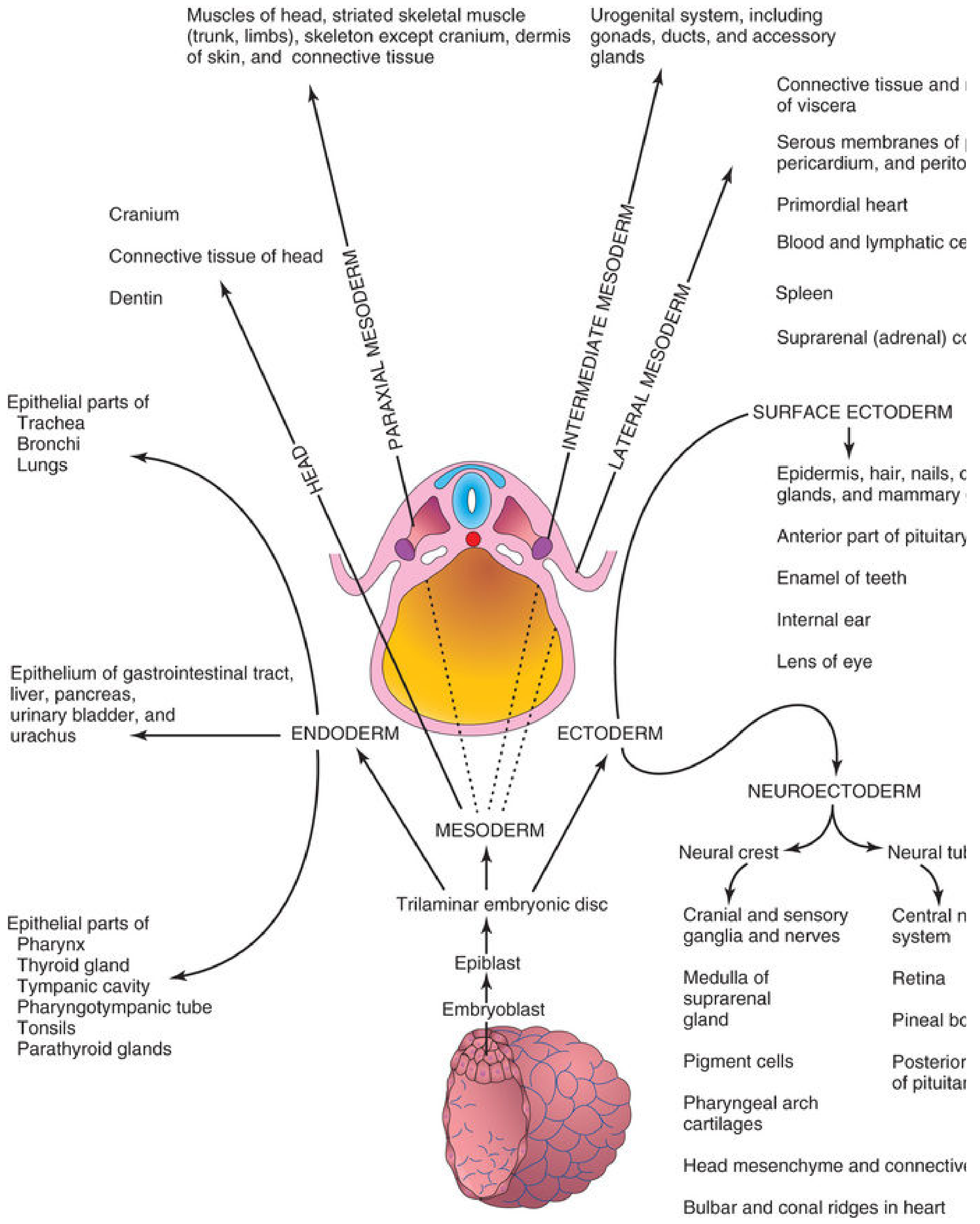
1. ECTODERM
Surface Ectoderm
| Structure |
|---|
| Epidermis (skin) and its appendages - hair, nails, sweat glands, sebaceous glands |
| Mammary glands |
| Anterior pituitary (adenohypophysis) |
| Enamel of teeth |
| Lens and corneal epithelium of the eye |
| Internal ear (otic vesicle) |
| Mucosa of oral cavity and lower anal canal |
Neuroectoderm - Neural Tube
| Structure |
|---|
| Brain and spinal cord (central nervous system) |
| Retina and optic nerve |
| Pineal body |
| Posterior pituitary (neurohypophysis) |
Neuroectoderm - Neural Crest Cells
| Structure |
|---|
| Peripheral nervous system (spinal and cranial ganglia) |
| Cranial nerves V, VII, IX, X |
| Autonomic ganglia |
| Schwann cells (ensheathing cells of PNS) |
| Melanocytes (pigment cells of skin/dermis) |
| Adrenal (suprarenal) medulla |
| Meninges of brain and spinal cord |
| Cartilages, bones, and connective tissue of pharyngeal arch origin |
| Head mesenchyme and connective tissue |
| Bulbar and conal ridges of the heart |
2. MESODERM
| Division | Derivatives |
|---|---|
| Paraxial mesoderm (somites) | Skeletal muscles of trunk and limbs; skeleton except cranium; dermis of skin; connective tissue of body wall |
| Head mesoderm | Cranium; connective tissue of head; dentin of teeth |
| Intermediate mesoderm | Urogenital system - kidneys, ureters, gonads (ovaries/testes), genital ducts and accessory glands |
| Lateral plate mesoderm | Heart (primordial heart); blood and lymphatic vessels and cells; spleen; serous membranes (pericardium, pleura, peritoneum); connective tissue and smooth muscle of viscera |
| General mesoderm | Adrenal (suprarenal) cortex |
3. ENDODERM
| System | Derivatives |
|---|---|
| GI tract | Epithelial lining of the entire digestive tract (esophagus to rectum) |
| Liver & Pancreas | Parenchyma of liver, gallbladder, pancreas (extramural digestive glands) |
| Respiratory | Epithelium of trachea, bronchi, and lungs |
| Urinary | Epithelial lining of urinary bladder and most of the urethra |
| Endocrine glands | Thyroid gland, parathyroid glands, thymus |
| Head & Neck | Pharynx epithelium, tonsil parenchyma, tympanic cavity, tympanic antrum, pharyngotympanic (Eustachian) tube |
Quick Reference Summary Table
| Germ Layer | Mnemonic | Key Derivatives |
|---|---|---|
| Ectoderm | "Outer layer - covers and senses" | Skin, CNS, PNS, sense organs, pituitary, adrenal medulla, tooth enamel |
| Mesoderm | "Middle layer - supports and pumps" | Muscle, bone, cartilage, heart, blood, kidneys, gonads, adrenal cortex |
| Endoderm | "Inner layer - lines and secretes" | GI lining, liver, pancreas, lung, bladder, thyroid, parathyroid, thymus |
- The Developing Human: Clinically Oriented Embryology (Moore & Persaud) - Germ Layer Derivatives, p. 247-249
- Histology: A Text and Atlas (Ross & Pawlina) - Histogenesis of Tissues, p. 309-310
i need thoda short mai cuz i its a short note
Gastrulation - Derivatives of 3 Germ Layers
1. ECTODERM - "Covers & Senses"
- Surface ectoderm: Epidermis, hair, nails, sweat/mammary glands, lens of eye, internal ear, tooth enamel, adenohypophysis
- Neural tube: Brain, spinal cord, retina, posterior pituitary
- Neural crest: PNS ganglia, Schwann cells, melanocytes, adrenal medulla, meninges, pharyngeal arch bones/cartilage
2. MESODERM - "Supports & Pumps"
- Muscles, bones, cartilage, dermis
- Heart, blood & lymphatic vessels
- Kidneys, gonads, genital ducts
- Serous membranes (pericardium, pleura, peritoneum)
- Spleen, adrenal cortex
3. ENDODERM - "Lines & Secretes"
- Epithelium of GI tract, respiratory tract
- Liver, pancreas, gallbladder
- Thyroid, parathyroid, thymus
- Urinary bladder & most of urethra
- Tympanic cavity, Eustachian tube, tonsils
| Layer | Key word | Main products |
|---|---|---|
| Ectoderm | Outside | Skin, CNS, sense organs |
| Mesoderm | Middle | Muscle, bone, heart, kidney |
| Endoderm | Inside | GI lining, glands, lungs |
placenta short note
Placenta - Short Note
Definition
- Fetal part - derived from the chorionic sac (chorion frondosum)
- Maternal part - derived from the decidua basalis (endometrium)
Structure
| Component | Details |
|---|---|
| Cotyledons | 15-30 lobes separated by placental septa; each has stem villi + branch villi |
| Chorionic villi | Finger-like projections bathed in maternal blood in the intervillous space |
| Placental membrane | Separates fetal & maternal blood - made of syncytiotrophoblast, cytotrophoblast, mesenchyme, fetal capillary endothelium |
| Decidua regions | Basalis (forms placenta), Capsularis (covers sac), Parietalis (rest of uterus) |
Villi Development
- Primary villi - syncytio + cytotrophoblast only
- Secondary villi - mesenchyme grows in
- Tertiary villi - blood vessels appear (functional)
Functions (PRENHE)
| Function | Detail |
|---|---|
| Protection | Acts as a barrier; passive immunity via IgG transfer (from 16 weeks) |
| Respiration | O₂ and CO₂ exchange by simple diffusion |
| Excretion | Fetal waste products (CO₂, urea) removed |
| Nutrition | Glucose (facilitated diffusion via GLUT-1), amino acids (active transport), vitamins, water |
| Hormones | Produces hCG, hCS (hPL), progesterone, estrogens |
| Endocrine | Takes over progesterone from corpus luteum after 1st trimester |
Hormones Produced
| Hormone | Function |
|---|---|
| hCG (human chorionic gonadotropin) | Maintains corpus luteum; basis of pregnancy test |
| hCS / hPL (human chorionic somatomammotropin) | Maternal metabolism; fetal growth |
| Progesterone | Maintains pregnancy (from maternal cholesterol) |
| Estrogens | Produced by syncytiotrophoblast in large quantities |
Placental Barrier
- Maternal and fetal blood do NOT mix
- Drugs, viruses (e.g., rubella, CMV), alcohol, and some bacteria can cross
- IgG crosses (passive immunity); IgM does NOT cross
- Placenta acts as an allograft - protected from maternal rejection by HLA-G expression on trophoblast
At Term
- Weight ~500 g, diameter ~15-20 cm
- Expelled as afterbirth shortly after delivery
- Fetal surface = smooth (covered by amnion); Maternal surface = lobulated (cotyledons)
Create a labeled diagram poster of placental structure
A detailed, fully labeled anatomical diagram poster of placental structure, cross-sectional view. Show the following labeled structures with clear annotation lines: Uterine wall, Decidua basalis (maternal part), Decidua parietalis, Myometrium, Spiral arteries (carrying oxygenated maternal blood in), Uterine veins (draining deoxygenated blood out), Intervillous space (filled with maternal blood), Placental septum, Cotyledon, Stem villus (anchoring villus), Branch villi (floating villi), Chorionic plate, Chorionic villi (tertiary - with fetal capillaries inside), Placental membrane layers (syncytiotrophoblast, cytotrophoblast, fetal capillary endothelium), Umbilical cord (with 2 umbilical arteries and 1 umbilical vein), Amnion, Chorion, Fetal surface (smooth), Maternal surface (lobulated). Use color coding: pink/red for maternal blood flow, blue for fetal deoxygenated blood, red for fetal oxygenated blood. Include arrows showing direction of blood flow. Title at top: "STRUCTURE OF THE PLACENTA". Clean medical illustration style, white background, educational poster format.
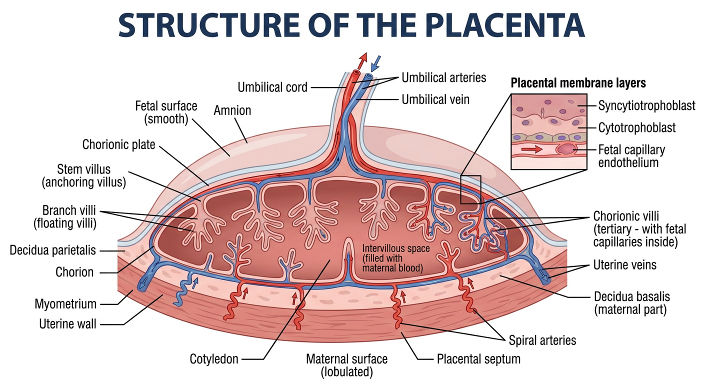
- Maternal side - uterine wall layers, decidua basalis, spiral arteries bringing oxygenated blood in, uterine veins draining out
- Intervillous space - where maternal blood bathes the villi
- Chorionic villi - stem and branch villi with internal fetal capillaries
- Placental membrane - the barrier separating maternal and fetal blood
- Cotyledons separated by placental septa
- Fetal side - chorionic plate, umbilical cord (2 arteries + 1 vein), amnion and chorion
- Blood flow arrows - color-coded for maternal vs. fetal circulation