
Thyroid gland anatomy
Reading File
Now let me search the medical library for thyroid anatomy content and images in parallel.
Finding Sources
Finding Sources
Excellent sources found. Let me now read the key anatomy sections from multiple books in parallel.
Reading File
Reading File
Reading File
Great content found. Let me now read more detailed surgical anatomy from Bailey & Love, plus fetch the key images in parallel.
Reading File
Loading Image
Loading Image
Loading Image
Excellent - I have rich content from multiple authoritative textbooks and excellent images. Now let me read more from Bailey & Love on surgical anatomy (blood supply, relations, lymphatics) and fetch the embryology image.
Reading File
Loading Image
I now have comprehensive content from multiple authoritative textbooks with excellent images. Let me compile the full answer.
Thyroid Gland Anatomy
Overview and Position
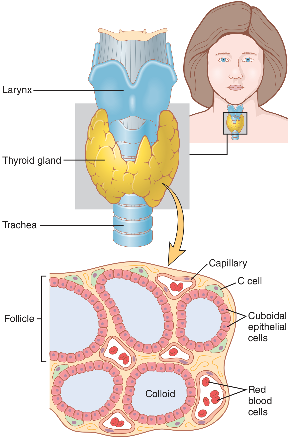
The thyroid gland is a brownish, firm endocrine gland located in the anterior neck, posterior to the strap muscles and anterior to the trachea, larynx, and oesophagus. It is the largest endocrine gland in the body. In adults it weighs 20-25 g and sits at the level of the 2nd-4th tracheal rings. It is bound to the trachea by Berry's ligament (a condensation of pretracheal fascia), which means it moves upward on swallowing - a key clinical sign for distinguishing thyroid swellings from other neck masses.
Gross Structure
The gland consists of:
- Two lateral lobes - connected by a narrow central isthmus overlying the 2nd and 3rd tracheal rings
- Pyramidal lobe - a small upward projection from the isthmus (present in ~50% of people), representing a remnant of the thyroglossal duct; it may extend as high as the hyoid bone
- Tubercle of Zuckerkandl - a posterolateral extension of each lobe, surgically important because the recurrent laryngeal nerve (RLN) runs just below it
Embryology
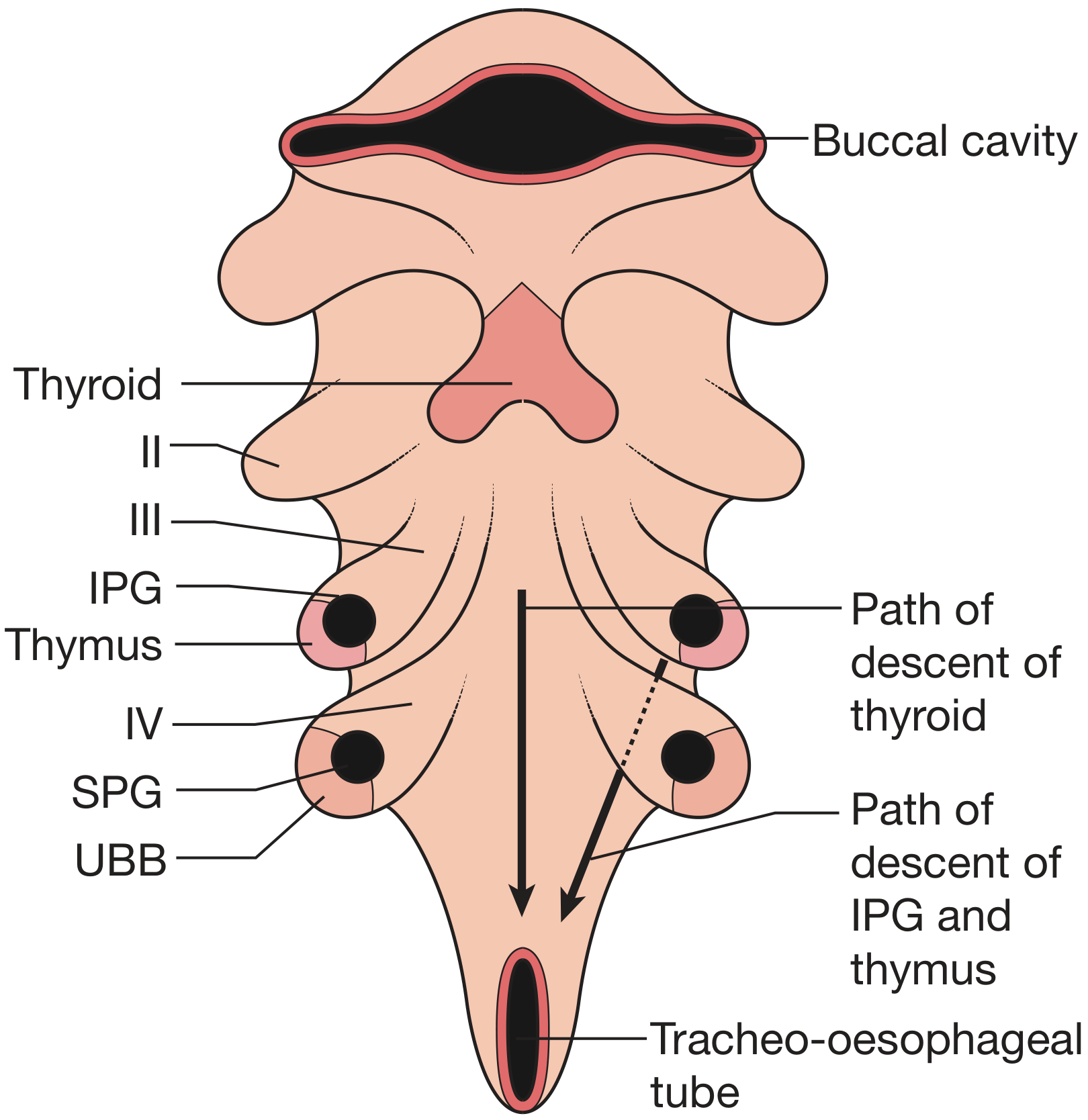
The thyroid originates from a median bud of the pharyngeal floor at the foramen caecum (junction of anterior two-thirds and posterior one-third of the tongue). It descends caudally via the thyroglossal duct, passing in close relation to (sometimes through) the developing hyoid bone. Failure of complete descent leads to a lingual thyroid or thyroglossal duct cyst along any point of this tract. Parafollicular C cells reach the thyroid from neural crest cells via the ultimobranchial body (4th pharyngeal pouch derivative).
Histological Structure
The functional unit of the thyroid is the follicle (100-300 μm in diameter), consisting of:
- Cuboidal follicular epithelial cells - synthesise thyroglobulin and thyroid hormones
- Colloid (centre of follicle) - stores thyroglobulin, the precursor to T3 and T4
- C cells (parafollicular cells) - scattered between follicles; produce calcitonin, which lowers plasma calcium
Each functional unit supplied by a single arteriole contains 24-40 follicles. Blood flow to the thyroid is among the highest per gram of any organ - approximately 5x the gland weight per minute.
Blood Supply
Arteries
| Artery | Origin | Entry Point |
|---|---|---|
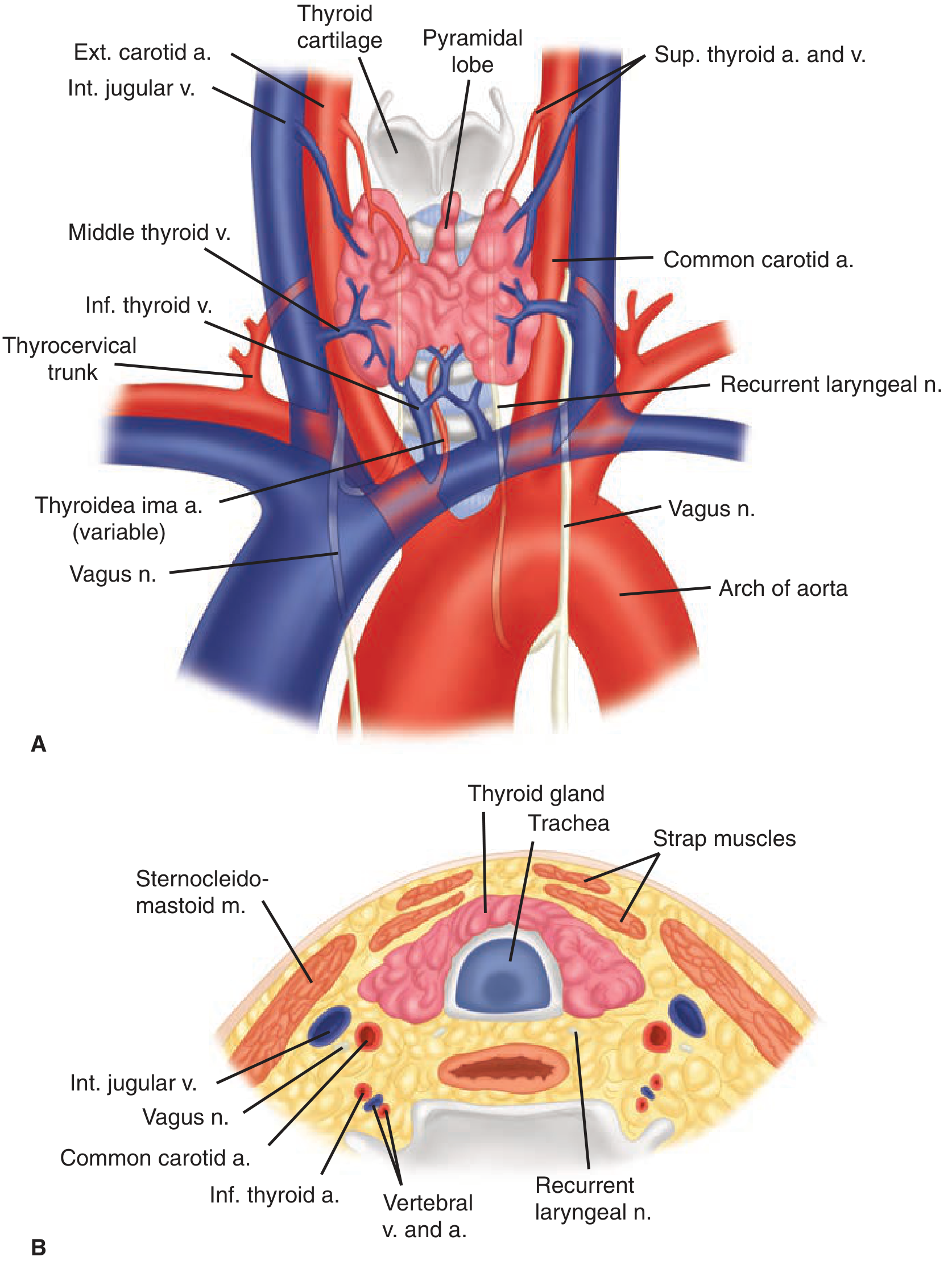
| Superior thyroid artery (×2) | External carotid artery | Upper pole of each lobe |
| Inferior thyroid artery (×2) | Thyrocervical trunk (from subclavian) | Mid-point of each lobe, posteriorly |
| Thyroidea ima artery | Aorta or innominate (variant, 1-4%) | Isthmus or replaces inferior thyroid artery |
Extensive anastomoses occur between main thyroid arteries and branches of tracheal and oesophageal arteries.
Veins
Three paired sets of thyroid veins drain into:
- Superior thyroid veins - run with superior thyroid arteries; drain into the internal jugular vein
- Middle thyroid veins - least consistent; drain into the internal jugular vein
- Inferior thyroid veins - form a plexus draining into the brachiocephalic veins
Posterior Relations and Nerve Supply
Recurrent Laryngeal Nerve (RLN)
The RLN is the most important nerve in thyroid surgery. It is a branch of the vagus nerve (CN X).
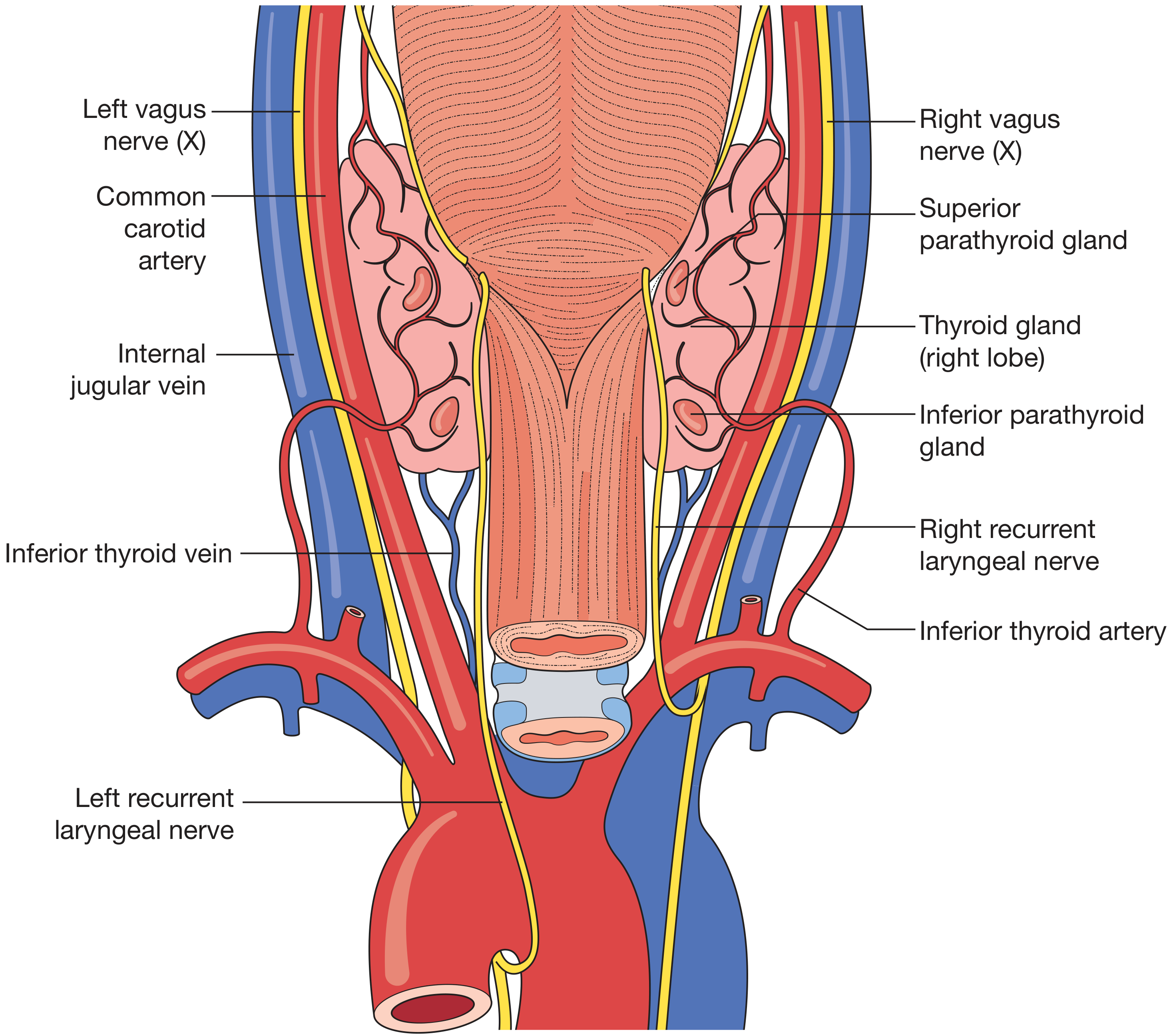
- Left RLN - loops around the arch of the aorta (at the ligamentum arteriosum), then ascends medially in the tracheo-oesophageal groove. Its longer course allows a more medial, predictable position.
- Right RLN - loops around the right subclavian artery, ascends more obliquely. ~2% are non-recurrent on the right (entering the larynx from above), usually associated with an aberrant right subclavian artery.
Both nerves cross the inferior thyroid artery (may be anterior, posterior, or interdigitate with its branches), enter the larynx at the cricothyroid joint, and are at greatest risk immediately beneath Berry's ligament. The nerve can be located in Behr's triangle (bounded by the common carotid artery, inferior thyroid artery, and the nerve itself).
Superior Laryngeal Nerve (External Branch)
The external branch of the superior laryngeal nerve (EBSLN) runs with the superior thyroid artery and innervates the cricothyroid muscle. It is at risk during ligation of the superior thyroid artery - high ligation close to the gland reduces this risk.
Lymphatic Drainage
The subcapsular lymphatic plexus drains via a hierarchical system:
- Central compartment (Level VI): juxtathyroid ("Delphian") nodes and paratracheal nodes, and nodes on superior/inferior thyroid veins
- Deep cervical nodes (Levels II, III, IV, V)
- Mediastinal nodes (Level VII)
Some channels pass directly to deep cervical nodes, bypassing level VI.
Fascial Relations (Cross-Section)
From anterior to posterior, the key layers are:
- Skin and platysma
- Investing layer of deep cervical fascia
- Strap muscles (sternohyoid, sternothyroid, thyrohyoid - innervated by the ansa cervicalis)
- Pretracheal fascia (envelopes the thyroid)
- Thyroid gland (lying on trachea/larynx)
- Tracheo-oesophageal groove (containing the RLN)
- Carotid sheath laterally (common carotid artery, internal jugular vein, vagus nerve)
Parathyroid Glands
Four parathyroid glands normally lie on the posterior surface of the thyroid within the pretracheal fascia:
- Superior parathyroids (from 4th pharyngeal pouch) - lie dorsal to the RLN, characteristically at the level of the cricoid; more constant in position
- Inferior parathyroids (from 3rd pharyngeal pouch) - lie ventral to the RLN; more variable position (may descend with the thymus as far as the anterior mediastinum)
Sources
- Guyton and Hall Textbook of Medical Physiology, p. 930
- Bailey and Love's Short Practice of Surgery, 28th Edition, pp. 872-873
- Schwartz's Principles of Surgery, 11th Edition, pp. 1655-1657
This is a shared conversation. Sign in to Orris to start your own chat.