
Everything about endotracheal tubes
endotracheal tube intubation airway
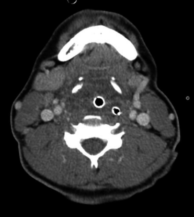
This axial computed tomography (CT) scan depicts the upper airway of a patient who has undergone endotracheal intubation. An endotracheal tube (ETT) is visible as a high-density, ring-like structure centrally located within the hypopharyngeal airway. The surrounding soft tissues demonstrate significant diffuse inflammatory changes, characterized by extensive edema and marked hypertrophy of the lymphoid tissues, including the lingual tonsils and adenoids. These enlarged structures contribute to substantial narrowing and compression of the anatomical airway space. The mandible is visible anteriorly, and a cervical vertebral body is located posteriorly, maintaining normal bony architecture. Bilateral cervical lymphadenopathy is suggested by the presence of prominent soft tissue nodes lateral to the airway. No discrete fluid collections or abscesses are visualized. This diagnostic image illustrates severe airway compromise secondary to inflammatory or infectious processes, such as complicated epiglottitis or severe viral pharyngitis, necessitating mechanical airway protection.
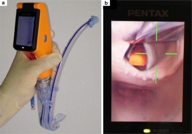
This composite image illustrates the design and clinical application of the Airway Scope® (AWS), a portable, battery-operated video-laryngoscope used for endotracheal intubation. Figure A shows the device's physical components: an orange handle with an integrated 6.1 cm LCD monitor and a single-use 'Intlock' blade. An endotracheal tube (ETT) with a blue radiopaque stripe is preloaded into the blade’s dedicated side-channel guide, which facilitates direct advancement into the trachea. Figure B demonstrates the device's functional output during indirect laryngoscopy. The LCD screen displays a real-time view of the upper airway, specifically focusing on the glottic opening. To assist the clinician, a green crosshair target is superimposed on the digital image; alignment of this target with the glottis indicates the correct path for ETT insertion. This visual aid is designed to simplify intubation by providing a wide viewing angle (180°) and clear anatomical visualization of the vocal cords, thereby potentially increasing first-pass success rates in both novice and expert practitioners.
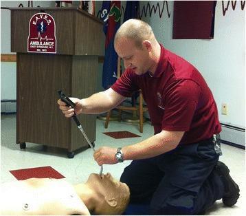
A clinical educational photograph depicting an Advanced Life Support (ALS) provider performing a simulated endotracheal intubation. The provider is kneeling on the floor, simulating an out-of-hospital medical emergency or field environment. The target is an anatomical airway management trainer (mannequin) positioned in a supine posture. The provider is utilizing a Clarus Video System (CVS), which consists of a semi-rigid, malleable fiberoptic stylet with an integrated handle and camera interface. An endotracheal tube is loaded onto the stylet and is shown being inserted into the mannequin's oral cavity using a midline approach. The image demonstrates a specialized video-assisted intubation technique used in difficult airway management and emergency medical services (EMS) training. The setup focuses on the tactile and visual coordination required for successful tube placement without the use of a standard laryngoscope.

This sequence of clinical images demonstrates the four steps of endotracheal intubation (ETI) using a polycarbonate barrier box on an airway trainer manikin. The setup is designed to minimize aerosol exposure during airway management. Step 1: An operator wearing personal protective equipment (PPE), including blue gloves, inserts hands through the rear access ports of the transparent box to begin laryngoscopy. Step 2: The operator uses a Macintosh blade laryngoscope to visualize the glottis while an assistant provides a 7.0 mm cuffed endotracheal tube through a side port. Step 3: The operator inserts the tracheal tube into the trachea. Step 4: The tube is secured, the cuff is inflated, and an Ambu-Bag (manual resuscitator) is connected to begin positive-pressure ventilation. The images highlight the ergonomic constraints and procedural adjustments required when performing direct laryngoscopy within a confined barrier enclosure, commonly utilized in protocols for highly infectious respiratory diseases such as COVID-19.
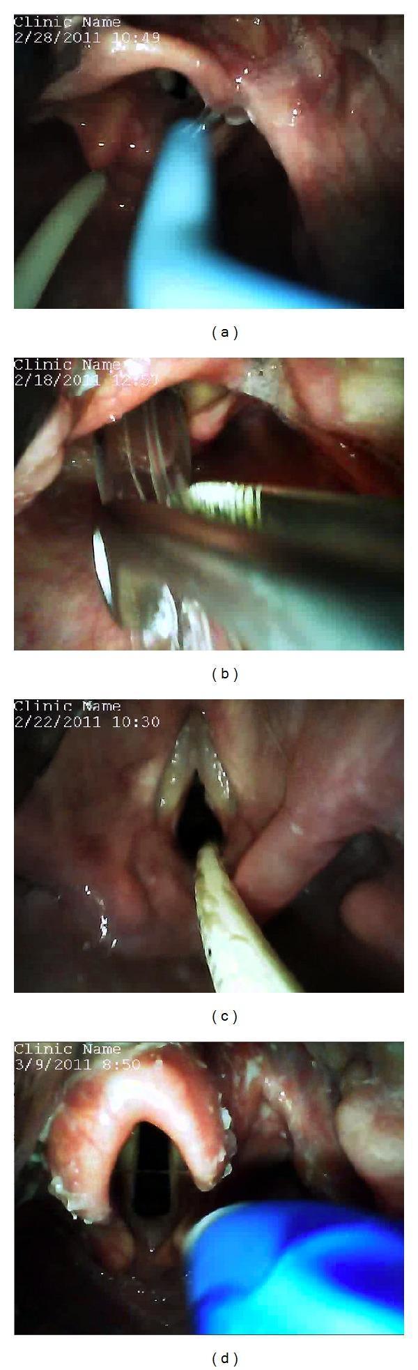
A series of four endolaryngeal clinical photographs (a-d) captured via video laryngoscopy (GlideScope Direct) during airway management, demonstrating various intubation adjuncts and specialized tubes. Panel (a) shows a blue gum elastic bougie being guided through the glottic opening for endotracheal intubation. Panel (b) illustrates nasotracheal intubation with the assistance of Magill forceps, which are seen grasping the distal end of the endotracheal tube to direct it into the trachea. Panel (c) displays an endotracheal tube exchange procedure utilizing a Cook Airway Exchange Catheter positioned within the glottis. Panel (d) shows the placement of a large-bore, blue-cuffed double-lumen endotracheal tube, typically used for independent lung ventilation. In all images, key anatomical landmarks including the epiglottis, vocal cords, and glottic aperture are visible. The images emphasize the compatibility of video-enabled Macintosh blades with standard airway adjuncts and specialized tubes in anesthesiology and emergency medicine.
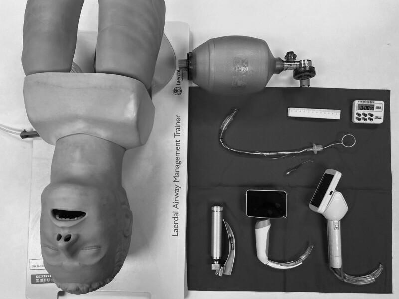
A high-angle clinical photograph displays standard and advanced airway management equipment for endotracheal intubation, positioned alongside a Laerdal Airway Management Trainer mannequin. The equipment includes three types of laryngoscopes: a conventional Macintosh-style laryngoscope with a metal handle and curved blade, and two video laryngoscopes featuring integrated digital screens and specialized curved blades for enhanced glottic visualization. Also present is a Bag Valve Mask (BVM) for manual ventilation. Centrally located is a clear cuffed endotracheal tube (ETT) with an internal stylet bent into a 'hockey-stick' configuration to facilitate insertion; the ETT includes a visible pilot balloon, connector, and depth markings. Ancillary procedural tools include a ruler for measurement and a digital timer for monitoring intubation time. This setup is typical for anesthesia training, emergency medicine simulation, or comparative studies between direct and video laryngoscopy techniques.
Endotracheal Tubes (ETTs) - A Complete Guide
1. Definition and Purpose
- General anesthesia (to control ventilation)
- Critically ill patients requiring mechanical ventilation
- Airway protection (e.g., altered consciousness, risk of aspiration)
- Resuscitation and CPR
2. Components and Design
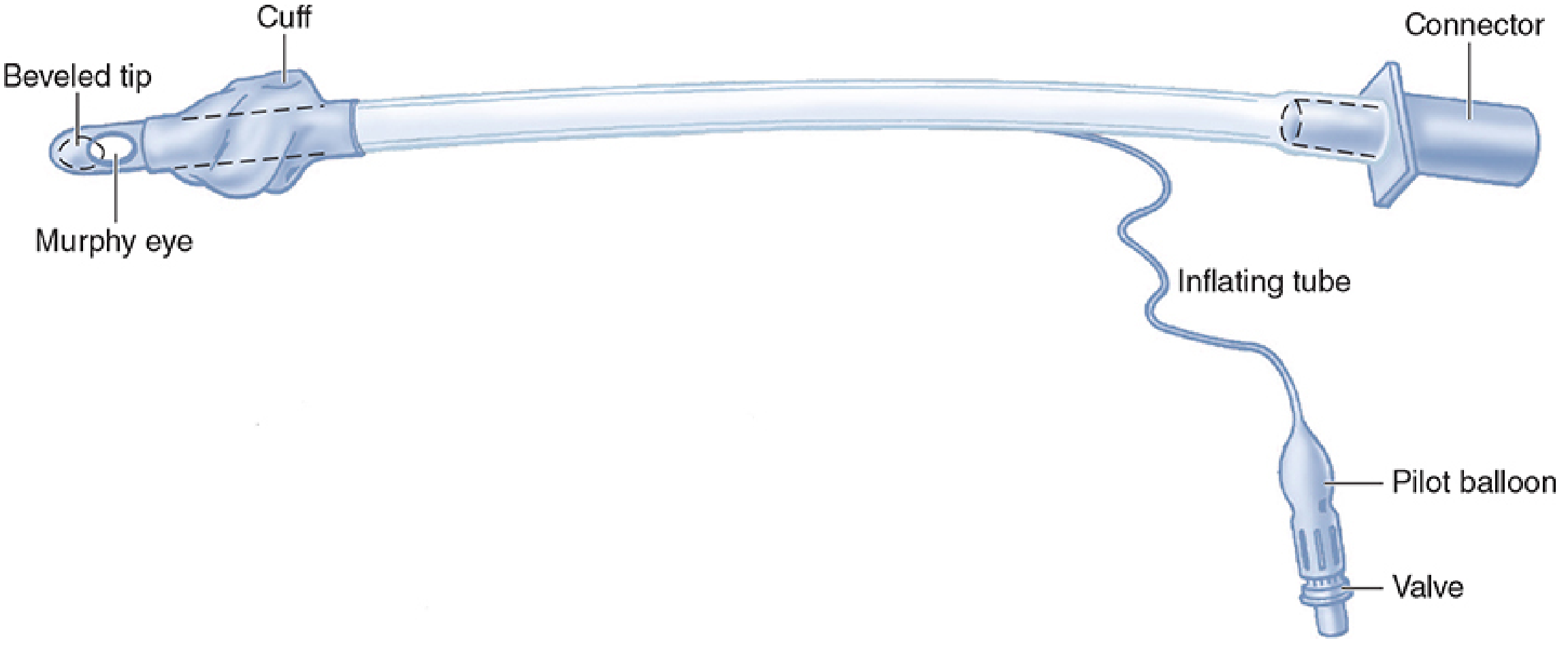
| Component | Function |
|---|---|
| Beveled tip | Angled patient end aids visualization and passage through vocal cords |
| Murphy eye | A lateral hole near the distal end - reduces risk of total occlusion if the tip abuts the carina or tracheal wall |
| Cuff | Inflatable balloon that seals the trachea, enabling positive-pressure ventilation and reducing aspiration risk |
| Inflating tube | Incorporated into the tube wall; connects cuff to pilot balloon |
| Pilot balloon | Provides gross indication of cuff inflation status |
| Valve | Prevents air loss after cuff inflation |
| Proximal connector | 15 mm standard adapter connecting ETT to breathing circuit or bag-valve |
| Depth markings | cm markings on tube body to guide insertion depth |
| Radiopaque line | Embedded to allow position confirmation on X-ray |
- Morgan and Mikhail's Clinical Anesthesiology, 7e, p. 588
3. Cuff Types
- Concentrated pressure at a small contact area
- More ischemic damage to tracheal mucosa
- Unsuitable for prolonged intubation
- Achieve a gas-tight seal over a broad area of tracheal mucosa at low pressure - the "high-volume, low-pressure" cuff
- Standard for modern ETTs
- Lower incidence of mucosal ischemia and necrosis
- Trade-offs: larger mucosal contact area may cause more sore throat, and a floppy cuff can be harder to insert
-
Cuff pressure should be kept below 20-25 cm H2O (tracheal mucosal capillary perfusion pressure is ~25 cm H2O)
-
Excessive cuff pressure causes mucosal injury, ischemia, and potentially tracheal stenosis or malacia
-
The cuff should be inflated until gas leak ceases during ventilation, then a manometer used to verify pressure
-
Do not perform intermittent deflation - this practice is now known to cause more harm; once inflated to a gas-tight seal, keep it inflated
-
Pye's Surgical Handicraft, p. 456
-
Fishman's Pulmonary Diseases and Disorders, p. 2597
4. Tube Sizing
Adults
| Patient | Internal Diameter | Cut Length (oral) |
|---|---|---|
| Female | 7.0-7.5 mm | 24 cm |
| Male | 7.5-9.0 mm | 24 cm |
Children
| Age | Internal Diameter | Blade |
|---|---|---|
| Premature (<30 wGA) | 2.5 mm uncuffed | 0 Straight |
| 30-34 wGA | 3.0 mm | 0 Straight |
| >35 wGA / Full-term | 3.5 mm | 1 Straight |
| 0-6 months | 3.5 mm cuffed | 1 Straight |
| 6-12 months | 4.0 mm cuffed | 1-1.5 Straight |
| 1-2 years | 4.5 mm cuffed | 1.5 Straight |
| 3-4 years | 4.5 mm cuffed | 1.5-2 |
| 5-6 years | 5.0 mm cuffed | 2.0 Curved |
| 7-8 years | 5.5 mm cuffed | 2.0 Curved |
| ≥11 years | 6.5 mm cuffed | 3.0 Curved |
- Children >1 year: ETT size (ID mm) = (age/4) + 4 (cuffed)
- Neonatal depth: Weight (kg) + 6 cm
- Tintinalli's Emergency Medicine, Chapter 113; Harriet Lane Handbook, 23rd ed.
- Morgan and Mikhail's Clinical Anesthesiology, 7e, p. 589
5. Routes of Intubation
Orotracheal (preferred route)
- Advantages: Less traumatic, lower bleeding risk, allows larger tube, more technique options
- Disadvantages: May damage teeth, stimulates gag reflex, requires denser airway anesthesia in awake patients
Nasotracheal
-
Indicated when: Mouth opening severely limited, or surgical access precludes oral route
-
Advantages: Bypasses gag reflex, better tolerated in awake patients; easier for blind, awake, and fiberoptic intubation techniques
-
Disadvantages: Risk of epistaxis, trauma to nasal turbinates, submucosal tunneling
-
Contraindications: Maxillary fractures, skull base fractures
-
Miller's Anesthesia, 10e, p. 5903
6. Intubation Techniques
Direct Laryngoscopy (DL)
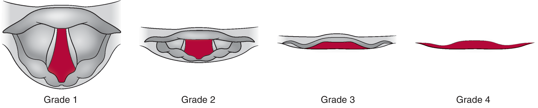
- Grade 1: Full view of glottis
- Grade 2: Partial view
- Grade 3: Only epiglottis visible
- Grade 4: Neither epiglottis nor glottis visible
Indirect/Video Laryngoscopy
Fiberoptic/Flexible Bronchoscopic Intubation (FSI)
ETT Introducers (Bougie)
-
Correct tracheal placement is confirmed by: (a) tracheal clicks as coudé tip passes over tracheal rings, and (b) distal hold-up as it reaches small bronchi
-
Esophageal placement is suggested by absence of clicks and smooth, unlimited advancement
-
Miller's Anesthesia, 10e, p. 5903
7. Confirming ETT Placement
Primary Method: Direct Visualization
Capnography (Most Reliable Objective Method)
- End-tidal CO2 (ETCO2) detection is the gold standard
- Waveform capnography: A normal capnogram for ≥3 breaths is the most important objective indicator of tracheal intubation. The "No Trace = Wrong Place" campaign (Royal College of Anaesthetists/Difficult Airway Society) highlights this principle
- Colorimetric ETCO2: pH-sensitive paper changes from purple to yellow with CO2 exposure. Highly reliable in patients with circulation. In cardiac arrest, CO2 may be low but capnography waveforms may still be detectable; absence of color change should prompt evaluation for esophageal intubation even in arrest
- False positives: Recent BMV or carbonated beverage ingestion may cause transient CO2 from esophagus - washout universally occurs within 6 breaths
Clinical Signs
- Chest rise (bilateral and symmetric)
- Visible condensation (fogging) in the ETT
- Bilateral breath sounds on auscultation, absence of gurgling over epigastrium
- Large exhaled tidal volumes, appropriate reservoir bag compliance
- Absence of CO2 suggests displacement
Esophageal Detection Device (EDD)
Ultrasound
Chest X-Ray
Flexible Bronchoscopy
- Rosen's Emergency Medicine, 10e, p. 1; Tintinalli's EM, p. 225; Miller's Anesthesia, 10e, p. 5921
8. Depth and Positioning
- Adult oral ETT tip: Should sit 3-5 cm above the carina; typical lip-to-teeth marking ~21-23 cm in women, 23-25 cm in men
- Neonatal: Weight (kg) + 6 cm
- After any patient repositioning, tube placement must be reconfirmed
- Neck flexion moves the tube toward the carina (risk of endobronchial intubation)
- Neck extension or lateral rotation moves the tube away from the carina (risk of accidental extubation)
9. Securing the ETT
- Most common: tape to the skin of the maxilla (preferred - less mobile than mandible)
- When tape cannot be used (severe tape allergy, facial burns, epidermolysis bullosa): surgical mask tied around the head; wire fixation to a tooth; suturing to cheek skin
- Secure before repositioning the patient
10. Errors in Positioning
Esophageal Intubation
- The most dangerous complication - can cause hypoxia, hypoxemia, hypercarbia, bradycardia, and cardiac arrest
- Unrecognized esophageal intubation can cause esophageal rupture and mediastinitis (severe sore throat, fever, sepsis, subcutaneous emphysema/crepitus)
- Prevention: direct visualization + CO2 confirmation
Right Mainstem Bronchial Intubation
- Occurs when the ETT is inserted too deeply (right bronchus forms a less acute angle with the trachea than the left)
- Signs: unilateral breath sounds (absent left), unexpected hypoxia, inability to palpate cuff in sternal notch, high peak airway pressures, decreased bag compliance
- Treatment: pull back 1-2 cm and reconfirm
Too-Shallow Insertion
-
Cuff in the larynx: predisposes to laryngeal trauma, cuff may be palpable over thyroid cartilage
-
Risk of extubation with position changes
-
Morgan and Mikhail's Clinical Anesthesiology, 7e, p. 624
11. ETT Malfunction
- Kinking: causes obstruction; use a stylet or reinforced (armored) tubes in high-risk positions
- Cuff/valve damage: must be excluded by testing before insertion (inflate cuff in air, check for leaks)
- Obstruction: from foreign body aspiration or thick/inspissated secretions in the lumen
- Fire hazard: PVC tubes can ignite with cautery or laser in O2/N2O-enriched environments; special laser-resistant ETTs exist (no tube is completely laser-proof)
12. Special ETT Types
Reinforced (Armored) ETT
- Wire-reinforced wall resists kinking
- Used in head, neck, and prone surgeries
Double-Lumen ETT (DLT)
- Two channels: a longer bronchial lumen (seated in a mainstem bronchus) and a shorter tracheal lumen
- Two cuffs: tracheal cuff and bronchial cuff
- Left-sided DLT is most commonly used (longer left mainstem bronchus provides safer margin)
- Right-sided DLT reserved for left pneumonectomy or left sleeve resections; harder to position (must align side hole with right upper lobe orifice)
- Placement must be confirmed bronchoscopically, both supine and after lateral positioning
- Murray & Nadel's Respiratory Medicine, p. 694
Laser ETTs
- Specially designed for use in laser airway surgery
- No currently available ETT is reliably laser-proof
- Cuff inflated with dyed saline to detect early cuff rupture
- Morgan and Mikhail's Clinical Anesthesiology, 7e
Pre-formed (RAE) Tubes
- Oral or nasal pre-formed curve (e.g., Ring-Adair-Elwyn tube)
- Moves the breathing circuit away from the surgical field (oral/maxillofacial surgery)
13. Physiological Responses to Intubation
- Hypertension and tachycardia from stimulation of pharyngeal/laryngeal structures
- Cardiac arrhythmias (especially premature ventricular beats) - may indicate light anesthesia
- Can be attenuated by: IV propofol, lidocaine, opioids, beta-blockers, deeper inhalation anesthesia, or vasodilators (esmolol, nicardipine, nitroprusside) before laryngoscopy
- Forceful involuntary spasm of laryngeal musculature
- Triggered by secretions or passing an ETT through the larynx during extubation
- Prevented by extubating patients fully awake or deeply asleep
- Treatment: gentle positive-pressure ventilation with 100% O2; IV lidocaine (1-1.5 mg/kg) or succinylcholine for refractory cases
14. Complications
Immediate/Short-term
- Esophageal intubation
- Endobronchial intubation
- Dental damage
- Lip/oral soft tissue laceration
- Vocal cord injury (from forceful attempts or high cuff pressure)
- Arytenoid cartilage dislocation (especially with rheumatoid arthritis affecting cricoarytenoid joint)
- Laryngospasm/bronchospasm
Intermediate
- Tube dislodgement (accidental extubation)
- Tube obstruction
- Aspiration
- Ventilator-associated pneumonia (VAP)
Long-term (from prolonged intubation)
-
Postextubation stridor: From glottic edema, laryngospasm, or laryngotracheal stenosis. Reintubation rates range 4-33%; "acceptable" rate 5-15%. Cuff leak test used to assess risk pre-extubation; pre-extubation glucocorticoids recommended in high-risk patients
-
Tracheal stenosis: At cuff level or tube tip, from overdistended/uneven cuff, local infection, or prolonged intubation
-
Tracheomalacia: From chronic cuff overdistension and pressure necrosis, can lead to tracheal erosion into surrounding structures (e.g., innominate vein)
-
Vocal cord paralysis: Unilateral or bilateral; presents as hoarseness or airway obstruction; usually resolves within 10 weeks
-
Vocal cord (contact) granuloma: Develops 4-6 weeks post-intubation; treated with antireflux therapy, corticosteroids, speech therapy
-
Subglottic stenosis
-
Fishman's Pulmonary Diseases, p. 2597; Pye's Surgical Handicraft, p. 456
15. Humidification
16. VAP Prevention and ETT Design
-
Subglottic secretion drainage ports (specialized ETT feature) allow suctioning above the cuff
-
Innovations in cuff materials and shape aim to reduce micro-leakage
-
Cuff pressure monitoring is the most consistent preventive measure
-
Head-of-bed elevation to 30-45°, oral hygiene protocols, and selective digestive decontamination (SDD) are complementary strategies
-
Murray & Nadel's Respiratory Medicine, p. 1148
17. ETT in COVID-19
- Video laryngoscopy over direct laryngoscopy
- Rapid sequence intubation (RSI) to minimize bag-mask ventilation
- Barrier devices (e.g., aerosol boxes)
- Avoiding awake fiberoptic intubation when possible
Summary Table: Quick Reference
| Parameter | Value/Note |
|---|---|
| Material | PVC (polyvinyl chloride) |
| Adult female ID | 7.0-7.5 mm |
| Adult male ID | 7.5-9.0 mm |
| Pediatric ID formula | (age/4) + 4 mm |
| Cuff pressure limit | < 20-25 cm H2O |
| Best placement confirmation | Waveform capnography (≥3 breaths) |
| Depth (adult oral) | ~21-25 cm at lip |
| Neonatal depth formula | Weight (kg) + 6 cm |
| Most common position error | Right mainstem bronchial intubation |
| Cuff type (standard) | High-volume, low-pressure |
| Preferred DLT | Left-sided (longer mainstem bronchus) |
- Morgan and Mikhail's Clinical Anesthesiology, 7e, pp. 588-627
- Miller's Anesthesia, 10e, pp. 5903-5923
- Tintinalli's Emergency Medicine, Chapters 29A, 113
- Rosen's Emergency Medicine, Chapter 1
- Fishman's Pulmonary Diseases and Disorders, Chapters 47, 146
- Murray & Nadel's Respiratory Medicine, Chapters 30, 11
- Harriet Lane Handbook, 23rd ed., Chapter 18
- Pye's Surgical Handicraft, Chapter 31