
Explain me a cerebral concussion Cerebral contusion Cerebral laceration For 10 marks answer
Reading File
Now let me search the medical library for all three conditions simultaneously.
Finding Sources
Finding Sources
Good sources found. Let me read the most detailed sections in parallel.
Reading File
Reading File
Finding Sources
Excellent! Let me read the contusion/laceration sections from the most comprehensive sources.
Reading File
Reading File
Let me read more on cerebral laceration and the rest of the concussion/contusion content, plus the S Das content on these injuries.
Reading File
Reading File
Let me now read the concussion content from S Das and also get the remaining laceration content:
Reading File
Reading File
Now let me get the S Das section specifically on cerebral concussion, contusion and laceration details:
Reading File
Reading File
I now have comprehensive information from multiple authoritative sources. Let me also fetch the key brain injury diagram and contusion image for visual support.
Loading Image
Reading File
I now have all the information needed to write a comprehensive 10-mark answer. Let me compose it.
Head Injuries: Cerebral Concussion, Cerebral Contusion, and Cerebral Laceration
(10-mark answer)
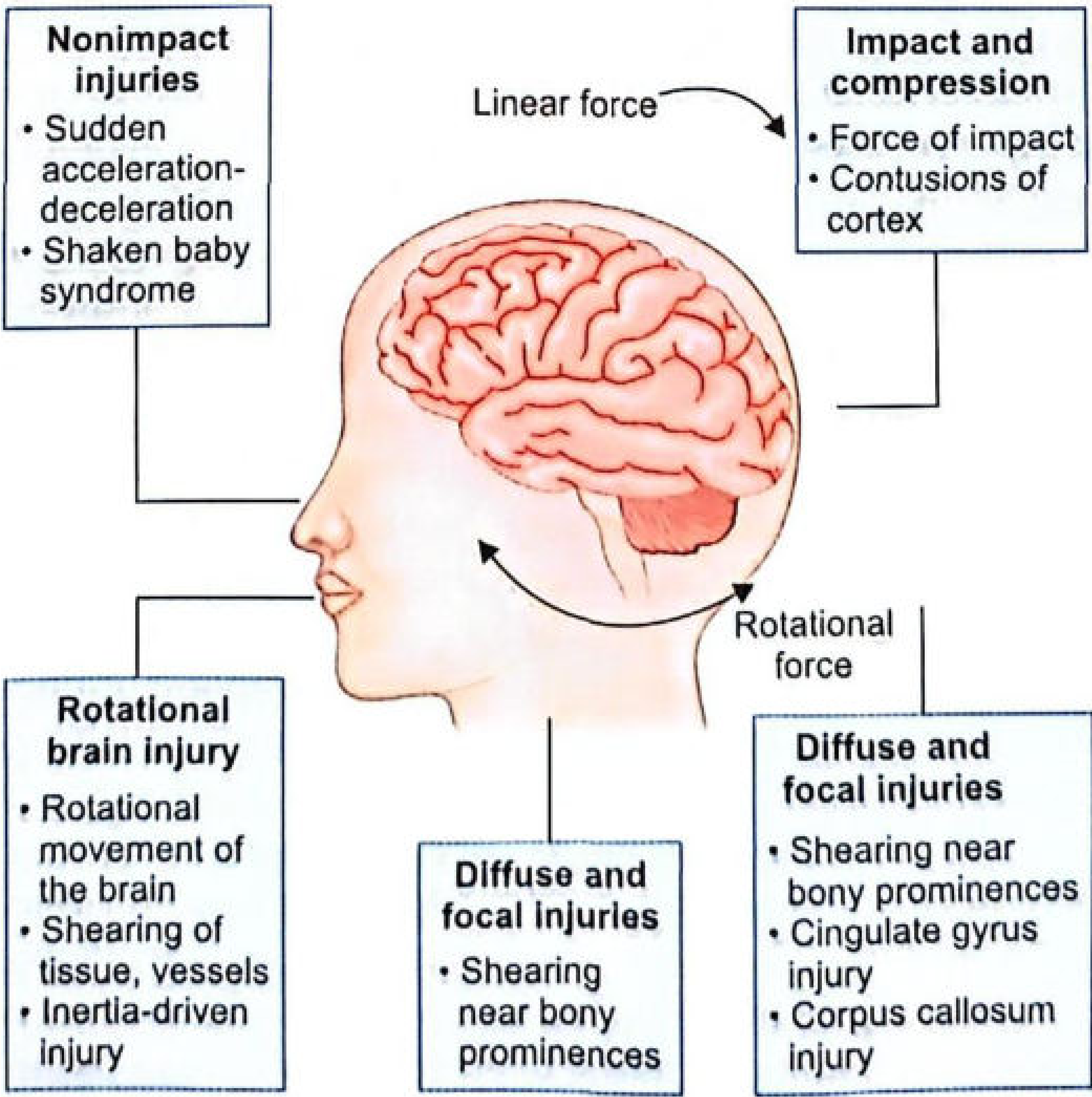
Fig: Mechanism of cerebral injury
All three conditions represent increasing degrees of traumatic brain injury produced by the same fundamental mechanism - displacement and distortion of cerebral tissues at the moment of impact.
1. CEREBRAL CONCUSSION
Definition:
Cerebral concussion (also called "stunning") is a state of temporary physiological paralysis of brain function due to diffuse neuronal injury (involving the brainstem), but with little or no noticeable anatomical/structural damage. It comes on immediately after injury, is always followed by amnesia, and tends towards spontaneous recovery.
Mechanism:
- Occurs due to sudden acceleration/deceleration of the head (the head must be freely movable at the time of injury).
- Violent head movement causes shearing or stretching of nerve fibres and axonal damage.
- Loss of consciousness is believed to be a transient electrophysiological dysfunction of the reticular activating system (RAS) in the upper midbrain, caused by rotation of the cerebral hemispheres on the relatively fixed brainstem.
- Severe injuries occur with coronal head motion; sagittal motion produces mild/moderate injury.
Causes:
- Direct violence to the head
- Indirect violence - a violent fall on feet/buttocks
- Traffic and industrial accidents (deceleration injuries are more severe than direct blows)
Clinical Features:
- Immediate loss of consciousness (lasts seconds to minutes)
- Muscles are flaccid; pupils are dilated and unreactive
- Pulse is rapid, thready; blood pressure falls
- Respiration is quick and shallow
- Temperature remains subnormal
- Amnesia (retrograde and post-traumatic) - duration proportional to severity of injury
- As consciousness returns, there is a period of automatism - the patient behaves automatically but not rationally (post-traumatic automatism)
- Mild concussion: no loss of consciousness, no confusion, amnesia may or may not be present
- Severe concussion: amnesia + loss of consciousness
Post-Concussion Syndrome: Headache, dizziness, nausea, vomiting, insomnia, mental irritability - recovery usually complete within 10 days.
Autopsy Findings:
- In mild/moderate cases: no macroscopic damage
- In fatal cases: no naked-eye lesions; only microscopic haemorrhages without disruption of tissue structure
- Histology shows diffuse neuronal injury with intracellular disturbances and conduction defects at synaptic junctions
Medicolegal Importance:
- Can be ruled out if unconsciousness does not occur immediately after the blow
- The automatism phase may be misused as a defence in criminal cases ("the person committed an act without conscious awareness")
2. CEREBRAL CONTUSION
Definition:
Cerebral contusion represents a more severe injury with actual circumscribed areas of brain tissue destruction accompanied by extravasation (haemorrhage) into the affected tissue, but without actual tearing/loss of continuity of the brain substance. The pia-arachnoid remains intact over surface contusions.
Mechanism:
- When a localised segment of the skull undergoes deformation at the moment of impact, shear strains develop in the underlying brain tissue, producing a zone of contusion in the surface layers.
- When the head is rotated by impact, layers of brain tissue slide over each other at different depths in the cortex, damaging blood vessels.
- Produced by blunt force; may occur with or without skull fracture.
Pathological Features:
- Contusions appear as streaks or groups of punctate haemorrhages in grey and white matter
- Haemorrhage is first seen in perivascular spaces (Robin-Virchow spaces) → ring haemorrhages
- Most often found in frontal and temporal lobes; also on lateral and ventral surfaces of cerebral hemispheres
- Deeper structures (basal ganglia, midbrain, brainstem) are contused especially from impacts to the forehead and vertex
- Haemorrhages are densely arranged and elongated, pointing radially towards the white matter
- If close to the cortical surface, may cause overlying focal subarachnoid haemorrhage
- Haemorrhages also noted in the corpus callosum, 3rd ventricle, and substantia nigra of the brainstem
Types of Contusions:
- Coup contusions - beneath the area of direct impact
- Contrecoup contusions - opposite the side of impact (due to shear strain from rotation)
- Intermediary coup contusions - along the midbrain/brainstem between coup and contrecoup points
- Fracture contusions - caused by skull fracture
- Gliding contusions - in frontal/central convolutions near the upper margin of hemispheres due to stretching/shearing during to-and-fro gliding of the brain
- Herniation contusions - in cerebellar tonsils/medulla, produced by momentary shifting of the brain toward the foramen magnum
Age of Contusions (Dating):
- Ischaemic changes in neurons: within 1 hour
- Capillary proliferation: begins at 5 days, maximum at 10-12 days
- Macrophages: present in first 2 weeks
- Astrocytic proliferation: within a few weeks
- Scar formation: approximately 2 months (pale/golden yellow, depressed)
Clinical Features:
- Unconsciousness is more prolonged than in concussion (30 minutes to several days)
- Low intracranial pressure initially
- Recovery is imperfect - post-contusional state with defective memory, change of personality, confusion, irritability, delirium
- Pulse: rapid, low volume; BP reduced; respiration shallow; muscles relaxed
- On recovery: headache, photophobia, vomiting
- Cerebral irritation (develops 2-3 days post-injury, due to cerebral oedema): patient curled up in attitude of flexion, resists interference, photophobia
3. CEREBRAL LACERATION
Definition:
Cerebral laceration is the most severe form of brain injury, defined as a traumatic lesion in which there is actual loss of continuity (tearing) of the substance of the brain. Unlike contusion, the pia mater is torn in lacerations.
Mechanism:
- Stretching and shearing forces produced by blunt force
- Brain surface torn against bony ridges and edges of dural septa (falx cerebri, tentorium cerebelli)
- All penetrating injuries produce lacerations
Sites:
- Inner aspect of the hemisphere (especially undersurface of frontal lobe and tip of temporal lobe)
- Lacerations often occur under skull fractures
- In depressed fractures, bone fragments physically tear into brain
- Corpus callosum and septum pellucidum (especially in younger individuals from blunt trauma without fracture)
- Pyramids at the pontomedullary junction - in severe hyperextension of the head (motor vehicle accidents), often associated with fractures of the base of skull
Types:
- Surface lacerations - accompanied by rupture of pia mater and subarachnoid haemorrhages; usually surrounded by contusions
- Deep lacerations - when parenchyma is completely disorganised, termed pulpyfaction; may involve ventricles
- Contrecoup lacerations - on the side opposite the impact
Clinical Features:
- Signs and symptoms similar to cerebral contusion but more severe
- Blood enters the CSF → subarachnoid haemorrhage (CSF becomes bloodstained)
- Lacerations of the undersurface of the frontal lobe may cause anosmia (damage to olfactory nerves) and change of personality
- Deeper lacerations involving ventricles may produce large glial cysts filled with CSF (traumatic porencephalic cysts)
- Healing of surface lacerations may form adhesions between brain and dura → secondary epilepsy (due to contracting fibrous scar)
- Infants up to 5 months are particularly susceptible (easily deformable skull + very soft brain)
COMPARATIVE TABLE
| Feature | Concussion | Contusion | Laceration |
|---|---|---|---|
| Structural damage | None (physiological) | Haemorrhagic areas, no tearing | Actual tearing of brain tissue |
| Pia-arachnoid | Intact | Intact over contusion | Torn |
| Unconsciousness | Seconds to minutes | 30 minutes to days | Prolonged |
| Recovery | Complete, perfect | Imperfect | Incomplete / permanent deficits |
| Subarachnoid bleed | Absent | May be present (focal) | Present (prominent) |
| Autopsy (naked eye) | Normal | Punctate haemorrhages | Visible tearing/pulpyfaction |
| CSF | Normal | May be slightly bloodstained | Bloodstained |
Sources: The Essentials of Forensic Medicine and Toxicology 36th Ed.; S Das A Manual on Clinical Surgery 13th Ed.; Parikh's Textbook of Medical Jurisprudence, Forensic Medicine and Toxicology; P C Dikshit Textbook of Forensic Medicine and Toxicology
Answer like bams students can write
Here is the same answer written in a clean, exam-ready style suitable for BAMS students:
Cerebral Concussion, Cerebral Contusion and Cerebral Laceration
These are three types of injury to the brain commonly seen in head injuries. They are produced by the same mechanism — displacement and distortion of cerebral tissues at the moment of impact. They represent increasing degrees of severity.
1. CEREBRAL CONCUSSION
Definition
Cerebral concussion is a condition of temporary physiological disturbance of brain function following head injury, with no gross structural or anatomical damage to the brain tissue. It comes on immediately after injury and tends towards complete spontaneous recovery.
Mechanism
- Caused by sudden acceleration/deceleration of the head
- Shearing and stretching of nerve fibres causes diffuse neuronal injury
- Loss of consciousness occurs due to transient dysfunction of the Reticular Activating System (RAS) in the upper midbrain
Causes
- Direct blow to the head
- Indirect violence — fall on feet or buttocks
- Road traffic accidents, industrial accidents
Clinical Features
- Immediate loss of consciousness (lasts seconds to minutes)
- Muscles are flaccid
- Pupils are dilated and unreactive
- Pulse is rapid and thready; BP falls
- Respiration is shallow
- Temperature subnormal
- Amnesia — both retrograde and post-traumatic
- Post-traumatic automatism — patient appears conscious but behaves irrationally
Types
- Mild concussion — no loss of consciousness, no confusion
- Severe concussion — loss of consciousness + amnesia
Post-Concussion Syndrome
Headache, dizziness, nausea, insomnia, irritability — usually recovers within 10 days.
Autopsy Findings
- No macroscopic lesions in the brain
- Microscopic haemorrhages may be seen without tissue disruption
- Diffuse neuronal injury with intracellular disturbances
Medicolegal Importance
- Unconsciousness must occur immediately after injury — if it is delayed, concussion can be ruled out
- Post-traumatic automatism may be used as a defence in criminal cases
2. CEREBRAL CONTUSION
Definition
Cerebral contusion is a condition where there are circumscribed areas of brain tissue destruction with extravasation of blood (haemorrhage into tissue), but without actual tearing of the brain substance. The pia-arachnoid remains intact.
Mechanism
- Blunt force causes shear strains in the brain underlying the point of impact
- Rotation of the head causes layers of brain tissue to slide over each other, damaging blood vessels
- May occur with or without skull fracture
Pathology
- Appears as punctate (pin-point) haemorrhages in grey and white matter
- Ring haemorrhages — bleeding into perivascular spaces of Robin-Virchow
- Haemorrhages seen in corpus callosum, 3rd ventricle, and substantia nigra of brainstem
- Most common in frontal and temporal lobes
Types
| Type | Description |
|---|---|
| Coup | Beneath the area of impact |
| Contrecoup | Opposite side to impact |
| Gliding | Upper margin of hemispheres due to to-and-fro brain movement |
| Fracture | Associated with skull fracture |
| Herniation | In cerebellar tonsils/medulla |
Clinical Features
- Unconsciousness — more prolonged (30 minutes to several days)
- Recovery is imperfect — defective memory, personality change, confusion, irritability
- Headache, photophobia, vomiting on recovery
- Cerebral irritation (2-3 days after injury): patient lies curled up, flexed posture, resists interference
Autopsy — Age of Contusion
- 1 hour — ischaemic neuronal changes
- 5 days — capillary proliferation begins
- 10-12 days — capillary proliferation maximum
- 2 weeks — macrophages with fat
- Few weeks — astrocytic proliferation
- 2 months — scar formation (pale/golden yellow, depressed)
3. CEREBRAL LACERATION
Definition
Cerebral laceration is the most severe form of brain injury where there is actual tearing or loss of continuity of the brain substance. The pia mater is torn, unlike in contusion.
Mechanism
- Shearing and stretching forces from blunt trauma
- Brain surface tears against bony ridges and edges of dural septa (falx cerebri, tentorium)
- All penetrating injuries produce laceration
- In depressed fractures, bone fragments are driven into brain
Common Sites
- Undersurface of frontal lobe
- Tip of temporal lobe
- Corpus callosum
- Pontomedullary junction — in severe hyperextension of head (road accidents)
Types
- Surface laceration — pia mater torn, subarachnoid haemorrhage, surrounded by contusions
- Deep laceration — complete disorganisation of parenchyma = pulpyfaction
- Contrecoup laceration — on the opposite side of impact
Clinical Features
- Symptoms similar to contusion but more severe
- Blood enters CSF → subarachnoid haemorrhage (bloodstained CSF)
- Laceration of frontal lobe → anosmia + change of personality
- Traumatic porencephalic cysts — deep lacerations involving ventricles heal to form large glial cysts filled with CSF
- Secondary epilepsy — adhesions between brain and dura form at healed laceration sites
Special Points
- Common in infants under 5 months (soft brain + easily deformable skull)
- Lacerations at the pontomedullary junction in hyperextension injuries are often fatal
COMPARISON TABLE (Important for Exam)
| Feature | Concussion | Contusion | Laceration |
|---|---|---|---|
| Structural damage | Nil (physiological only) | Haemorrhage, no tearing | Actual tearing |
| Pia-arachnoid | Intact | Intact | Torn |
| Unconsciousness | Seconds-minutes | Minutes-days | Prolonged |
| Recovery | Complete | Imperfect | Incomplete/permanent |
| Subarachnoid bleed | Absent | Focal (if near surface) | Present |
| CSF | Normal | May be normal | Bloodstained |
| Naked eye autopsy | Normal | Punctate haemorrhages | Visible tearing |
Key Line to Remember: Concussion = No damage, Contusion = Bruising (haemorrhage, no tear), Laceration = Tearing Pia intact in contusion; Pia torn in laceration.
We don't need autopsy finding we need management and investigation
Finding Sources
Finding Sources
Finding Sources
Finding Sources
Finding Sources
Finding Sources
Reading File
Reading File
Reading File
Finding Sources
Reading File
Finding Sources
Reading File
I now have all the investigation and management content needed. Here is the complete rewritten answer for BAMS students:
Cerebral Concussion, Cerebral Contusion and Cerebral Laceration
(10-mark BAMS exam answer)
INTRODUCTION
These are three types of primary brain injuries caused by head trauma. They represent increasing degrees of severity. All are produced by the same fundamental mechanism - displacement and distortion of cerebral tissues at the moment of impact.
1. CEREBRAL CONCUSSION
Definition
A state of temporary physiological paralysis of brain function following head injury with no structural damage to brain tissue. Recovery is complete and spontaneous.
Mechanism
- Sudden acceleration/deceleration of head
- Shearing of nerve fibres → diffuse neuronal injury
- Reticular Activating System (RAS) in upper midbrain is transiently disrupted → loss of consciousness
Clinical Features
- Immediate loss of consciousness (seconds to minutes)
- Flaccid muscles, dilated unreactive pupils
- Pulse rapid and thready, BP falls
- Respiration shallow, temperature subnormal
- Retrograde and post-traumatic amnesia
- Post-traumatic automatism (acts without awareness)
Investigations
| Investigation | Findings |
|---|---|
| Skull X-ray | Usually normal; may show fracture |
| CT scan (brain) | Normal - no structural damage |
| MRI brain | May show subtle white matter changes |
| CSF analysis | Normal (no blood) |
| Blood investigations | CBC, blood sugar, electrolytes (baseline) |
| GCS assessment | Mild impairment, recovers quickly |
Management
Immediate (First Aid):
- Maintain clear airway
- Control breathing and circulation (ABC)
- Do not move the patient without cervical spine precautions
- Control scalp bleeding with pressure bandage
In Hospital:
- Admit for observation (minimum 24 hours)
- Monitor: GCS, pulse, BP, respiration, pupils every hour
- CT scan of brain to rule out intracranial haemorrhage
- IV line with normal saline (avoid free water/hypotonic fluids)
- Oxygen supplementation
- Analgesics for headache - paracetamol or NSAIDs
- No sedatives (mask neurological signs)
- Instruct patient/family to return if: worsening headache, vomiting, confusion, or difficulty arousing
Discharge advice:
- Physical and cognitive rest
- Gradual return to activity
- Avoid contact sports until fully recovered
- Written instruction sheet with warning signs
2. CEREBRAL CONTUSION
Definition
Circumscribed areas of brain tissue destruction with haemorrhage (extravasation of blood) into the tissues, without actual tearing of brain substance. Pia-arachnoid remains intact.
Pathology
- Punctate/ring haemorrhages in grey and white matter
- Perivascular bleeding (Robin-Virchow spaces)
- Most common in frontal and temporal lobes
- May involve corpus callosum, substantia nigra of brainstem
Clinical Features
- Unconsciousness - more prolonged (30 min to several days)
- Pulse rapid, low volume; BP reduced; respiration shallow
- Recovery is imperfect - memory loss, personality change, confusion, irritability
- On recovery: headache, photophobia, vomiting
- Cerebral irritation (2-3 days later): patient curled up, resists interference, photophobia
Investigations
| Investigation | Findings |
|---|---|
| CT scan brain (Gold standard) | Hyperdense (white) areas = haemorrhagic contusions; surrounding oedema |
| MRI brain | More sensitive; shows exact extent of contusions and white matter injury |
| Skull X-ray | May show fracture |
| CSF analysis | May be slightly bloodstained; raised pressure |
| ICP monitoring | If GCS 3-8 with CT abnormality - monitor ICP |
| Blood tests | CBC, coagulation profile (PT/INR), blood sugar, electrolytes |
| GCS scoring | Serial monitoring every hour |
Management
Emergency:
- ABC - Airway, Breathing, Circulation
- Cervical spine immobilisation
- Endotracheal intubation if necessary (GCS < 8)
- IV access - infuse normal saline (avoid hypotonic fluids)
- Maintain systolic BP > 90 mmHg (hypotension doubles mortality)
- Oxygen - maintain arterial oxygen saturation at normal levels
Specific Medical Management:
- Head elevation at 30 degrees (reduces ICP)
- Hyperventilation - lowers PaCO2 → decreases intracranial oedema (short-term)
- Mannitol 20% (0.25-1 g/kg IV) - hyperosmolar therapy to reduce cerebral oedema
- Hypertonic saline - alternative to mannitol for reducing ICP
- Steroids - dexamethasone (action is delayed; limited role in trauma)
- Anti-epileptics - prophylaxis to prevent post-traumatic seizures (phenytoin)
- Urinary catheterisation if needed; monitor urine output
- Temperature, pulse, BP, respiration, pupils charted every hour
Surgical Management:
- If haematoma or mass effect develops → surgical evacuation
- Decompressive craniectomy in refractory raised ICP
3. CEREBRAL LACERATION
Definition
Most severe form of brain injury. There is actual tearing/loss of continuity of brain substance. The pia mater is torn, unlike contusion.
Mechanism
- Shearing forces; brain torn against bony ridges and dural septa
- Depressed skull fracture driving bone into brain
- All penetrating injuries
- Severe hyperextension of head (pontomedullary junction tears)
Sites
- Undersurface of frontal lobe, tip of temporal lobe
- Corpus callosum
- Pontomedullary junction - in hyperextension injuries
Clinical Features
- Most severe - signs similar to contusion but worse
- Subarachnoid haemorrhage - blood enters CSF
- Bloodstained CSF on lumbar puncture
- Laceration of frontal lobe → anosmia + personality change
- Deep lacerations → traumatic porencephalic cysts
- Healed lacerations → secondary epilepsy (brain-dura adhesions)
Investigations
| Investigation | Findings |
|---|---|
| CT scan brain (essential) | Hyperdense areas of haemorrhage; visible tearing; subarachnoid blood; oedema; midline shift |
| MRI brain | Best for assessing extent of laceration and white matter damage |
| Skull X-ray | Fracture, depressed fragment |
| Lumbar puncture | Bloodstained CSF (subarachnoid haemorrhage) - do only after CT rules out raised ICP |
| ICP monitoring | Essential if GCS 3-8 with CT abnormality |
| Blood tests | CBC, coagulation profile, blood grouping and cross-matching (for surgery) |
| EEG | If post-traumatic seizures develop |
Management
Emergency:
- Full ATLS protocol - Airway, Breathing, Circulation
- Endotracheal intubation (GCS usually < 8)
- IV normal saline; avoid hypotonic fluids (worsens cerebral oedema)
- Immediate CT brain to assess extent of laceration and haematoma
Medical Management:
- Same as contusion + more aggressive ICP control
- Mannitol / Hypertonic saline for raised ICP
- Avoid: hypoxia, hypercarbia, hyperthermia, awkward neck positions (all raise ICP)
- Anti-epileptics (phenytoin or levetiracetam) - mandatory
- Nutritional support (NGT feeding in unconscious patients)
- Bladder catheterisation; monitor fluid balance
Surgical Management:
- Evacuation of haematoma if significant (epidural, subdural, intracerebral)
- Debridement of wound in penetrating/depressed fractures
- Decompressive craniectomy if ICP is refractory to medical management
- Repair of dural tear to prevent CSF leakage and infection
COMPARISON TABLE
| Feature | Concussion | Contusion | Laceration |
|---|---|---|---|
| Structural damage | None | Haemorrhage, no tear | Actual tearing |
| Pia mater | Intact | Intact | Torn |
| Unconsciousness | Seconds-minutes | Minutes-days | Prolonged |
| Recovery | Complete | Imperfect | Incomplete |
| CT scan | Normal | Hyperdense spots | Haemorrhage + tearing |
| CSF | Normal | Slightly abnormal | Bloodstained |
| Management | Observation + rest | Medical ± surgical | Surgical + intensive medical |
Key line to remember for exam: Concussion = No damage → rest and observe Contusion = Bruise (haemorrhage without tear) → CT + medical management Laceration = Tear (pia torn, subarachnoid bleed) → CT + surgery often needed
This is a shared conversation. Sign in to Orris to start your own chat.