
Diagram of denis brown spkint
Denis Browne splint diagram clubfoot
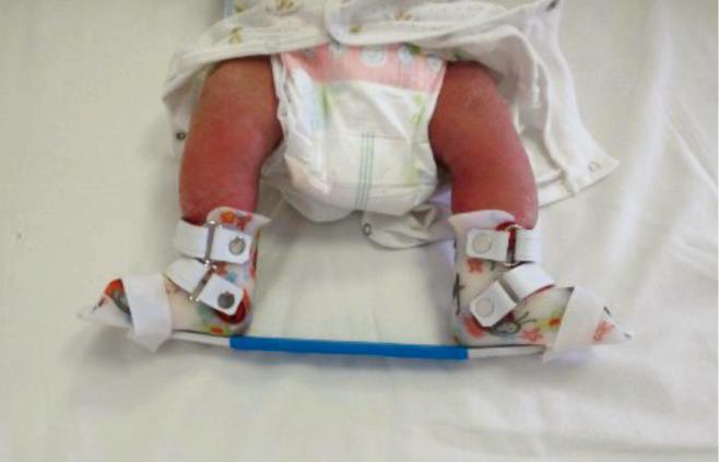
This clinical photograph displays a foot abduction orthosis (FAO), also known as a Denis Browne bar, applied to an infant following the correction phase of the Ponseti method for congenital talipes equinovarus (clubfoot). The device consists of two open-toed, high-top shoes with patterned rigid shells, secured around the infant's feet and ankles using multiple white leather straps with buckle closures. The shoes are mounted onto a horizontal blue abduction bar, which maintains the feet in a fixed position of external rotation and abduction. Asymmetrical positioning is visible, consistent with clinical protocols where the affected foot is abducted to 60–70 degrees while a non-affected or 'normal' side is held at a lower angle of 30–40 degrees. This orthotic intervention is critical in pediatric orthopedics to prevent relapse of the clubfoot deformity by stretching the medial soft tissues and maintaining the corrected alignment of the tarsal bones. The photograph illustrates a common post-corrective maintenance phase in clubfoot management.
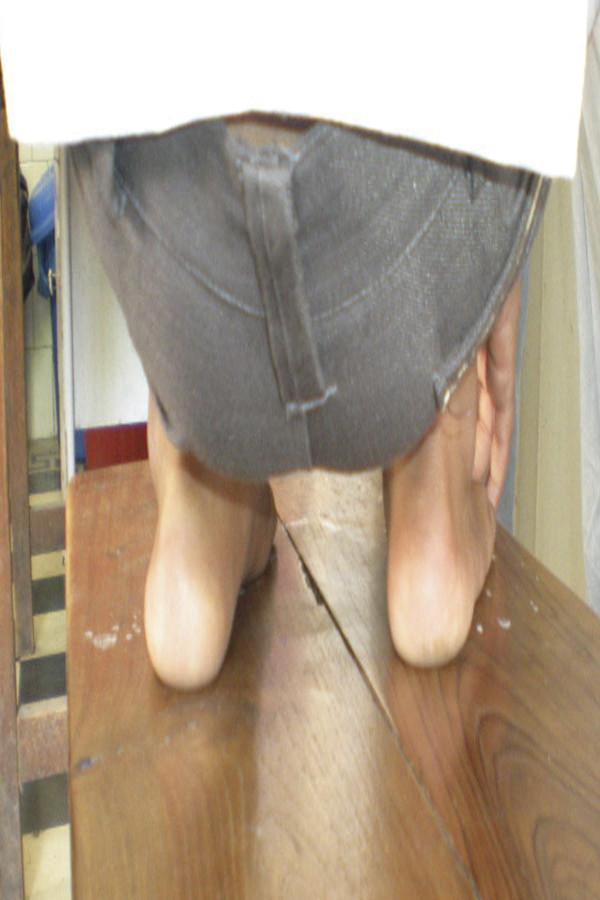
This clinical photograph provides a posterior view of a pediatric patient's lower extremities, specifically focusing on the feet and ankles. The image documents the morphological outcome of bilateral clubfoot (talipes equinovarus) at a five-year follow-up following treatment, potentially via the Ponseti method. Visually, both heels exhibit a mild residual varus deformity, characterized by an inward tilting of the calcaneus relative to the tibia. There is evidence of forefoot adduction, where the distal portion of the feet deviates toward the midline. The patient is shown in a weight-bearing or functional stance on a wooden surface, which helps assess the alignment of the hindfoot and midfoot. This image is clinically relevant for orthopedists and physical therapists evaluating long-term corrective outcomes, assessing for recurrence of equinus, varus, or adductus components of clubfoot, and determining the necessity for further orthotic intervention or maintenance such as the Denis-Browne splint.
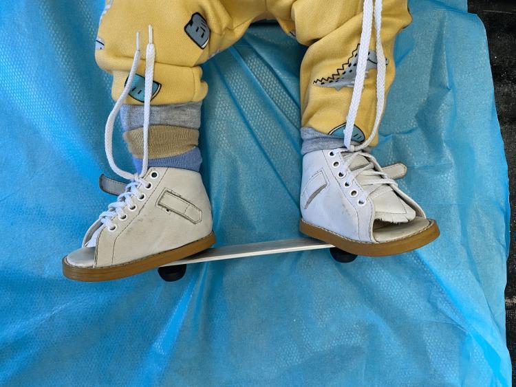
This clinical photograph shows a pediatric patient wearing a standard foot abduction brace, commonly known as a Denis Browne bar. The device is used in the orthopedic management of Congenital Talipes Equinovarus (CTEV), typically following the serial casting phase of the Ponseti method. The brace consists of two white, high-top, open-toe leather shoes with brown rubber soles and white laces, attached to a rigid, adjustable metallic horizontal bar (likely aluminum). The shoes are positioned on the bar to maintain the feet in a specific degree of abduction and external rotation to prevent relapse of the clubfoot deformity. The patient is shown from the waist down, wearing a yellow patterned sweatshirt and multiple layers of socks, positioned on a light blue clinical drape. This visual serves as a medical education example of postoperative or post-corrective maintenance therapy in pediatric orthopedics, highlighting the mechanical components required for maintaining corrected foot alignment.
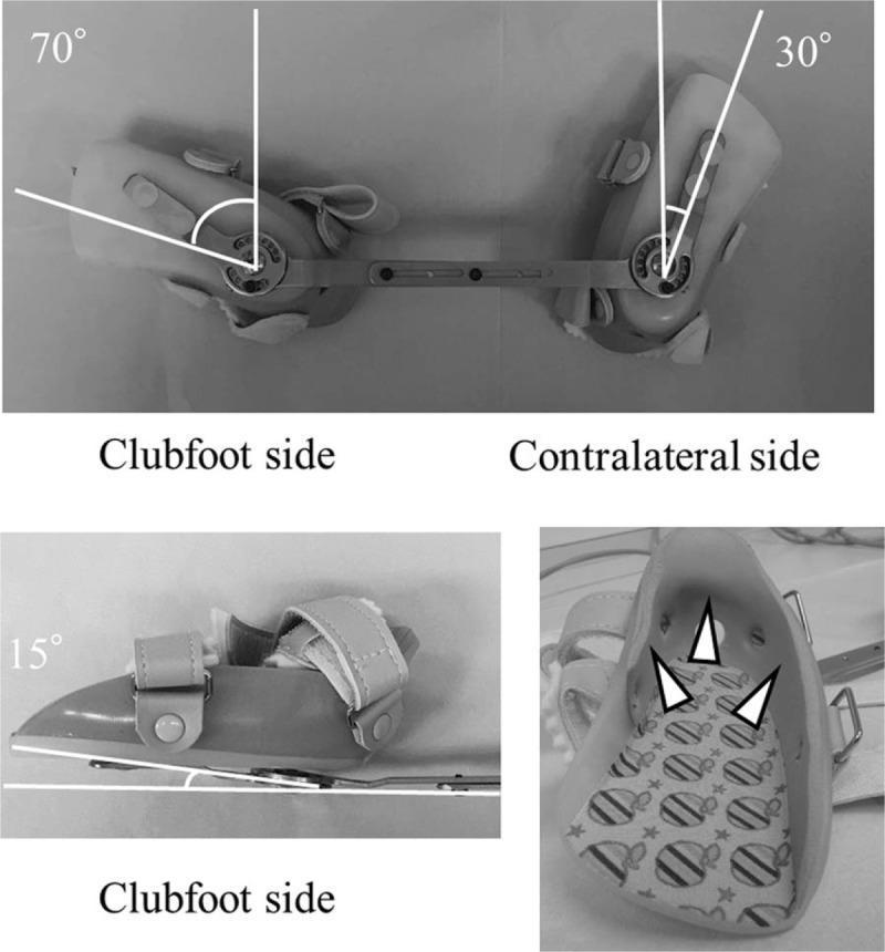
This composite clinical photograph illustrates a Foot Abduction Brace (FAB) used in the Ponseti method for treating idiopathic clubfoot. The top panel shows a superior view of the brace, consisting of two molded shoe inserts connected by a Denis Browne bar. The affected 'clubfoot' side is set at 70 degrees of external rotation, while the unaffected 'contralateral' side is set at 30 degrees. The bottom-left panel provides a lateral view of the clubfoot insert, highlighting a 15-degree dorsiflexion angle. The bottom-right panel displays the interior of a shoe insert, where arrowheads indicate molded thermoplastic ethylene-vinyl acetate (EVA) integrated into the heel section to prevent hindfoot slippage. The inserts feature adjustable straps with buckles for secure fixation and a padded, patterned interior for patient comfort. This medical device is designed to maintain the corrected position of the foot following serial casting or tenotomy, preventing relapse by maintaining specific abduction and dorsiflexion angles during growth.
Denis Browne Splint - Clinical Images

- Superior view of both shoes on the bar
- Affected (clubfoot) side: set at 70° external rotation
- Unaffected (normal) side: set at 30° external rotation
- Lateral view: shows 15° dorsiflexion angle
- Interior of shoe insert showing thermoplastic EVA heel support
Key Components of Denis Browne Splint
| Component | Description |
|---|---|
| Shoes/boots | High-top, open-toe, rigid shell |
| Connecting bar | Horizontal rigid bar (metal or plastic) |
| Affected foot angle | 60-70° external rotation |
| Normal foot angle | 30-40° external rotation |
| Dorsiflexion | ~15° built into the shoe |
| Purpose | Maintains correction after Ponseti casting for CTEV (clubfoot) |
To draw
Medical diagram of Denis Browne Splint (Denis Browne Bar) for clubfoot treatment. Clean labeled medical illustration showing: two high-top open-toe orthopedic shoes/boots connected by a horizontal rigid metal bar. Labels pointing to: "Open-toe high-top boot", "Horizontal connecting bar", "Affected foot: 60-70° external rotation", "Normal foot: 30-40° external rotation", "15° dorsiflexion angle". Show a front/superior view of both feet in the boots mounted on the bar, with angle indicators showing the abduction positions. Include a small side-view inset showing the dorsiflexion angle. White background, clear anatomical medical illustration style with blue and grey tones.
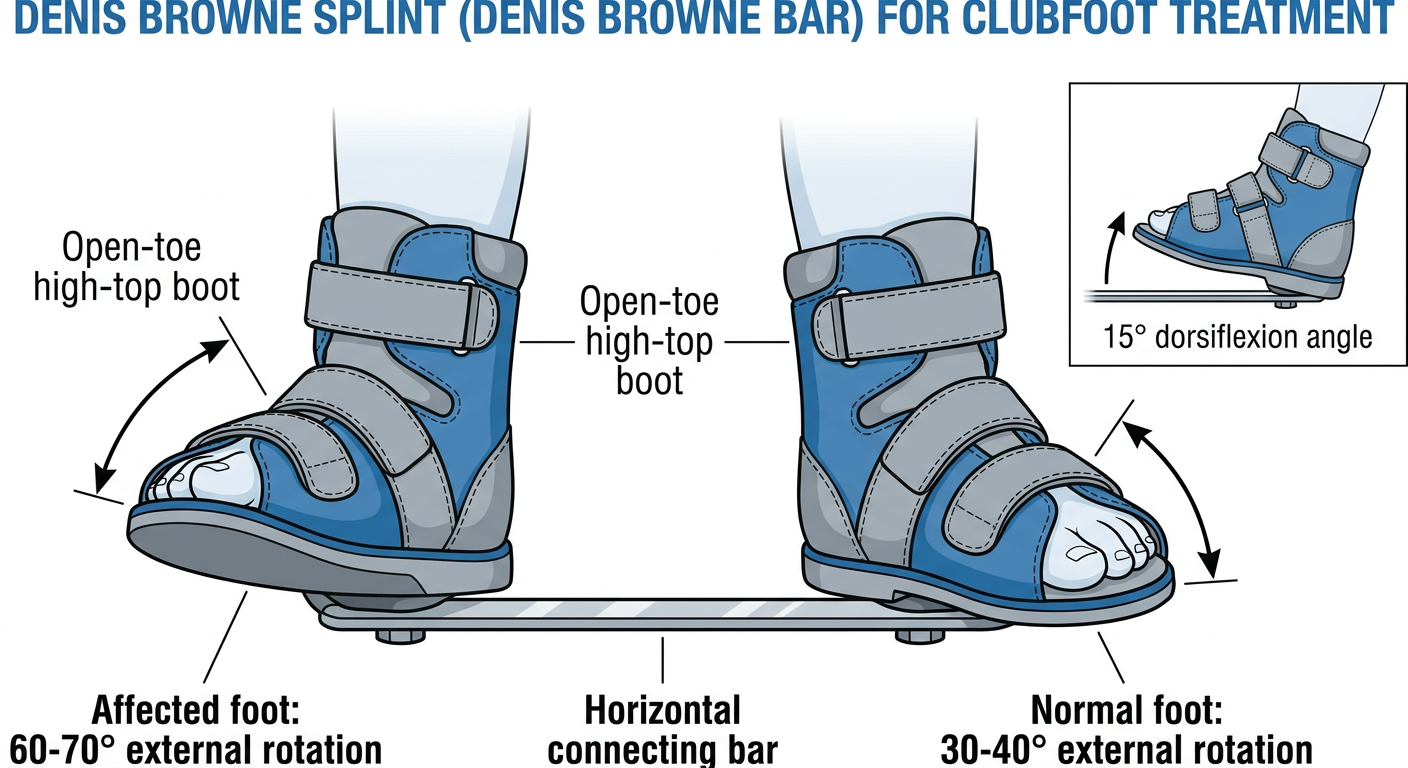
- Two open-toe high-top boots connected by a rigid horizontal bar
- Affected (clubfoot) side - 60-70° external rotation
- Normal side - 30-40° external rotation
- 15° dorsiflexion angle built into the boot
- The bar keeps both feet in abduction to maintain correction after Ponseti casting for CTEV (clubfoot)
Draw adigaram of night splint
Which type of night splint would you like?
Just night splint
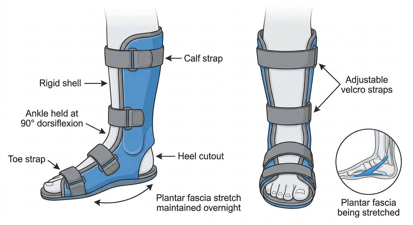
Medical diagram of a plantar fasciitis night splint (ankle foot orthosis). Clean labeled medical illustration showing: a rigid or semi-rigid boot-style splint worn on the foot and lower leg, holding the ankle at 90 degrees (neutral dorsiflexion). Show lateral/side view and front view. Labels pointing to: "Rigid shell", "Ankle held at 90° dorsiflexion", "Toe strap", "Calf strap", "Heel cutout", "Plantar fascia stretch maintained overnight", "Adjustable velcro straps". Include a small anatomical inset showing the plantar fascia being stretched. White background, clear medical illustration style with blue and grey tones, professional orthopedic diagram.
| Feature | Detail |
|---|---|
| Type | Ankle-Foot Orthosis (AFO) |
| Ankle position | Held at 90° dorsiflexion |
| Purpose | Keeps plantar fascia and Achilles tendon gently stretched overnight |
| Straps | Adjustable velcro straps at calf, ankle, and toes |
| Shell | Rigid or semi-rigid posterior shell |
| Worn | During sleep (6-8 hours) |