
Visual pathway
Reading File
Now let me search the medical library for visual pathway content in parallel.
Finding Sources
Finding Sources
Excellent sources found. Let me read the most relevant ones in parallel.
Reading File
Reading File
Reading File
Great content gathered. Now let me fetch the key images and also get the Kandel neuroscience content on the geniculostriate pathway.
Loading Image
Loading Image
Loading Image
Reading File
Now let me get the visual field defects diagram and the LGN/optic radiation content.
Loading Image
Reading File
Now let me fetch the visual field defects diagram and the LGN layers content.
Loading Image
Finding Sources
I now have comprehensive material from multiple authoritative textbooks and excellent diagrams. Let me compile the full answer.
The Visual Pathway
The visual pathway is the series of neural structures that carry visual information from the retina to the primary visual cortex. It is organized to ensure that all information from one side of the visual world (one hemifield) is processed by the contralateral occipital lobe.
1. Overview of the Pathway
The complete route, in order:
Retina → Optic nerve → Optic chiasm → Optic tract → Lateral geniculate nucleus (LGN) → Optic radiations → Primary visual cortex (V1)
A small branch at the level of the optic tract also projects to the pretectal area and superior colliculus for the pupillary light reflex and eye movement control.
2. Retina and Optic Nerve
- Photoreceptors (rods and cones) transduce light into signals.
- These signals are processed through bipolar cells to retinal ganglion cells (RGCs), whose axons converge at the optic disc (blind spot - no photoreceptors here).
- Axons leave the eye as the optic nerve, which is considered part of the CNS (myelinated by oligodendrocytes, covered by cranial meninges).
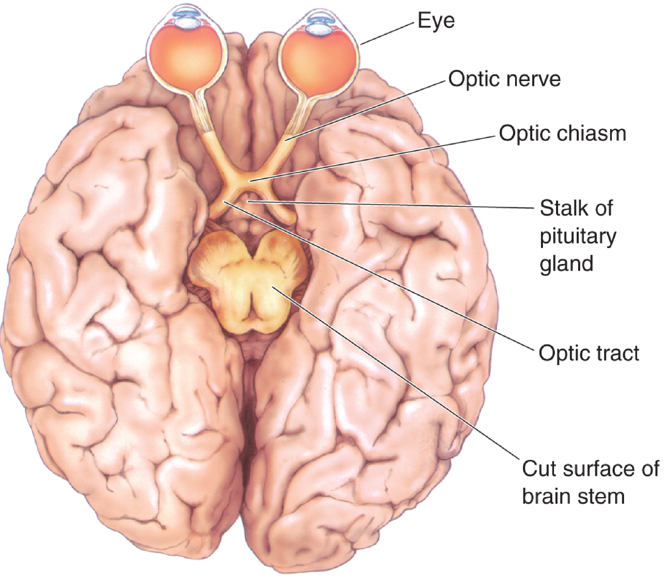
From Neuroscience: Exploring the Brain, 5th ed. - The retinofugal projection viewed from the base of the brain.
3. Optic Chiasm - The Key Crossing Point
The optic chiasm sits at the base of the brain, just anterior to the pituitary stalk (infundibulum). Here occurs a partial decussation:
| Fiber origin | At the chiasm | Destination |
|---|---|---|
| Nasal retina (sees temporal visual field) | Crosses to opposite side | Contralateral optic tract |
| Temporal retina (sees nasal visual field) | Stays ipsilateral | Ipsilateral optic tract |
Result: Each optic tract carries information from the contralateral visual hemifield of both eyes. The left optic tract carries information about the right visual hemifield (from the right nasal retina + left temporal retina), and vice versa.
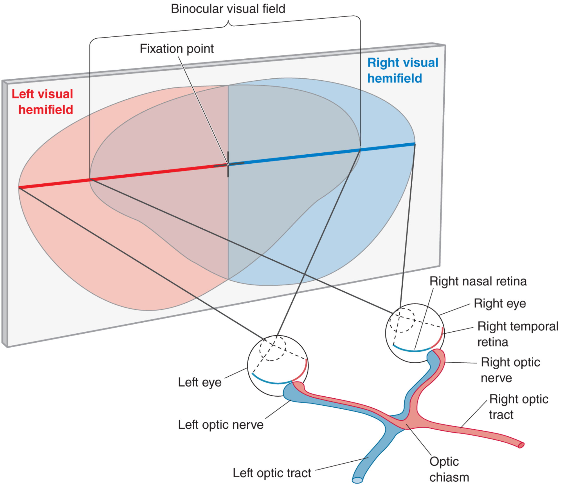
From Neuroscience: Exploring the Brain, 5th ed. - The right optic tract receives input from the right temporal retina (blue) and the left nasal retina (pink), representing the left visual hemifield.
4. Optic Tract
The optic tracts course posteriorly around the midbrain (lateral surfaces of the diencephalon) to reach the lateral geniculate nucleus (LGN) of the thalamus.
A small proportion of fibers leave the optic tract here to synapse in:
- Pretectal nucleus - pupillary light reflex (afferent limb of PLR)
- Superior colliculus - orienting eye and head movements
5. Lateral Geniculate Nucleus (LGN)
The LGN is the thalamic relay station for vision. It has 6 layers with distinct functional roles:
| Layers | Cell type | Input | Function |
|---|---|---|---|
| 1, 2 (ventral) | Magnocellular (M) | M-type RGCs | Motion, depth, low spatial frequency |
| 3, 4, 5, 6 (dorsal) | Parvocellular (P) | P-type RGCs | Color, fine detail, high spatial frequency |
Layers 1, 4, 6 receive input from the contralateral eye; layers 2, 3, 5 from the ipsilateral eye. This strict eye-specific layering is maintained here but merges in V1.
The visual field representation flows through the LGN retinotopically (preserving spatial map):
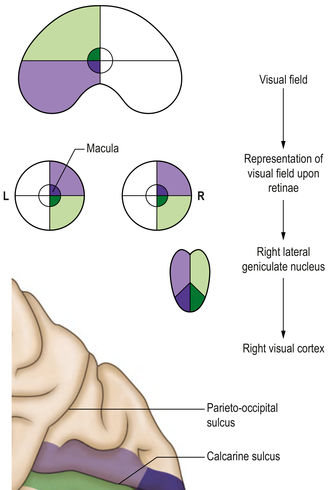
From Gray's Anatomy for Students - visual field representation at retinae, LGN, and visual cortex.
6. Optic Radiations (Geniculocalcarine Tract)
Axons from the LGN form the optic radiations, traveling to V1. They split into two divisions:
Upper division (parietal pathway)
- Carries lower visual field information
- Runs through the parietal lobe (superior pathway)
- Terminates on the lower bank of the calcarine sulcus
Lower division - Meyer's loop
- Carries upper visual field information
- Sweeps anteriorly over the temporal horn of the lateral ventricle in a wide arc (Meyer's or Archambault loop)
- Then runs posteriorly to the upper bank of the calcarine sulcus
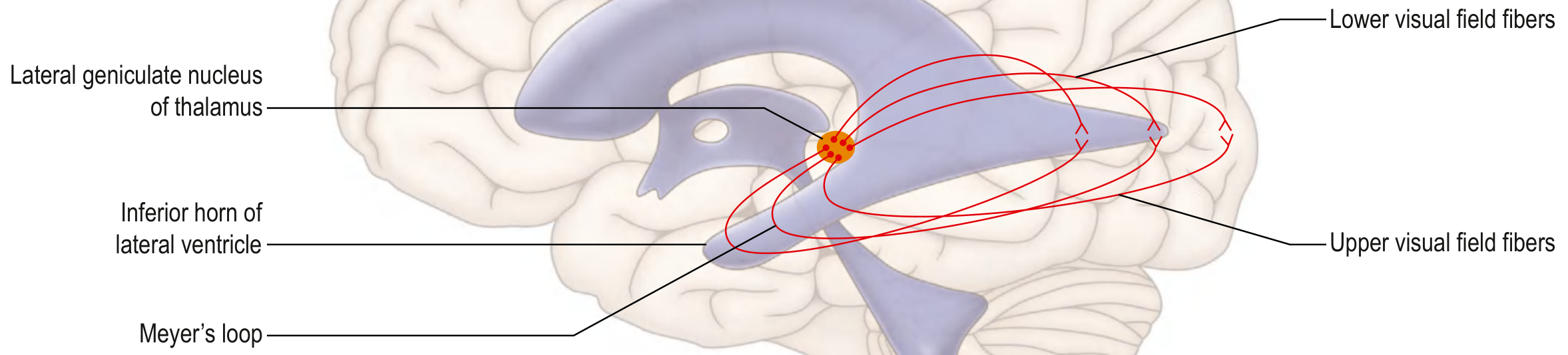
From Gray's Anatomy for Students - thalamocortical fibers projecting to primary visual cortex showing Meyer's loop, lower and upper visual field fibers.
7. Primary Visual Cortex (V1 / Striate Cortex / Brodmann Area 17)
- Located in the occipital lobe, on both banks of the calcarine sulcus
- Upper bank processes the lower visual field; lower bank processes the upper visual field
- Foveal (central) vision has a disproportionately large representation (cortical magnification)
- The fovea is represented most posteriorly; peripheral vision more anteriorly
8. Higher Visual Processing - Dorsal and Ventral Streams
From V1, visual information diverges into two major processing streams:
| Stream | Route | Function |
|---|---|---|
| Ventral ("What") stream | V1 → V2 → V4 → Inferior temporal cortex (IT) | Object recognition, color, face identification |
| Dorsal ("Where/How") stream | V1 → V2 → MT/V5 → Parietal cortex | Spatial location, motion, visually guided movement |
9. Lesions and Visual Field Defects
This is clinically essential - the level of the lesion determines the pattern of visual loss:
| Site of lesion | Visual field defect |
|---|---|
| Optic nerve (before chiasm) | Monocular blindness (same eye) |
| Optic chiasm (crossing fibers) | Bitemporal hemianopia (heteronymous) |
| Optic tract (behind chiasm) | Contralateral homonymous hemianopia (incongruous) |
| Meyer's loop (temporal lobe) | Contralateral superior quadrantanopia ("pie in the sky") |
| Parietal optic radiations | Contralateral inferior quadrantanopia |
| Occipital cortex (V1) | Contralateral homonymous hemianopia (congruous) with macular sparing |
Key rules:
- Retrochiasmal lesions (behind the chiasm) always cause homonymous defects (same side in both eyes)
- More posterior lesions produce more congruous defects (same shape in both eyes)
- More anterior (optic tract) lesions produce incongruous defects
- Occipital lesions often spare the macula because the foveal cortical representation is large and has dual vascular supply
10. Pupillary Light Reflex - The Side Branch
This is not strictly the visual (perception) pathway but uses the first part of it:
- Afferent limb: retina → optic nerve → optic chiasm → optic tract → pretectal nucleus (midbrain)
- Efferent limb: pretectal nucleus → bilateral Edinger-Westphal nuclei → CN III → ciliary ganglion → pupilloconstrictor muscle
This explains why a unilateral optic nerve lesion causes a relative afferent pupillary defect (RAPD/Marcus Gunn pupil) but a lesion behind the chiasm does not, since both eyes project to both pretectal nuclei via crossing fibers.
Sources:
- Gray's Anatomy for Students, Central Visual Pathway, p. 1346-1347
- Neuroscience: Exploring the Brain, 5th ed., The Optic Nerve, Optic Chiasm, and Optic Tract, p. 919-921
- Adams and Victor's Principles of Neurology, 12th ed., Lesions of the Chiasm, Optic Tract, and Geniculocalcarine Pathway, p. 270
- Kandel's Principles of Neural Science, 6th ed., Visual Processing - Geniculostriate Pathway, p. 547-550
This is a shared conversation. Sign in to Orris to start your own chat.