
Conjuctiva
Reading File
The query "Conjunctiva" is a broad anatomical/clinical topic. Let me search the library for comprehensive coverage.
Searching sources...
Finding Sources
Finding Sources
The best sources are Kanski's Clinical Ophthalmology and Junqueira's Histology. Let me read from all the key sources in parallel.
Reading File
Reading File
Reading File
Excellent content from Kanski's. Let me now read the full anatomy section and the Kanski histology/clinical sections, plus fetch relevant images.
Reading File
Reading File
Loading Image
Now let me fetch the Kanski histology image and read more about conjunctival reactions and disorders.
Loading Image
Reading File
I now have very comprehensive material. Let me compile the full, detailed answer on the conjunctiva.
The Conjunctiva
A complete overview covering anatomy, histology, immunology, clinical features of disease, and major disorders.
Anatomy
The conjunctiva is a transparent mucous membrane that lines the inner surface of the eyelids and covers the anterior surface of the globe, terminating at the corneoscleral limbus. It is richly vascular, supplied by the anterior ciliary and palpebral arteries. There is a dense lymphatic network draining to the preauricular and submandibular nodes.
Anatomically, it is divided into three parts:
| Region | Description |
|---|---|
| Palpebral conjunctiva | Starts at the mucocutaneous junction of the lid margin; firmly attached to the posterior tarsal plates; tarsal blood vessels run vertically |
| Fornical conjunctiva | Loose and redundant; provides mobility |
| Bulbar conjunctiva | Covers the anterior sclera; continuous with corneal epithelium at the limbus; loosely attached to the underlying Tenon capsule (fuses at the limbus) |
Special structures at the medial canthus:
- Plica semilunaris - the semilunar fold, present nasally
- Caruncle - a fleshy nodule medial to the plica, composed of modified cutaneous tissue
Palisades of Vogt - radial ridges at the limbus, the likely reservoir of corneal epithelial stem cells.
- Kanski's Clinical Ophthalmology, p. 184
Histology

(Fig. 6.1 - Histology of the conjunctiva, Kanski's Clinical Ophthalmology)
The conjunctival epithelium is:
- Non-keratinizing, approximately 5 cell layers deep
- Basal cuboidal cells mature into flattened polyhedral cells that are shed from the surface
- Goblet cells (mucus-secreting) are interspersed throughout the epithelium; they are most dense inferonasally and in the fornices
The stroma (substantia propria) consists of richly vascularized loose connective tissue. Embedded deep within it are the accessory lacrimal glands:
- Glands of Krause - in the upper and lower fornices
- Glands of Wolfring - near the upper tarsal border
Secretions from these accessory glands form essential components of the aqueous layer of the tear film.
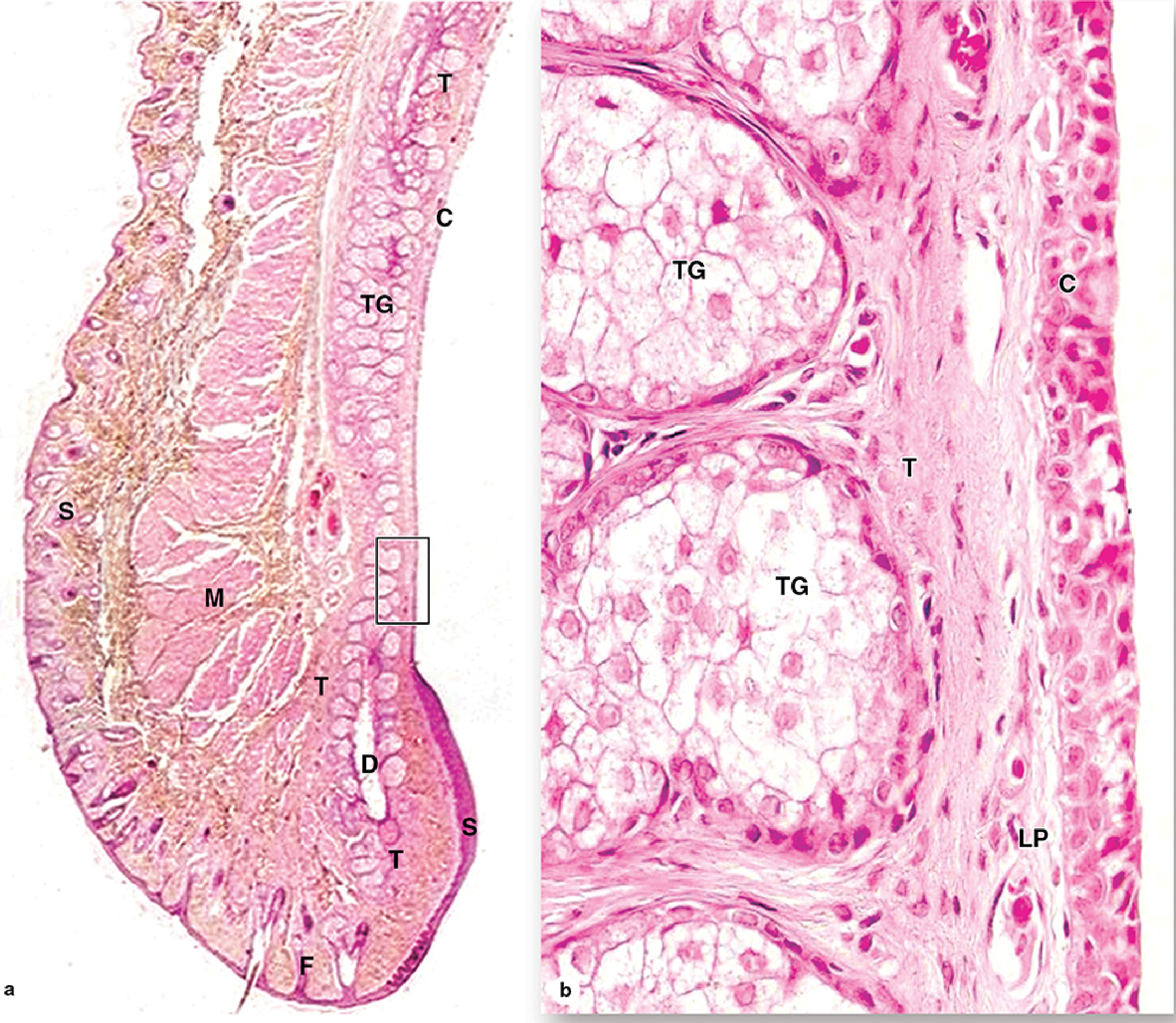
(Junqueira's Basic Histology - Eyelid section showing conjunctiva at low and high magnification)
- Kanski's Clinical Ophthalmology, p. 184; Junqueira's Basic Histology, p. 1214-1215
Immunology - Conjunctiva-Associated Lymphoid Tissue (CALT)
CALT plays a key role in the initiation and regulation of ocular surface immune responses. It consists of:
- Lymphocytes within the epithelial layers
- Lymphatics and associated blood vessels
- A stromal component of lymphocytes and plasma cells, including follicular aggregates
CALT mediates both passive and active (adaptive) immunity at the ocular surface.
- Kanski's Clinical Ophthalmology, p. 184
Clinical Features of Conjunctival Inflammation
Symptoms
- Non-specific: lacrimation, grittiness, stinging, burning
- Itching is the hallmark of allergic disease
- Visual acuity is usually unaffected
- Significant pain or photophobia suggests corneal involvement
Discharge - Diagnostic Significance
| Type | Cause |
|---|---|
| Watery (serous) | Acute viral or acute allergic conjunctivitis |
| Mucoid | Chronic allergic conjunctivitis, dry eye |
| Mucopurulent | Chlamydial or acute bacterial infection |
| Moderately purulent | Acute bacterial conjunctivitis |
| Severely purulent | Gonococcal infection |
Conjunctival Reactions
-
Hyperaemia: Diffuse, beefy-red, more intense away from the limbus = bacterial. Distinguish conjunctival injection from ciliary injection (iridocyclitis - deeper ring at limbus)
-
Haemorrhages: Petechial (multiple, small) in viral; larger and diffuse in severe bacterial
-
Chemosis: Translucent conjunctival oedema; acute = hypersensitivity; chronic = thyroid eye disease, venous/lymphatic obstruction, hypoalbuminaemia
-
Papillae: Vascular tufts in the stroma with a central feeding vessel; seen in allergic and chronic conjunctivitis
-
Follicles: Lymphoid aggregates without a central vessel; seen in viral conjunctivitis and chlamydial disease
-
Membranes:
- Pseudomembranes - coagulated exudate adherent to epithelium; peels away leaving epithelium intact
- True membranes - involve superficial epithelial layers; attempted removal causes tearing
- Causes: severe adenoviral, gonococcal, Streptococcus, C. diphtheriae, ligneous conjunctivitis, Stevens-Johnson syndrome
-
Kanski's Clinical Ophthalmology, p. 185-186
Major Conjunctival Disorders (Overview from Kanski's)
Infective Conjunctivitis
| Type | Key Features |
|---|---|
| Bacterial (acute) | Purulent discharge, diffuse hyperaemia |
| Viral (adenoviral) | Watery discharge, follicles, petechial haemorrhages, preauricular lymphadenopathy |
| Chlamydial | Mucopurulent, chronic follicular; trachoma leads to scarring and blindness |
| Gonococcal | Hyperacute, profuse purulent discharge, risk of corneal perforation |
Allergic Conjunctivitis
- Vernal keratoconjunctivitis (VKC): Young males, cobblestone papillae on upper tarsal plate, limbal involvement, shield ulcers
- Atopic keratoconjunctivitis (AKC): Adults with atopic eczema, lower lid involvement, forniceal scarring
- Seasonal/perennial allergic conjunctivitis: IgE-mediated, watery itchy discharge
Cicatrising (Scarring) Conjunctival Disorders
- Mucous membrane pemphigoid (ocular cicatricial pemphigoid): Subepithelial fibrosis, symblepharon (fusion of bulbar and palpebral conjunctiva), dry eye due to goblet cell destruction, forniceal shortening, ankyloblepharon
- Stevens-Johnson syndrome / toxic epidermal necrolysis: Acute mucosal blistering, severe scarring
- Trachoma: Leading infectious cause of blindness globally
Degenerations
- Pinguecula: Yellowish deposits on the bulbar conjunctiva nasally/temporally; elastotic degeneration of stroma
- Pterygium: Wing-shaped fibrovascular growth from the bulbar conjunctiva onto the cornea, usually nasally; associated with UV exposure
- Concretions: Calcium deposits in the palpebral conjunctiva; may erode and cause foreign body sensation
- Conjunctivochalasis: Redundant bulbar conjunctiva, especially inferiorly; may cause symptoms of dry eye
Superior Limbic Keratoconjunctivitis (SLK)
- Associated with thyroid disease in ~50% of cases
- Hyperaemia of the superior bulbar conjunctiva, limbal papillary hypertrophy, superior tarsal papillae
- Treated with lubricants, acetylcysteine, cyclosporin
Subconjunctival Haemorrhage
- Bright red, well-demarcated haemorrhage; usually spontaneous or from Valsalva manoeuvre
- Rule out hypertension, bleeding diathesis, trauma
Conjunctivitis in Medical Practice (Junqueira note)
- Conjunctivitis ("pink eye"): Inflammation causing redness, mucus discharge; bacterial and viral forms are contagious but rarely affect vision permanently
The Tear Film and Goblet Cells
Goblet cells in the conjunctival epithelium secrete mucins that are critical for the innermost mucus layer of the tear film. Destruction of goblet cells (e.g., in cicatricial disease, chemical burns, Stevens-Johnson syndrome) leads to:
- Mucin deficiency
- Instability of the tear film
- Severe dry eye despite adequate aqueous secretion
Summary Table
| Feature | Details |
|---|---|
| Type of membrane | Transparent mucous membrane |
| Epithelium | Non-keratinizing stratified columnar, ~5 layers |
| Key cells | Goblet cells (most in infernasal fornix) |
| Stroma | Vascular loose connective tissue (substantia propria) |
| Accessory glands | Glands of Krause (fornix) and Wolfring (tarsal border) |
| Immune tissue | CALT (lymphocytes, follicles, plasma cells) |
| Blood supply | Anterior ciliary + palpebral arteries |
| Lymph drainage | Preauricular + submandibular nodes |
| Key landmarks | Plica semilunaris, caruncle, palisades of Vogt |
Sources: Kanski's Clinical Ophthalmology: A Systematic Approach, 10th ed. | Junqueira's Basic Histology, 17th ed.
Conjuctiva
I already covered the Conjunctiva in detail in my previous response just above. Here's a quick recap of what was covered:
Conjunctiva - Summary
Anatomy
- Transparent mucous membrane lining inner eyelids + anterior globe up to the limbus
- Three parts: Palpebral (firmly attached to tarsus) → Fornical (loose, redundant) → Bulbar (over sclera, fuses at limbus)
- Special structures: Plica semilunaris, Caruncle, Palisades of Vogt (corneal stem cell reservoir)
Histology
- Non-keratinizing stratified columnar epithelium, ~5 layers deep
- Goblet cells (mucus-secreting) densest inferonasally and in the fornices
- Stroma = substantia propria (loose vascular connective tissue)
- Accessory lacrimal glands: Krause (fornices) and Wolfring (near upper tarsal border)
Immunology
- CALT (Conjunctiva-Associated Lymphoid Tissue) - lymphocytes, plasma cells, follicular aggregates
Clinical Reactions
- Hyperaemia, chemosis, follicles, papillae, membranes/pseudomembranes, scarring
Key Disorders
- Infective: bacterial, viral, chlamydial, gonococcal
- Allergic: VKC, AKC, seasonal
- Cicatricial: OCP (mucous membrane pemphigoid), Stevens-Johnson syndrome
- Degenerations: Pinguecula, Pterygium, Concretions, Conjunctivochalasis
Is there a specific aspect of the conjunctiva you'd like me to go deeper on - such as a particular disease, its histopathology, management, or something else? Just let me know!
This is a shared conversation. Sign in to Orris to start your own chat.