
Give me a surgical histroy taking of a man having hernia .
inguinal hernia anatomy diagram direct indirect
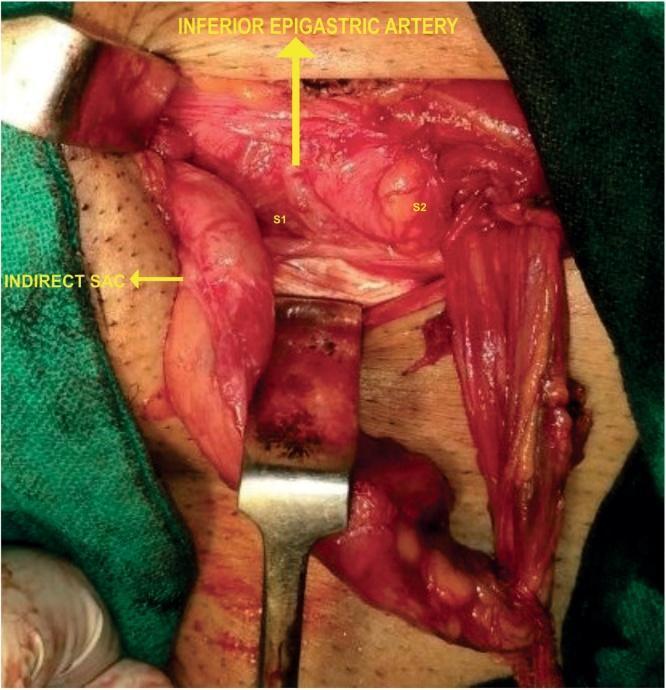
This clinical photograph captures an intraoperative view of an inguinal hernia repair, illustrating complex regional anatomy and multiple hernia sacs. The surgical field displays a rare anatomical variation where the inferior epigastric artery (IEA) follows a superficial course along the posterior wall of the inguinal canal. Two distinct direct inguinal hernia sacs are visible: S1, located lateral to the IEA, and S2, located medial to the IEA. Sac S2 appears larger with a wider base compared to the more elongated S1. Additionally, an indirect hernia sac is identified lateral to the direct components. Surgical retractors are in place to expose the posterior wall and cord structures. This image serves as a high-level educational resource for surgical trainees, demonstrating the critical importance of identifying the inferior epigastric vessels to differentiate between direct and indirect hernia components and to recognize vascular anomalies during Lichtenstein or other inguinal hernioplasty procedures.
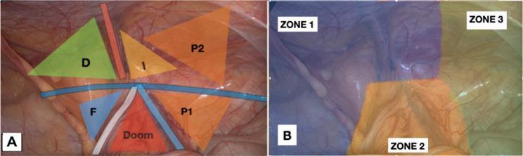
This composite intraoperative clinical photograph illustrates the surgical anatomy of the posterior inguinal region during a laparoscopic hernia repair. Image A displays essential anatomical landmarks and triangles: a green triangle (D) representing the direct hernia area; a yellow triangle (I) for the indirect hernia area; a blue triangle (F) for the femoral hernia area; and the red 'Triangle of Doom' containing the external iliac vessels. Two orange regions (P1 and P2) designate 'Triangles of Pain,' with P2 highlighting a revised superior boundary (2 cm above the iliopubic tract) to account for variations in nerve branches (lateral cutaneous nerve of the thigh and genitofemoral nerve). Image B divides the surgical field into three zones for procedural standardization: Zone 1 (medial/inferior) focusing on Cooper's ligament and epigastric vessels, Zone 2 (central) involving the spermatic cord and vas deferens parietalization, and Zone 3 (lateral) encompassing the pain triangles. The images demonstrate the preperitoneal view necessary for TAPP/TEP procedures, emphasizing safe zones for mesh fixation to avoid neurovascular injury.
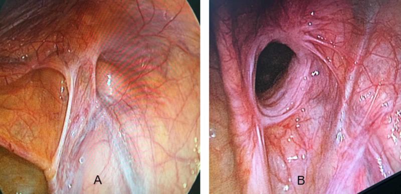
This diagnostic comparison uses laparoscopic (endoscopic) imaging to demonstrate the internal anatomical differences between an indirect and a direct inguinal hernia. Image A displays an indirect inguinal hernia, characterized by a rounded protrusion located lateral to the inferior epigastric vessels. The defect is clearly defined with a localized network of fine vasculature concentrated near the base of the herniated sac. Image B depicts a direct inguinal hernia, presenting as a more diffuse, wide-based bulge through the abdominal wall, located medial to the epigastric vessels in Hesselbach's triangle. The borders of the direct hernia are less distinct than the indirect type, and the overlying peritoneum shows a more prominent, dense, and tortuous vascular network. These images illustrate the surgical perspective during a Transabdominal Preperitoneal (TAPP) repair, emphasizing the importance of anatomical landmarks, specifically the inferior epigastric vessels, in differentiating hernia types based on their exit point relative to the deep inguinal ring.
Surgical History Taking: Adult Male with Hernia
1. PRESENTING COMPLAINT
- "Swelling / lump in the groin" (patients often call it a "rupture")
- Site: right groin / left groin / bilateral / scrotal
- Duration: how long the swelling has been noticed
2. HISTORY OF PRESENTING COMPLAINT
Swelling
- Onset: sudden or gradual?
- Progression: getting larger over time?
- Character: soft/hard, smooth/irregular
- Site: groin, scrotum, umbilical region, epigastric area, or previous scar
- Reducibility:
- Does it disappear when lying down? (spontaneously reducible)
- Can the patient push it back himself? (manually reducible)
- Is it always present and cannot be pushed back? (irreducible/incarcerated)
- Cough impulse: does the swelling enlarge or bulge when the patient coughs or strains? A positive cough impulse is characteristic of a hernia (- Yamada's Textbook of Gastroenterology, p. 763)
- Relationship to straining/exertion: does it appear or worsen with lifting, coughing, defecating, or prolonged standing?
Pain / Discomfort
- Location, character (aching, dragging, sharp)
- Timing: constant or only when swelling appears
- Aggravating factors: standing, straining, walking, exertion
- Relieving factors: lying down, manual reduction
- Severe, sudden pain in the swelling - red flag for strangulation (Yamada's, p. 759)
Associated Symptoms
- Nausea / vomiting: suggests intestinal obstruction from incarceration
- Constipation / absolute constipation: bowel in the sac
- Abdominal distension
- Fever: late sign of strangulation and peritonitis
- Urinary symptoms: if bladder is involved (sliding hernia - "en glissade")
- Testicular pain or swelling (may indicate compromised blood supply to testis)
3. HERNIA-SPECIFIC SYSTEMIC REVIEW
| System | Ask About |
|---|---|
| Respiratory | Chronic cough (COPD, asthma, smoking, TB) |
| Gastrointestinal | Constipation, straining at stool, colorectal malignancy |
| Urinary | Prostatism - urinary hesitancy, straining to void, poor stream, nocturia (BPH) |
| Abdominal | Ascites, abdominal distension |
| Nutritional | Rapid weight loss, malnutrition (tissue weakening) |
4. PAST SURGICAL HISTORY
- Previous hernia repair on either side (recurrent hernia - particularly relevant since laparoscopic repairs may make it hard to remember the correct side; a CT may be needed per Sabiston, p. 747)
- Previous abdominal or pelvic surgery (risk for incisional hernia)
- Previous scrotal or inguinal surgery (orchidopexy, vasectomy)
5. PAST MEDICAL HISTORY
- Connective tissue disorders (Marfan's, Ehlers-Danlos) - weakened fascia
- COPD / chronic bronchitis / asthma
- Benign prostatic hypertrophy
- Constipation / irritable bowel disease
- Ascites (liver cirrhosis, malignancy)
- Diabetes mellitus (surgical risk, wound healing)
- Hypertension / cardiac disease (anaesthetic risk)
- Bleeding disorders or anticoagulant use
6. DRUG HISTORY
- Anticoagulants (warfarin, NOACs, aspirin, clopidogrel) - perioperative management
- Steroids (impair wound healing)
- Immunosuppressants
- Laxatives / straining medications
- Drugs causing chronic cough (e.g., ACE inhibitors)
- Allergies (especially latex, antibiotics, anaesthetic agents)
7. FAMILY HISTORY
- Family history of hernia (genetic predisposition: collagen defect, processus vaginalis anomaly)
- Family history of colorectal cancer (may cause straining/obstruction)
- Connective tissue diseases
8. SOCIAL HISTORY
- Occupation: heavy manual labour, prolonged standing (key risk factor for hernia formation and recurrence)
- Smoking: chronic cough, impaired wound healing, anaesthetic risk
- Alcohol use: risk of liver cirrhosis / ascites
- Recreational activities: heavy lifting, contact sports
- Exercise tolerance / functional status (fitness for surgery and anaesthesia)
- Social support: who will care for patient post-operatively?
9. REVIEW OF SYSTEMS (Pre-operative Assessment Focused)
- Cardiorespiratory fitness: chest pain, dyspnoea, exercise tolerance (MET equivalent)
- Known cardiac disease, previous MI, stents
- Any bleeding tendency, bruising
- Previous reactions to anaesthesia or family history of malignant hyperthermia
- Nil-by-mouth compliance, last meal time (if emergency)
10. DIFFERENTIATING FEATURES ON HISTORY (Inguinal vs Femoral vs Other)
| Feature | Inguinal Hernia | Femoral Hernia |
|---|---|---|
| Sex | Men (10x more common) | Women (relatively) |
| Location | Above & medial to pubic tubercle | Below & lateral to pubic tubercle |
| Reducibility | Usually reducible | More often irreducible |
| Risk of strangulation | Lower (but still occurs) | Higher (narrow neck) |
11. EMERGENCY FEATURES - RED FLAGS
- Irreducible, tense, tender swelling - incarceration
- Overlying skin changes (erythema, oedema) - strangulation
- Vomiting + absolute constipation - bowel obstruction
- Fever + tachycardia + peritonism - strangulated bowel with ischaemia/peritonitis
- Inability to reduce despite sedation - emergency surgery needed
SUMMARY BOX
- PC - swelling, site, duration
- HPC - reducibility, cough impulse, pain character, bowel/urinary symptoms
- Systemic - causes of raised IAP (cough, constipation, prostatism)
- PMHx - previous repairs, abdominal surgery, connective tissue disease
- DHx - anticoagulants, steroids, ACE inhibitors
- FHx - hernia, collagen disorders
- SHx - occupation, smoking, lifting, social support
- Red flags - irreducibility, tenderness, vomiting, fever
Give me in this order, g3neral information, cheif compaint, histroy of presnt illnes, past history , personal histroy, treatm3nt history , medical histroy, family history, investigation, general examination and summary
SURGICAL HISTORY TAKING
Adult Male Patient - Inguinal Hernia
1. GENERAL INFORMATION
| Field | Details |
|---|---|
| Name | Mr. _______________ |
| Age | e.g., 45 years |
| Sex | Male |
| Occupation | e.g., labourer / farmer / office worker |
| Address | _______________ |
| Date of Admission | _______________ |
| Informant | Patient himself / relative (reliability: good/fair/poor) |
| Religion / Marital Status | _______________ |
Note: Inguinal hernia occurs in ~27% of men over their lifetime and is 10 times more common in men than women. - Bailey and Love's Short Practice of Surgery, 28e
2. CHIEF COMPLAINT
Written in the patient's own words, with duration.
- Painless groin swelling (most common)
- Groin swelling with dragging discomfort
- Groin swelling that goes down when lying flat
- Sudden painful, irreducible groin swelling (emergency)
3. HISTORY OF PRESENT ILLNESS (HPI)
A. Swelling
- Onset: When did the patient first notice it? Was it sudden or gradual?
- Site: Right groin / left groin / bilateral / extending into the scrotum
- Size: Size at onset, and has it been increasing?
- Progression: Slowly enlarging over months/years, or static?
- Character: Soft and compressible, or firm and tense?
- Reducibility:
- Does it disappear on lying down? (spontaneously reducible)
- Can the patient push it back himself? (manually reducible)
- Is it always present, cannot be reduced? (irreducible/incarcerated)
- Cough impulse: Does the swelling enlarge when the patient coughs, strains, or stands? A positive cough impulse confirms a hernia. - Yamada's Textbook of Gastroenterology, 7e, p.763
- Relationship to activity: Appears or worsens with lifting, prolonged standing, defecation, or exercise; reduces with rest or recumbency
B. Pain / Discomfort
- Site and radiation
- Character: dull aching, dragging, sharp
- Timing: only when swelling appears, or constant
- Severity: mild background discomfort vs. severe acute pain
- Aggravating factors: straining, standing, walking
- Relieving factors: lying down, manually reducing the hernia
C. Associated Symptoms (MUST ASK)
| Symptom | Significance |
|---|---|
| Nausea / vomiting | Bowel in sac; incarceration / obstruction |
| Absolute constipation | Complete bowel obstruction |
| Abdominal distension | Intestinal obstruction |
| Fever / chills | Strangulation, peritonitis |
| Urinary hesitancy / frequency | Sliding hernia containing bladder |
| Testicular pain or swelling | Compromised blood supply to testis |
| Sudden severe pain in swelling | Strangulation - RED FLAG |
"Exquisite local tenderness and pain should raise concern for ischemia/strangulation. The risk of strangulation is inversely proportional to the size of the neck." - Yamada's Textbook of Gastroenterology, 7e, p.759
4. PAST HISTORY
- Previous hernia: Any prior hernia on either side? (recurrence is common - patients with one hernia have a 50% lifetime risk of developing one on the opposite side - Bailey and Love's, p.2024)
- Previous abdominal surgery: Appendicectomy, bowel surgery, prostatectomy, laparotomy (risk factor for incisional hernia)
- Previous scrotal surgery: Orchidopexy, varicocelectomy, vasectomy
- Previous hernia repair: Open (Lichtenstein) or laparoscopic? Which side? Any mesh used? Any complications?
- Hospitalizations: Any major illness requiring admission
- Trauma: Blunt abdominal trauma
5. PERSONAL HISTORY
- Diet: Vegetarian / non-vegetarian; adequate nutrition (malnutrition weakens abdominal wall)
- Bowel habits: Constipation (chronic straining is a key causative factor)
- Urinary habits: Difficulty initiating micturition, poor stream, frequency, nocturia (suggests BPH causing straining)
- Sleep: Normal / disturbed
- Appetite: Normal / decreased (if decreased - suspect malignancy causing obstruction)
- Weight loss: Significant weight loss weakens the abdominal wall fascia
- Physical activity: Sedentary vs. heavy manual labour
6. TREATMENT HISTORY
- Any previous treatment for the current hernia:
- Use of a truss or support belt (temporary measure - does not cure hernia)
- Any previous attempts at surgical repair (failed / recurrence)
- Current medications:
- Anticoagulants (warfarin, rivaroxaban, aspirin, clopidogrel) - must be managed perioperatively
- Steroids / immunosuppressants (impair wound healing)
- Laxatives (suggests chronic constipation - predisposing factor)
- ACE inhibitors (cause chronic cough - raises intra-abdominal pressure)
- Alpha-blockers for BPH
- Drug allergies: Medications, latex, antiseptics, anaesthetic agents - document clearly
7. MEDICAL HISTORY
| Condition | Relevance |
|---|---|
| COPD / Chronic bronchitis / Asthma | Chronic cough raises IAP |
| Benign Prostatic Hypertrophy (BPH) | Straining at micturition raises IAP |
| Chronic constipation | Straining at defecation raises IAP |
| Liver cirrhosis / Ascites | Raised IAP; also affects wound healing |
| Diabetes mellitus | Impaired wound healing; surgical risk |
| Hypertension / IHD | Anaesthetic and perioperative risk |
| Connective tissue disorders (Marfan's, Ehlers-Danlos) | Weakened fascia; hernia predisposition |
| Malignancy (colorectal, prostate) | Obstruction causing straining; also direct cause |
| Obesity | Raised IAP; poor wound healing |
| Previous MI / stroke | Perioperative risk; anticoagulation |
8. FAMILY HISTORY
- Family history of hernia: First-degree relatives (genetic predisposition via collagen defects)
- Connective tissue disorders in family (Marfan's syndrome, Ehlers-Danlos)
- Colorectal cancer: Family history may be relevant if patient has altered bowel habit causing straining
- Bleeding disorders in family
9. INVESTIGATIONS
Routine Pre-operative Investigations
| Investigation | Purpose |
|---|---|
| Complete Blood Count (CBC) | Anaemia, infection (raised WBC in strangulation) |
| Blood Group & Cross-match | Pre-operative |
| Serum Electrolytes (Na, K, Cl) | Derangement in vomiting / obstruction |
| Renal Function Tests (BUN, Creatinine) | Pre-operative baseline; renal disease |
| Liver Function Tests | If cirrhosis / ascites suspected |
| Blood Glucose (FBS / RBS) | Diabetes screen |
| Coagulation profile (PT, APTT, INR) | If on anticoagulants or liver disease |
| ECG | Cardiac disease; age >40 |
| Chest X-Ray | Cardiorespiratory assessment; chronic lung disease |
| Urine Routine & Microscopy | Baseline; exclude UTI |
Hernia-Specific Investigations
| Investigation | Indication |
|---|---|
| Ultrasound (USG) of groin | First-line imaging; confirms diagnosis in obese patients or when examination is equivocal; detects bowel / omentum in sac - Bailey and Love's, p.2042 |
| CT Abdomen & Pelvis | Best in acute/emergency setting; shows incarceration, strangulation, bowel obstruction; also evaluates opposite side - Yamada's, p.822 |
| MRI Groin | When sportsman's hernia or musculoskeletal cause is differential diagnosis - Yamada's, p.834 |
| Herniography (fluoroscopic) | When hernia clinically absent but symptoms strong; rarely needed |
| PSA / Digital Rectal Exam | If BPH/prostate malignancy suspected |
"Most cases require no diagnostic tests but ultrasonography, CT and MRI are occasionally used." - Bailey and Love's Short Practice of Surgery, 28e, p.2042
10. GENERAL EXAMINATION
Build and Nutrition
- Built: thin / average / obese
- Nourished: well / poorly nourished
- Obesity is both a risk factor and a complication risk for surgery
Vital Signs
| Parameter | Finding |
|---|---|
| Pulse | Rate, rhythm, volume, character (tachycardia in strangulation/sepsis) |
| Blood Pressure | Hypertension (common comorbidity); hypotension (septic shock in strangulation) |
| Temperature | Fever suggests strangulation / peritonitis |
| Respiratory Rate | Tachypnoea in bowel obstruction / sepsis |
| SpO2 | Baseline, especially in COPD patients |
Signs to Note
- Pallor: Anaemia
- Icterus (Jaundice): Liver disease / cirrhosis
- Cyanosis: Respiratory / cardiac disease
- Clubbing: Chronic lung/GI disease
- Lymphadenopathy: Generalised nodes - malignancy? (also a differential for groin swelling)
- Oedema: Pitting oedema of legs - cardiac / hypoproteinaemic state
- Dehydration: Skin turgor, dry mucous membranes (if vomiting / obstruction)
Abdominal Observation (from general inspection)
- Abdominal distension (intestinal obstruction)
- Visible peristalsis (obstruction)
- Visible groin swelling at rest or on coughing
SUMMARY
(Written as a brief clinical summary at the end of history-taking)
Genral infirmation in senstence
GENERAL INFORMATION
Inguinal hernia Give me in this order, g3neral information, cheif compaint, histroy of presnt illnes, past history , personal histroy, treatm3nt history , medical histroy, family history, investigation, general examination and summary