

Explain in easy to write, do not change scientific terms, give flowchart also
Lymphoid System - Simple Explanation
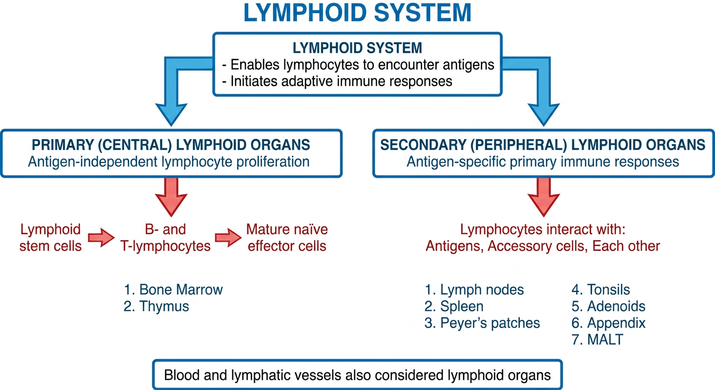
1. Primary (Central) Lymphoid Organs
- These are where B- and T-lymphocytes are born and mature from lymphoid stem cells
- The process happens without needing antigens - called antigen-independent lymphocyte proliferation
- The cells become mature naïve effector cells
- Organs: Bone marrow + Thymus
2. Secondary (Peripheral) Lymphoid Organs
- These give lymphocytes a place to meet antigens, talk to accessory cells, and interact with each other
- This is where antigen-specific primary immune responses begin
- Organs: Lymph nodes, Spleen, Peyer's patches, Tonsils, Adenoids, Appendix, MALT (Mucosa-Associated Lymphoid Tissue)
Blood and lymphatic vessels that carry lymphocytes can also be considered lymphoid organs.
A clean medical educational flowchart titled "Lymphoid System" showing the following hierarchy: Top box: "LYMPHOID SYSTEM - Enables lymphocytes to encounter antigens - Initiates adaptive immune responses" Split into TWO branches: LEFT BRANCH - Box: "PRIMARY (CENTRAL) LYMPHOID ORGANS" - subtitle: "Antigen-independent lymphocyte proliferation" - Process arrow: "Lymphoid stem cells → B- and T-lymphocytes → Mature naïve effector cells" - Organs listed: "1. Bone Marrow 2. Thymus" RIGHT BRANCH - Box: "SECONDARY (PERIPHERAL) LYMPHOID ORGANS" - subtitle: "Antigen-specific primary immune responses" - Process: "Lymphocytes interact with: Antigens, Accessory cells, Each other" - Organs listed: "1. Lymph nodes 2. Spleen 3. Peyer's patches 4. Tonsils 5. Adenoids 6. Appendix 7. MALT" Bottom note box: "Blood and lymphatic vessels also considered lymphoid organs" Use a white background, blue and red color scheme, clear arrows, medical textbook style, well-spaced boxes
Quick Summary Table
| Feature | Primary Lymphoid Organs | Secondary Lymphoid Organs |
|---|---|---|
| Also called | Central | Peripheral |
| Function | Lymphocyte maturation | Immune response initiation |
| Antigen needed? | NO | YES |
| Cell process | Stem cell → Naïve effector cell | Lymphocyte activation |
| Examples | Bone marrow, Thymus | Lymph nodes, Spleen, Tonsils, Peyer's patches, Adenoids, Appendix, MALT |
- Primary organs = where lymphocytes are MADE and TRAINED (no antigen needed)
- Secondary organs = where lymphocytes FIGHT (antigen is present here)
- The whole system connects via blood and lymphatic vessels which also transport lymphocytes

Flowchart
A clean, detailed medical educational flowchart titled "LYMPH NODES - Anatomical Structure" on white background with pink and purple color scheme, medical textbook style. TOP BOX (pink header): "LYMPH NODES" - subtitle: "Numerous, disseminated all over the body | Size: 1-25 mm diameter | Role: Initial/inductive states of immune response" SECOND BOX: "STRUCTURE: Circumscribed by a connective tissue capsule" with two inputs shown as arrows: "← Afferent lymphatics (draining peripheral interstitial spaces)" and "← Blood from systemic circulation (via hilar arteriole)" Then splits into TWO MAIN REGIONS side by side: LEFT REGION BOX (blue): "CORTEX" - Contains: Follicles / Nodules (roughly spherical, densely packed lymphocytes) - Sub-box: "FOLLICLES (T-independent area)" containing: B-lymphocytes (predominate), Macrophages, Follicular dendritic cells, Some T-lymphocytes RIGHT REGION BOX (green): "MEDULLA" - (deep region of lymph node) BELOW CORTEX, add connected box: "DEEP CORTEX (Paracortical area)" - Contains: T-lymphocytes (predominate) - Note: T-dependent area BOTTOM NOTE BOX: "Cortex + Deep cortex: Densely populated by lymphocytes in constant traffic between lymphatic and systemic circulation" "Key: T- and B-lymphocytes occupy DIFFERENT areas in the cortex" Use clear directional arrows, well-spaced boxes, labels bold, clean medical diagram style
429 RESOURCE_EXHAUSTED. {'error': {'code': 429, 'message': 'Resource exhausted. Please try again later. Please refer to https://cloud.google.com/vertex-ai/generative-ai/docs/error-code-429 for more details.', 'status': 'RESOURCE_EXHAUSTED'}}
We encountered an error while processing your request. Please try again or reach out to us for help.
Flowchart
A clean medical educational flowchart titled "LYMPH NODES - Anatomical Structure" on white background with pink and purple color scheme. TOP BOX (pink): "LYMPH NODES" - "Size: 1-25 mm | Found all over the body | Role: Initial and inductive states of immune response" SECOND BOX: "Circumscribed by a connective tissue capsule" - Two input arrows: "Afferent lymphatics (draining peripheral interstitial spaces)" and "Blood from systemic circulation via hilar arteriole" Splits into TWO MAIN REGIONS: LEFT BOX (blue): "CORTEX" - contains sub-box: "Follicles / Nodules (densely packed lymphocytes)" - inside follicles: B-lymphocytes predominate, Macrophages, Follicular dendritic cells, some T-lymphocytes - labeled: T-INDEPENDENT AREA CENTER-LEFT BOX (purple): "DEEP CORTEX (Paracortical area)" - T-lymphocytes predominate - labeled: T-DEPENDENT AREA RIGHT BOX (green): "MEDULLA" BOTTOM NOTE: "T- and B-lymphocytes occupy DIFFERENT areas in the cortex" and "Cortex and deep cortex are in constant lymphocyte traffic between lymphatic and systemic circulation" Clear arrows, bold labels, well-spaced, clean medical diagram
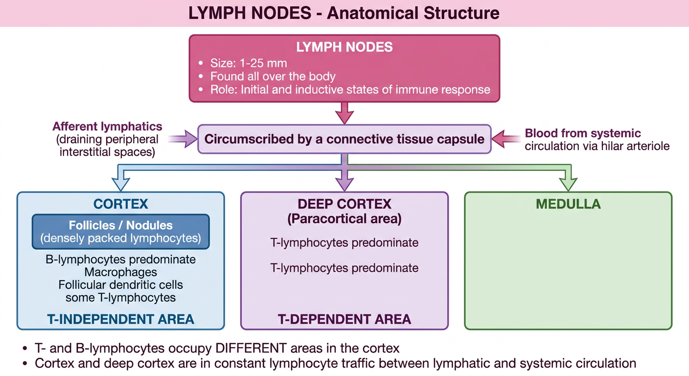
- Size: 1-25 mm, found all over the body
- Role: initial/inductive states of immune response
- Enclosed by a connective tissue capsule
- Receive: afferent lymphatics (from peripheral interstitial spaces) + blood (via hilar arteriole)
| Region | Also Called | Main Cells | Type |
|---|---|---|---|
| Cortex (Follicles/Nodules) | T-independent area | B-lymphocytes, Macrophages, Follicular dendritic cells, some T-lymphocytes | B-cell zone |
| Deep Cortex | Paracortical area | T-lymphocytes | T-dependent area |
| Medulla | - | Mixed | Efferent zone |